J. Exp. Biomed. Sci. 2010, 16(1): 10~18
Detection of Waterborne Pathogens by PCR-reverse Blot Hybridization
Yeonim Choi, Gyusang Lee, Hyeeun Bang, Jong-Bae Kim and Hyeyoung Lee
†Department of Biomedical Laboratory Science, College of Health Sciences, Yonsei University,
Wonju-si, Gangwon Province, 220-710, Republic of Korea
The present study was set to develop comprehensive system for assessing the safety of drinking water using PCR-reverse blot hybridization assay (REBA). The REBA developed in this study can detect waterborne pathogens such as Shigella spp., Salmonella spp., Citrobacter spp., Enterobacter spp., Klebsiella spp., Yersinia spp., Mycobacterium spp., Listeria spp. at the genus level, and Escherichia coli, Citrobacter freundii, Klebsiella pneumoniae, Pseudomonas aeruginosa, Yersinia enterocolitica, Y. pseudotuberculosis, Mycobacterium avium complex, M. marinum, Enterococcus faecalis, and Staphylococcus aureus at the species level, and E. coli O157:H7 at the strain level.
Key Words: Waterborne pathogen, Reverse blot hybridization assay, Nested PCR, Water contamination
INTRODUCTION
Currently, monitoring microbial parameters in water employs microbiological analysis which includes process of sampling and filtration of water followed by cultivation of the chosen microorganism using enrichment and selective media. The whole process usually takes 24 to 72 hours and still may not be able to pick up a number of microorganisms, if appropriate selective media and growth conditions are not used. For that reason, in routine monitoring process, not all of possible water microbial contaminants, but coliform bacteria including Escherichia coli that may represent a degree of water contamination have been routinely used for monitoring. However, there are many other species of bacteria that should not be present in the water as water contaminants. Therefore, there have been extensive efforts to develop a method more comprehensive for monitoring all the microbial water contaminants (Bej et al., 1990; Bej et al., 1991; Call et al., 2003).
Recently, molecular methods have been applied for the rapid detection of pathogens in food, soil, and water. These methods have shown high degrees of sensitivity and specificity and do not require the need for complicated cultivation and additional identification process (Bej et al., 1990; Bej et al., 1991; Wilson et al., 2001; Bej AK, 2003;
Call et al., 2003). Some of these methods allow detection of specific culturable and/or non-culturable bacteria within serial hours, instead of the days required with the traditional microbiological methods (Tsai et al., 1992; Josephson et al., 1993; Cooper et al., 1997; Mittelman et al., 1997; Jackson et al., 2001; Reynolds et al., 2001; Baudart et al., 2002;
Bayardelle et al., 2002; Field et al., 2003).
Most of molecular methods which have been developed so far have been mainly based on the molecular analysis of bacteria with respect to their 16S rRNA or its genes (Clarridge, 2004). These approaches take advantage of usefulness of 16S rRNA, which is a stable taxonomic marker for microorganisms, since genetic variations in 16S rRNA are intergenus and interspecies. Thus, the variation in 16S rRNA can be utilized for the designing of species- and genus-specific genetic markers (Amann et al., 1995; Te Giffel et al., 1997).
Recently, PCR primers and 16S rRNA-based oligo- nucleotide probes specific for some bacterial pathogens such as E. coli (Tsen et al., 1998), Listeria monocytogenes,
*Received: 11 February, 2010 / Revised: 10 March, 2010 Accepted: 15 March, 2010
†Corresponding author: Hyeyoung Lee, Ph. D. Department of Biomedical Laboratory Science College of Health Sciences, Yonsei University, Wonju-si, Gangwon Province, 220-710, Korea.
Tel: 82-33-760-2740, Fax: 82-33-760-2561 e-mail: [email protected]
Salmonella spp. (Gendel, 1996; Lin and Tsen, 1996; Tsen and Lin 1999), Staphylococcus spp. (Couzinet et al., 2005), and Bacillus species (Ash et al., 1991) have been reported to be successful in the PCR amplification and the DNA microarray. DNA microarray is especially powerful since it can simultaneously detect multiple targets by one assay (Warsen et al., 2004; Loy et al., 2005). Therefore, DNA hybridization techniques have been reported to be successful for the detection and identification of waterborne pathogens (Kim et al., 2003; Chiang et al., 2006). However, even though DNA microarray is an attractive method, it is still too expensive for many practical settings.
On the other hand, reverse blot hybridization assay (REBA) employing multiple target probes is affordable in many practical settings, since it is relatively simple, cheap, and yet still as informative as DNA microarray. That is, REBA is readily applicable to small clinical laboratories.
Several studies have reported the development of REBA, but those studies targeted no more than 6 bacterial species (Kim et al., 2003; Chiang et al., 2006) contrary to the need for more targets.
Therefore, the present study was set up to develop PCR- REBA which can be more comprehensive for detection of waterborne pathogens. Here we report development of new PCR-REBA targeting 1 strain (E. coli O157:H7), 10 species (E. coli, Citrobacter freundii, Klebsiella pneumoniae, Pseudomonas aeruginosa, Yersinia enterocolitica, Y.
pseudotuberculosis, Mycobacterium avium complex and M.
marinum, Enterococcus faecalis, Staphylococcus aureus) and 8 genus (Shigella spp., Salmonella spp., Citrobacter spp., Enterobacter spp., Klebsiella spp., Yersinia spp., Mycobacterium spp., Listeria spp.) of waterborne pathogens simultaneously. New PCR-REBA assays developed by this approach and evaluation of the new method for the potential usefulness in diagnosing waterborne pathogens in environmental samples are described.
MATERIALS AND METHODS Bacterial strains and cultivation
The bacterial strains used in this study were E. coli O157:H7 (ATCC 35150, ATCC 43894, DML 411),
Shigella dysenteriae, S. sonnei, S. flexneri, Citrobacter freundii, Salmonella typhi, Klebsiella pneumoniae, Enterobacter aerogenes, Pseudomonas aeruginosa, Yersinia pseudotuberculosis, Y. enterocolitica, Mycobacterium avium complex, M. marinum, Enterococcus faecalis, L.
monocytogenes, Staphylococcus aureus. The bacteria were obtained from clinical microbiology laboratory of Department of Biomedical Laboratory Science, Yonsei University. The bacterial strains were grown overnight at 37℃ in brain heart infusion broth and agar plate (Difco Laboratories, MI, USA). The bacterial genomic DNA was prepared using cethyltrimethyl ammonium bromide method as previously described (Lemarchand et al., 1996). The quality and the quantity of the extracted DNA were determined by electrophoresis using a 1% agarose gel and spectrophotometer.
Sample preparation
A total of seven water samples from diverse sources including tap water, spring water, mineral water, lake, river, effluent and sewage from Gangwon Province in Korea were collected, and handled according to guideline suggested by the Korea Ministry of Environment (Korea Ministry of Environment. 2002).
Within less than 4 hrs after sampling, microorganisms in water samples were harvested by filtration of 200 ml of water on a sandwich filter consisting of a 0.45 μm pore size nitrocellulose membrane (Millipore Corp., Bedford, MA, USA). After filtration, the membrane containing the bacteria was placed on a selective and differential medium (MacConkey agar, Mannitol salt agar, Bile esculin azide agar, Middlebrook 7H11 agar) and incubated at 37℃ for 18 hrs. Standard methods were used for the enrichment, isolation, identification, and biochemical confirmation of waterborne pathogens (Lemarchand et al., 1996). Organisms were identified using appropriate API
®strips (BioMerieux SA, Lyon, France).
The bacterial genomic DNA was prepared using
cethyltrimethyl ammonium bromide method as previously
described (Lemarchand et al., 1996).
PCR primers and oligonucleotide probes for PCR- REBA assay
To design specific probe molecules, database of 16S rRNA sequences of diverse types of bacteria were compiled from GenBank database of National Center for Biotechnology Information web site (http://www.ncbi.nlm.- nih.gov), and the sequence data for the target bacteria were aligned by using the Clustral method (http://www.- cmbi.kun.nl/bioinf/tools/clustalws.html).
Partial 16S rRNA sequences flanking variable regions, position 40 to 500 relative to the published E. coli 16S rRNA sequence, were used. All primers were synthesized by Bioneer (Daejeon, Korea). While the conserved regions were used to design the PCR primers universal for all the bacterial, divergent regions of 16S rRNA genes were used to design the probe molecules to gram-negative, gram- positive and to each species or genus of bacteria targeted.
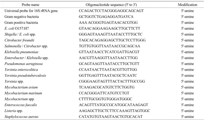
The probes designed in this study are listed in Table 1.
PCR amplification for waterborne pathogens
1For the 1st PCR amplification, 16S-SF2 (position 42 to 65, 5'-CR
†K
†GCY
†TAAY
†ACATGCAAGTCGA-3') and 16S-RSH-A5 (positions 498 to 520, 5'-TGGCACGD
†- AGTTR
†GCCGK
†K
†GCTT-3') were used, and the 16S-SF (positions 50 to 68, 5'AAY
†ACATGCAAGTCGAR
†CK-3') and 16s-R5H-A (positions 498 to 516, 5'-biotin-ACR
†D
†- AK
†TTRGCCGKK
†GCTT-3') were used for 2nd PCR amplification. The amplification reactions were performed with a thermal cycler (GeneAmp PCR
®System 2700). The 5' end of reverse primers was biotin labeled. PCR amplification was carried out in a 50 μl reaction containing 10 mM Tris-HCl, pH 9.0, 2 mM MgCl
2, 50 mM KCl, 200 μM dNTPs, 20 pmole of each primers, 1 units of Taq DNA polymerase (Bioneer Co., Daejeon, Korea), and 200 pg of genomic DNA extracted from bacterial pure culture. The PCR reaction was carried out as either single PCR using one set of primers, or one-tube nested PCR using two sets of primers. For single PCR, 35 cycles were done using program consisted of denaturation at 95℃ for 30 s, annealing at 52℃ for 30 s, and extension at 72℃ for 1 min.
Table 1. List of oligonucleotide probes for identification of waterborne pathogens
Probe name Oligonucleotide sequence (5' to 3') Modification
Universal probe for 16S rRNA gene CCAGACTCCTACGGGAGGCAGCAGT 5'-amine Gram negative bacteria GCTGGTCTGAGAGGATGAYCA 5'-amine
Gram positive bacteria AAA ACGGGTGAGTAACACGTGG 5'-amine
E. coli O157:H7 GTAACAGGAAGAAGCTTGCTTCTT 5'-amine Shigella / E. coli spp. GGGAGTAAAGTTAATACCTTTGCTC 5'-amine
Citrobacter freundii TAGCACAGAGGAGCTTGCTCCTTGGG 5'-amine Salmonella / Citrobacter spp. TGTTGTGGTTAATAACCGCAGCAA 5'-amine Klebsiella pneumoniae GTTAATAACCTCATCGATTGACGT 5'-amine Enterobacter / Klebsiella spp. AACGTTAAGGTTAATAACCTTGG 5'-amine
Pseudomonas aeruginosa GCAGTAAGTTAATACCTTGCTGTT 5'-amine Yersinia enterocolitica CCAATAACTTAATACGTTGTTGG 5'-amine Yersinia pseudotuberculosis GGTTGAGTTTAATACGCTCAATC 5'-amine Yersinia spp. CGGGAAGTAGTTTACTACTTTGCCGG 5'-amine Mycobacterium avium TCAAGACGCATGTCTTCTGGTG 5'-amine Mycobacterium marinum CCACGGGATTCATGTCCTGT 5'-amine Mycobacterium spp. CTTTTGCGGTGTGGGATGGGC 5'-amine Enterococcus faecalis ACAGTTTATGCCGCATGGCATAAGAGT 5'-amine Listeria spp. AAGAGCTTGCTCTTCCAAAGTTAGTGGC 5'-amine Staphylococcus aureus CATATGTGTAAGTAACTGTGCACAT 5'-amine
1 + Y=C:T, M=C:A, R=G:A, D=G:A:T, K=T:G +
For one-tube nested PCR, the first 15 cycles of PCR were done at the condition as follows: the initial denaturation at 95℃ for 30 s, annealing at 62℃ for 30 s, and extension at 72℃ for 1 min. Subsequently, the 35 cycles of the second PCR were done by the program consisted of denaturation at 95℃ for 30 s, annealing at 52℃ for 30 s, and extension at 72℃ for 1 min.
PCR-reverse blot hybridization assay
The preparation of the membrane containing probe molecules and the reverse blot hybridization was performed using a system that has been described previously (Xiang et al., 2002; Zwart et al., 2003). Optimal probe concentrations were determined by binding varying amounts of the probe in such a way that all the probes resulted in equally intense signals relative to the catchall at a concentration ranging from 1 to 40 pmoles.
For PCR-REBA, hybridization was carried out at 50℃
for 90 min in hybridization solution, and the membrane was washed twice with 2X SSPE/ 0.5% SDS at 62℃ for 10 min. Diluted streptavidin-alkaline phosphatase conjugate (1/2000) was then added and incubated at 42℃ within rolling bottle for 60 min. Finally the membrane was washed twice with 2X SSPE/ 0.5% SDS at 42℃ for 10 min, and with 2X SSPE at RT for 5 min. For detection, the membrane was incubated with CDP-star (Amersharm, Uppsala, Sweden) for 4 min and exposed to Hyperfilm (Amersharm, Uppsala, Sweden) for 60 min.
RESULTS
Development of PCR-REBA for detection of waterborne pathogens
For detection of waterborne pathogens, PCR can be used to amplify DNA from environmental samples, and then the PCR-REBA can be used to identify the amplified products.
E. coli O157:H7 (ATCC 35150) E. coli O157:H7 (ATCC 43894) E. coli O157:H7 (DML 411) Shigella dysenteriae Shigella flexneri Shigella sonnei Citrobacter freundii Salmonella typhi Klebsiella pneumoniae Enterobacter aerogenes Pseudomonas aeruginosa Yerisinia pseudotuberculosis Yersinia enterocolitica Mycobacterium avium Mycobacterium marinum Enterococcus faecalis Staphylococcus aureus Listeria monocytogenes
Pseudomonas aeruginosa
Citrobacter freundii Enterobacter / Klebsiella spp.
Universal for 16S rRNA gene Shigella spp. / E. coli spp. Yersiniapseudotuberculosis Mycobaterium spp.
Mycobacterium avium Mycobacterium marinum
Klebsiella pneumoniae Enterococcus faecalis
Yersinia spp.
Yersinia enterocolitica
E. coli O157:H7
Gram negative control Gram positive control Listeria spp. Staphylococcus aureus
Samples
Salmonella spp. / Citrobacter spp.
Probe
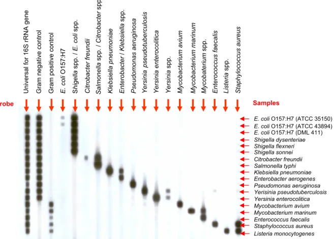
Fig. 1. The specificity of the PCR-REBA. PCR amplicons obtained from amplification of the reference strains of bacterial DNA were hybridized to the membrane. PCR and the hybridization condition are described in the text.
In other words, highly conserved primer sequences (universal 16S rRNA primers) can be used to amplify bacterial DNA followed by hybridization to the membrane composed of pathogen-specific probes. The probes themselves are located within the polymorphic region that is flanked by the conserved primer sequences.
To design specific probe molecules, database of 16S rRNA sequences of diverse types of bacteria were compiled from GenBank database of National Center for Biotechnology Information web site (http://www.ncbi.- nlm.nih.gov), and the sequence data for the target bacteria were aligned by using the Clustral method (http://www.- cmbi.kun.nl/bioinf/tools/clustalws.html).
While the conserved region was used to design the universal PCR primers for all the bacterial, divergent regions of 16S rRNA genes were used to design the probe to gram-negative, gram-positive and to each species or genus of bacteria targeted.
In order to develop PCR-REBA useful for comprehensive monitoring of microbial water contaminants, 18 waterborne pathogens were selected based on their significance as waterborne pathogens. Target bacterial species selected in this study include strains of common waterborne pathogens that may cause diarrhea or vomiting such as E. coli, E. coli O157:H7, Shigella spp., Salmonella spp., C. freundii, K.
pneumoniae, E. aerogenes, P. aeruginosa, Yersinia spp., Y.
enterocolitica, Y. pseudotuberculosis, E. faecalis, Listeria spp., S. aureus. M. avium complex and M. marinum that are major mycobacteria causing diseases by contaminated water and extremely difficult to culture were also targeted.
In brief, the probes designed in this study are listed in the Table 1, and results showing the specificity of the probes for detecting each target bacteria are shown in the Fig. 1.
Specificity of the PCR-REBA
Different parameters for PCR-REBA hybridization, such
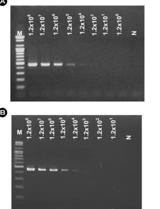
Fig. 2. The sensitivity of the PCR-REBA. (A-B) The sensitivity of the PCR using single and one-tube PCR was compared. (A) single PCR, PCR using one set of primers (16S-SF, 16S-R5H-A) and (B) one-tube nested PCR, PCR using two sets of PCR primers (16S-SF, 16S-R5H-A and 16S-SF2, 16S-R5H-A5). (C-D) The sensitivity of the PCR-REBA using PCR amplicons from single (C) and one-tube nested PCR (D) was compared. In order to compare the sensitivity of PCR with that of REBA, PCR amplicons obtained from (A) and (B) were subsequently hybridized to the REBA membrane containing probe for E. coli O157:H7. M; 100bp DNA ladder (Bioneer. Co., Daejeon., Korea). (C) shows the PCR-REBA result with (A), and (D) shows the PCR-REBA results with (B).
1.2x10⁶ 1.2x10⁵ 1.2x10³
1.2x10⁸ 1.2x10⁷ 1.2x10²
1.2x10⁴ 1.2x101
M N 8 1.2x10 7 1.2x10 6 1.2x10 5 1.2x10 4 1.2x10 3 1.2x10 1 1.2x10 0 1.2x10
E. coli O157:H7 probe
1.2x10⁸ 1.2x10⁷ 1.2x10⁶ 1.2x10⁵ 1.2x10⁴ 1.2x10³ 1.2x101 1.2x100
E. coli O157:H7 probe
M N
1.2x10⁸ 1.2x10⁷ 1.2x10³
1.2x10⁶ 1.2x10⁵ 1.2x10⁴ 1.2x101 1.2x100
M N
A C
D B
as temperature, incubation time, salt composition and shaking speed, were optimized for achieving the desired specificity of the PCR-REBA.
As shown in the Fig. 1, hybridization results were in precise agreement with those predicted from the probe sequences. The 9 species-specific probes (C. freundii, K.
pneumoniae, P. aeruginosa, Y. enterocolitica, Y. pseudotu- berculosis, M. avium complex, M. marinum, E. faecalis, S.
aureus), 1 strain-specific probe (E. coli O157:H7), and 3 genus-specific probes (Yersinia spp., Listeria spp., and Mycobacterium spp.) hybridized to their targets and did not show any cross reactivity.
On the other hand, some probes were not specific to the relevant genus. For example, Shigella spp. and E. coli, Salmonella spp. and Citrobacter spp., Klebsiella spp. and Enterobacter spp. hybridized to the same probe, since their 16S rRNA sequences are the same. Therefore, all probes demonstrated no cross-reaction with other DNA.
Sensitivity of PCR-REBA
The sensitivity of PCR-REBA using single PCR using one set of primers (16S-SF, 16S-R5H-A) and of one-tube nested PCR using two sets of PCR primers (16S-SF, 16S-R5H-A and 16S-SF2, 16S-R5H-A5) were compared
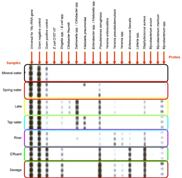
Fig. 3. Application of the PCR-REBA for detection and identification of waterborne pathogens from diverse water samples. PCR and the hybridization condition are described in the text.
(Fig. 2A and 2B). For this, the sensitivity of PCR was estimated by DNA obtained from serially diluted E. coli O157:H7 cultured cells. The results showed that the sensitivities of both PCRs were between 1.2×10
5cfu/ml and 1.2×10
4cfu/ml.
Subsequently, the sensitivity of the PCR-REBA using each PCR amplicons were compared (Fig. 2C and 2D). For that purpose, PCR amplicons from amplification of target DNA using 10 fold diluted E. coli O157: H7 chromosomal DNAs (Fig. 2A and 2B) were hybridized to the REBA membrane containing probe for E. coli O157:H7.
Unlike PCR results which showed similar sensitivity of the single PCR to the one-tube nested PCR, the sensitivity of the hybridization ranged between 1.2×10
4cfu/ml and 1.2×10
3cfu/ml (Fig. 2C and 2D) giving at least 10 times higher sensitivity of the PCR-REBA than PCR only. Thus, it seems that PCR method followed by hybridization of the products to the REBA would improve the detection sensitivity.
Application of PCR-REBA for detection of waterborne pathogens from diverse water samples
A total of seven water samples from diverse sources including tap water, spring water, mineral water, lake, river, effluent and sewage from Gangwon Province in Korea were collected, and handled according to guideline suggested by the Korea Ministry of Environment (Korea Ministry of Environment. 2002).
Subsequently, the water samples were used both for
isolation of waterborne pathogens using conventional culturing method and for identification of waterborne pathogens using PCR-REBA (Fig. 3).
Waterborne pathogens isolated and identified from this study is summarized in the Table 2. As shown in the Table 2, diverse waterborne pathogens including C. freundii, E.
coli, E. faecalis, K. pneumoniae, P. aeruginosa, Shigella spp., S. aureus, Y. pseudotuberculosis were isolated using conventional culture method. Meanwhile, most of bacteria isolated and identified by conventional methods were also identified by PCR-REBA. Moreover, some of bacteria such as Mycobacteria which are usually difficult to cultivate by using routine media which are often used for screening typical waterborne pathogens were also identified. The presence of mycobacteria which were not be able to be isolated by conventional method in this experiment were confirmed by isolating mycobacteria using specific media for mycobacteria. Therefore, it seems that the presence of waterborne pathogens in water samples can be monitored by using PCR-REBA.
DISCUSSION
At present, the conventional method for screening waterborne pathogens from environment samples requires 3~7 days and also has a low sensitivity. In this, study, we developed PCR-REBA for detection of waterborne patho- gens, and subsequently the sensitivity and the specificity of the PCR-REBA for detecting waterborne pathogens from
Table 2. Comparison of microorganisms detected using conventional culture-API tests and PCR-REBALake River Effluent Sewage Tap water Spring
water
Mineral water
Conventional method
E. coli Shigella spp.
C. freundii Pseudomonas spp.
Y. pseudotuberculosis S. aureus
Pseudomonas spp.
Y. pseudotuberculosis E. faecalis S. aureus
E. coli Shigella spp.
Pseudomonas spp.
Y. pseudotuberculosis S. aureus
E. coli Shigella spp.
Pseudomonas spp.
Y. pseudotuberculosis E. faecalis S. aureus
Shigella spp.
Pseudomonas spp.
K. pneumoniae
- -
PCR-REBA
E. coli / Shigella spp.
C. freundii
Salmonella/Citrobacter P. aeruginosa Y. pseudotuberculosis S. aureus
Mycobacterium spp.
P. aeruginosa Y. pseudotuberculosis Yersinia spp.
E. faecalis S. aureus
Salmonella/Citrobacter Mycobacterium spp.
E. coli / Shigella spp.
P. aeruginosa Y. pseudotuberculosis S. aureus E. faecalis Mycobacterium spp.
E. coli / Shigella spp.
P. aeruginosa Y. pseudotuberculosis Yersinia spp.
E. faecalis S. aureus Mycobacterium spp.
E. coli / Shigella spp.
P. aeruginosa K. pneumoniae Salmonella/
Citrobacter Mycobacterium spp.
K. pneumoniae P. aeruginosa -
environment sources were compared to those of culture.
The PCR-REBA certainly satisfies requirements for simultaneous detection system of microbial contaminants in water than other molecular methods. It is more convenient, less expensive, and easier to perform because it uses commonly available reagents, less expensive equipment, and can analyze 45 samples within a single run. The PCR- REBA format uses a non precipitating ECL substrate;
hence, it has the advantage in that nylon filters can be stripped and successfully reused at least 5 times and has significant impact in reducing the cost to minimal (Kohara et al., 2002).
Although relatively simple in concept, PCR-REBA is a powerful tool for detection and characterization of pathogen (Xiang et al., 2002; Zwart et al., 2003). Direct detection of nucleic acids from bacteria is feasible, but may lack the level of sensitivity needed for routine screening of environmental samples. When the amount of nucleic acid is not limiting, however, microarrays may prove very valuable as a fingerprinting tool and as a tool for marker discovery. When coupled to PCR, PCR-REBA have detection sensitivity equal to conventional methods with the added flexibility needed for discriminating multiple PCR reactions and for pathogen detection based on 16S rRNA sequences.
Acknowledgements
This work was supported by grant no. (R01- 2006-000-10250-0) from the Basic Research Program of the Korea Science & Engineering Foundation and by the Eco-Technopia 21 Project (102-061-046) of Ministry of Environment, Republic of Korea and authors appreciate the support.
REFERENCES
American Public Health Association. Standard Methods for the Examination of Water and Wastewater, 20th ed. 1998.
American Public Health Association, Washington, DC.
Ash C, Farrow JA, Wallbanks S, Collins MD. Phylogenetic heterogeneity of the genus Bacillus revealed by comparative analysis of small subunit-ribosomal RNA sequences. Lett
Appl Microbiol. 1991.13: 202-206.
Amann RI, Ludwig W, Schleifer KH. Phylogenetic identification and in situ detection of individual microbial cells without cultivation. Microbiol Rev. 1995. 59: 143-169.
Baudart J, Coallier J, Laurent P, Prevost M. Rapid and sensitive enumeration of viable diluted cells of members of the family Enterobacteriaceae in freshwater and drinking water. Appl Environ Microbiol. 2002. 68: 5057-5063.
Bayardelle P, Zafarullah M. Development of oligonucleotide primers for the specific PCR-based detection of the most frequent Enterobacteriaceae species DNA using wec gene templates. Can J Microbiol. 2002. 48: 113-122.
Bej AK, Steffan RJ, DiCesare JL, Haff L, Atlas RM. Detection of coliform bacteria in water by polymerase chain reaction and gene probes. Appl Environ Microbiol. 1990. 56: 307-314.
Bej AK, DiCesare JL, Haff L, Atlas RM. Detection of Escherichia coli and Shigella spp. in water by using the polymerase reaction and gene probes for uid. Appl Environ Microbiol.
1991. 57: 1013-1017.
Bej AK. Molecular based methods for the detection of microbial pathogens in the environment. J Microbiol Methods. 2003.
53: 139-140.
Call DR, Borucki MK, Loge FJ. Detection of bacterial pathogens in environmental samples using DNA microarrays. J Microbiol Methods. 2003. 53: 235-243.
Chiang YC, Yang CY, Li C, Ho YC, Lin CK, Tsen HY.
Identification of Bacillus spp., Escherichia coli, Salmonella spp., Staphylococcus spp. and Vibrio spp. with 16S ribosomal DNA-based oligonucleotide array hybridization. Int J Food Microbiol. 2006. 107: 131-137.
Clarridge JE. 3rd. Impact of 16S rRNA Gene Sequence Analysis for Identification of Bacteria on Clinical Microbiology and Infectious Diseases. Clin Microbiol Rev. 2004. 17: 840-862.
Cooper RC, Danielson RE. Detection of bacterial pathogens in wastewater and sludge. Manual of Environmental Microbiology. American Society of Microbiology, Washington, D.C., 1997. pp. 222-230.
Couzinet S, Jay C, Barras C, Vachon R, Vernet G, Ninet B, Jan I, Minazio MA, Francois P, Lew D, Troesch A, Schrenzel J.
High-density DNA probe arrays for identification of staphy- lococci to the species level. J Microbiol Methods. 2005. 61:
201-208.
Field KG, Bernhard AE, Brodeur TJ. Molecular approaches to microbiological monitoring: fecal source detection. Environ Monit Assess. 2003. 81: 313-326.
Gendel SM. Computational analysis of the specificity of 16S rRNA derived signature sequences for identifying food-related in microbes. Food Microbiol. 1996. 13: 1-15.
Jackson CR, Churchill PF, Roden E. Successional changes in bacterial assemblage structure during epilithic biofilm development. Ecology. 2001. 82: 555-566.
Josephson KL, Gerba CP, Pepper IL. Polymerase chain reaction detection of nonviable bacterial pathogens. Appl Environ Microbiol. 1993. 59: 3513-3515.
Kim MS, Shin KC, Lee HG, Han MA, Min BR, Choi YK. Using Reverse Dot Hybridization Method and 16S rRNA Gene (16S rDNA) for Identifying the Food Poisoning Microorganism in Foods.Korean J. Food SCI. Technology. 2003. 35: 470-474.
Kohara Y, Noda H, Okano K, Kambara H. DNA probes on beads arrayed in a capillary, 'Bead array', exhibited high hybridization performance. Nucleic Acids Res. 2002. 30: 16, e87.
Korea ministry of Environment. Bacterial contamination guidelines for drinking water in Korea. 2002.
Lemarchand K, Berthiaume F, Maynard C, Harel J, Payment P, Bayardelle P, Masson L, Brousseau R. Brousseau. Optimization of microbial DNA extraction and purification from raw wastewater samples for downstream pathogen detection by microarrays. J Microbiol Methods. 2005. 63: 115-126.
Lin CK, Tsen HY. Use of two 16S DNA targeted oligonucleotides as PCR primers for the specific detection of Salmonella in foods. J Appl Bacteriol. 1996. 80: 659-666.
Loy A, Schulz C, Lücker S, Schöpfer-Wendels A, Stoecker K, Baranyi C, Lehner A, Wagner M. 16S rRNA gene-based oligonucleotide microarray for environmental monitoring of the betaproteobacterial order "Rhodocyclales". Appl Environ Microbiol. 2005. 71: 1373-1386.
Mittelman MW, Habash M, Lacroix JM, Khoury AE, Krajden M.
Rapid detection of Enterobacteriaceae in urine by fluorescent 16s rRNA in situ hybridization on membrane filters. J Microbiol Methods. 1997. 30: 153-160.
Reynolds KA, Gerba CP, Abbaszadegan M, Pepper LL. ICC/PCR detection of enteroviruses and hepatitis A virus in environ- mental samples. Can J Microbiol. 2001. 47: 153-157.
Te Giffel MC, Beumer RR, Klijn N, Wagendorp A, Rombouts FM.
Discrimination between Bacillus cereus and Bacillus thuringiensis is using specific DNA probes based on variable regions of 16S rRNA. FEMS Microbiol Lett. 1997. 146: 47 -51.
Tsai YL, Olson BH. Detection of low numbers of bacterial cells in soils and sediments by polymerase chain reaction. Appl Environ Microbiol. 1992. 58: 754-757.
Tsen HY, Lin CK, Chi WR. Development and use of 16S rRNA gene targeted PCR primers for the identification of Escherichia coli cells in waters. J Appl Microbiol. 1998. 85: 554-560.
Tsen HY, Lin CK. Comparison of the partial 16S rRNA gene sequences and development of oligonucleotide probes for the detection of Escherichia coli cells in water and milk.
Food Microbiol. 1999. 16: 551-562.
Warsen AE, Krug MJ, LaFrentz S, Stanek DR, Loge FJ, Call DR.
Simultaneous discrimination between 15 fish pathogens by using 16S ribosomal DNA PCR and DNA microarrays. Appl Environ Microbiol. 2004. 70: 4216-4221.
Wilson WJ, Strout CL, DeSantis TZ, Stilwell LJ, Carrano AV, Andersen GL. Sequence-specific identification of 18 patho- genic microorganisms using microarray technology. Mol Cell Probes. 2002. 16: 119-127.
Xiang H, Xiong L, Liu X, Tu Z. Rapid simultaneous detection and identification of six species Candida using polymerase chain reaction and reverse line hybridization assay. J Microbiol Methods. 2007. 69: 282-287.
Zwart G, van Hannen EJ, Kamst-van Agterveld MP, Van der Gucht K, Lindstrom ES, Van Wichelen J, Lauridsen T, Crump BC, Han SK, Declerck S. Rapid Screening for Freshwater Bacterial Groups by Using Reverse Line Blot Hybridization.
Appl Environ Microbiol. 2003. 61: 5875-5883.