ISSN 2234-3806 • eISSN 2234-3814
http://dx.doi.org/10.3343/alm.2014.34.6.446
Evaluation of PCR-Reverse Blot Hybridization Assay, REBA Sepsis-ID Test, for Simultaneous Identification of Bacterial Pathogens and mecA and van Genes from Blood Culture Bottles
Soon Deok Park, Ph.D.1,*, Gyusang Lee, Ph.D.2,*, Hye-young Wang, M.S.3, Min Park, B.S.2, Sunghyun Kim, Ph.D.2, Hyunjung Kim, B.S.2, Jungho Kim, B.S.2, Young Keun Kim, M.D.4, Hyo Youl Kim, M.D.4, Hyeyoung Lee, Ph.D.2, Young Uh, M.D.1, and Jong Bae Kim, Ph.D.2
Departments of Laboratory Medicine1 and Internal Medicine4, Yonsei University Wonju College of Medicine; Department of Biomedical Laboratory Science2, College of Health Sciences, Yonsei University; M&D, Inc.3, Wonju Eco Environmental Technology Center, Wonju, Korea
Background: The aim of this study was to evaluate a newly developed PCR-based reverse blot hybridization assay (PCR-REBA), REBA Sepsis-ID (M&D, Wonju, Korea), to rapidly de- tect the presence of bacteremia and antimicrobial resistance gene in blood culture samples.
Methods: One thousand four hundred consecutive blood culture samples from patients with a delta neutrophil index greater than 2.7% were selected from March to July in 2013.
Three hundred positive and 1,100 negative for bacterial growth in blood culture bottles samples were tested by conventional and real-time PCR-REBA, respectively.
Results: The overall agreement between the conventional identification test and the REBA Sepsis-ID test was 95.3% (286/300). Agreement for gram-positive bacteria, gram-negative bacteria, fungi, and polymicrobials was 94.5% (190/201), 97.3% (71/73), 100% (14/14), and 91.7% (11/12), respectively. The detection rate of the mecA gene from methicillin-re- sistant Staphylococcus isolates was 97.8% (90/92). The vanA gene was detected in one blood culture sample from which vancomycin-resistant Enterococcus was isolated. When the cycle threshold for real-time PCR was defined as 30.0, 2.4% (26/1,100) of negative blood culture samples tested positive by real-time PCR.
Conclusions: The REBA Sepsis-ID test is capable of simultaneously and quickly detecting both causative agents and antimicrobial resistance genes, such as mecA and van, in blood culture positive samples.
Key Words: Real-time PCR, Blot, Hybridization, Bacteremia, mecA, Vancomycin resis- tance, Blood Culture
Received: March 17, 2014 Revision received: April 13, 2014 Accepted: August 27, 2014 Corresponding author: Young Uh
Department of Laboratory Medicine, Yonsei University Wonju College of Medicine, 20 Ilsan-ro, Wonju 220-701, Korea Tel: +82-33-741-1592
Fax: +82-33-731-0506 E-mail: [email protected] Corresponding author: Jong Bae Kim Department of Biomedical Laboratory Science, College of Health Sciences, Yonsei University, 1 Yonseidae-gil,
Wonju 220-710, Korea Tel: +82-33-760-2423 Fax: +82-33-760-2561 E-mail: [email protected]
*Soon Deok Park and Gyusang Lee con- tributed equally to this article.
© The Korean Society for Laboratory Medicine This is an Open Access article distributed under the terms of the Creative Commons Attribution Non-Commercial License (http://creativecom- mons.org/licenses/by-nc/3.0) which permits unrestricted non-commercial use, distribution, and reproduction in any medium, provided the original work is properly cited.
INTRODUCTION
Blood stream infections (BSIs) are associated with high rates of morbidity and mortality ranging from 20% to 70% worldwide [1- 4]. BSIs are the 10th leading cause of death in the United
States, accounting for 6% of all deaths [5]. An estimated 135,000 patients die each year of sepsis-associated complica- tions in Europe [6]. Blood culture systems, which detect viable microorganisms in blood, are the current gold standard for BSI diagnosis. Patients with sepsis, defined as a clinical infection re-
sulting in a systemic inflammatory response, account for only about one third of the total positive cultures [7]. Although blood cultures are currently performed with continuous-monitoring blood culture systems (CMBCSs), several factors such as poor timing of collection, insufficient blood volumes, and the presence of antibiotics in the samples reduce the sensitivity of blood cul- tures [7, 8]. The main limitation of utilizing blood culture method is the vast amount of time required, as treating patients with em- pirical therapies before the blood culture analysis is complete may not serve to cure the illness. Kumar et al. [9] reported that the mean survival rate decreased by 7.6% every hour that effec- tive antibiotic therapy was delayed following the onset of sepsis- related hypotension.
Recently, several PCR-based commercial assays that target a panel of clinically relevant bacterial and fungal bloodstream pathogens have been developed. Two types of commercial PCR- based assays are most common. The first type is designed for culture-positive samples. Examples of this type of assay are the peptide nucleic acid fluorescence in situ hybridization-based as- say (AdvanDx, Woburn, MA, USA) [10], Hyplex Blood Screen (BAG, Lich, Germany) [11], and Prove-it Sepsis (Mobidiag, Hel- sinki, Finland) [12]. The second type is designed for direct blood samples. Examples of this type of assay are SepsiTest (Molzym, Bremen, Germany) [13] and Vyoo (SIRS-Lab, Jena, Germany) [14]. The PCR-based reverse blot hybridization assay (PCR- REBA, REBA Sepsis-ID; M&D, Wonju, Korea) was developed to rapidly detect bacterial and fungal pathogens and antimicrobial resistance genes in blood culture samples [15]. It uses pan- probes to distinguish gram-positive bacteria (GPB), gram-nega- tive bacteria (GNB), and fungi. In addition, it uses probes for an- tibiotic resistance genes (i.e., the mecA gene of methicillin-resis- tant Staphylococcus spp. and the vanA and vanB genes of van- comycin-resistant enterococci).
The aim of this study was to evaluate the REBA Sepsis-ID test for rapid and accurate detection of pathogens and antimicrobial resistance genes in blood.
METHODS
This study was approved by the Institutional Review Board (CR312055) of Yonsei University Severance Hospital.
1. Collection of blood culture bottles
Three-hundred positive blood culture (PBC) and 1,100 negative blood culture (NBC) samples from patients with a delta neutro- phil index (DNI) greater than 2.7% [16] were consecutively col-
lected at Wonju Severance Christian Hospital from March to July in 2013. To avoid the redundancy of enrolled samples, only one blood culture sample per patient was allowed. The enrolled blood culture samples were simultaneously tested with the PCR- REBA and conventional microbiological tests. The overall posi- tive rate of blood culture in this study period was 7.46% (1,640/
21,979).
The PBC samples were eligible for enrollment if they had been flagged positive by BACTEC FX (Becton Dickinson, Sparks, MD, USA) or BacT/ALERT 3D (bioMérieux, Durham, NC, USA) with a positive Gram stain. The PBC bottles were then removed from the CMBCS and a 1,000 μL aliquot of the cul- ture-broth mixture was aseptically collected by using a syringe and needle. The aliquot was then divided into halves. The first half (500 μL) was used to perform a Gram stain and subcul- tured on sheep blood agar and MacConkey agar, which were in- cubated at 35˚C for 24-48 hr in 5% CO2, and the second half (500 μL) was kept at -20˚C for subsequent DNA extraction.
NBC samples were used if culture results were negative for five days of incubation in CMBCSs. After the blood culture bot- tles were removed from the CMBCS, 500 μL of blood suspen- sion was collected and kept at -20˚C for subsequent DNA ex- traction. All the NBC bottles were incubated at 35˚C until PCR results were obtained. For the NBC samples with a positive real- time PCR result, 1,000 μL of blood suspension was used to in- oculate routine subculture media (sheep blood agar, chocolate agar, and Sabouraud dextrose agar), and incubated at 35˚C un- der 5% CO2 for five days. For slow-growing bacteria, another 1,000 μL of blood suspension was used to inoculate special sub- culture media (plating count agar media [Becton Dickinson] [17]
and Reasoner’s 2A agar media [Becton Dickinson] [18]), which was incubated at 20˚C low temperature incubator. Additionally, the remaining blood suspension was used to inoculate Luria- Bertani [19] and brain heart infusion broths (Becton Dickinson), which were incubated at 37˚C incubator. Colonies isolated from the special subculture media and NBC samples that were posi- tive by real-time PCR were confirmed by bacterial 16S rRNA and fungal 18S to 5.8S internal transcribed sequence analysis. The amplicons were sequenced by Xenotech Company (Daejeon, Korea). The conventional identification test and antimicrobial susceptibility test were done by using the MicroScan system (Siemens Healthcare Diagnostics, Sacramento, CA, USA).
2. DNA preparation
To prepare DNA templates from the 300 PBC and 1,100 NBC samples, DNA was extracted by using the following procedure. A
200 μL aliquot of the blood was mixed with 1,000 μL of erythro- cyte lysis buffer (ELB) (Sigma, St. Louis, MO, USA) at room tem- perature for 10 min to disrupt erythrocytes. The supernatant was then removed after centrifugation at 13,000 g for 5 min. The pellet was washed with 1,000 μL of ELB to completely remove the erythrocytes and centrifuged under the same conditions.
One hundred microliters of ELB was added to the pellet, which was then frozen and thawed twice. One hundred microliters of DNA extraction solution (M&D) was added to the mixture, and it was boiled for 15 min. After centrifugation at 13,000 g for 10 min, the supernatant was used as a DNA template for PCR.
3. PCR amplification
Conventional PCR amplification was performed according to the manufacturer’s instructions to evaluate the PBC samples. Taq- Man real-time PCR assays were carried out by using Real-GP (gram-positive), -GN (gram-negative), and -CAN (Candida) real- time PCR kits (M&D) according to the following procedure to evaluate the NBC samples. The reaction mixture contained 10 μL of real-time PCR mixture, 5 μL of primer and probe mixture, 0.04 μL of 50× ROX reference dye, 5 μL of sample DNA, and sterile distilled water to give a final volume of 20 μL. The thermal cycling conditions were: 10 min at 94˚C, followed by 40 cycles of 30 sec at 94˚C and 30 sec at 60˚C. Each TaqMan real-time PCR assay included a positive control and an internal control, which was used to control for the effect of PCR inhibitors in the reaction. The cycle threshold (CT) values resulting from reac- tions in the master mix with and without specimen were com- pared. The bacterial load was quantified by determining the CT, the number of PCR cycles required for the fluorescence to ex- ceed a value significantly higher than the background fluores- cence. All reactions were performed by using an ABI 7500 FAST instrument (Applied Biosystems, Foster City, CA, USA).
The CT value was analyzed by using 7500 Software version 2.0.4 (Applied Biosystems). Fifteen PBC samples were evaluated by using real-time PCR TaqMan assay as a pilot study before eval- uating the NBC samples. The CT values resulting from this pilot study ranged from 11 to 20.94 cycles. The real-time PCR assay was defined as positive if the CT value was below 30.0.
4. PCR-reverse blot hybridization assay
The REBA Sepsis-ID test was performed according to the stan- dard protocol provided by the manufacturer [15]. The mem- brane used in the REBA Sepsis-ID test contained DNA probes for GPB, including Staphylococcus aureus, Staphylococcus spp., Streptococcus pneumoniae, Streptococcus spp., Entero-
coccus spp., and Mycobacterium spp.; DNA probes for GNB including Escherichia coli, Klebsiella pneumoniae, Citrobacter freundii, Salmonella spp., Shigella spp., Haemophilus influen- zae, Pseudomonas aeruginosa, and Acinetobacter baumannii;
DNA probes for Candida species including C. albicans, C. tropi- calis, C. glabrata, C. parapsilosis, and C. krusei; and DNA probes for antimicrobial resistance genes including mecA, vanA, and vanB. Indicator lines on the REBA strips were evaluated by us- ing a template provided with the kit. A universal control band was used to evaluate the intensity of faint bands (only the bands with color intensity equal to or greater than that of the control band were considered positive). The results indicated by band patterns on the developed strips specific for each species and resistance gene were compared to the conventional microbio- logic results.
5. 16S rRNA sequence analysis
All the isolates and PCR amplicons with discrepant results be- tween the REBA Sepsis-ID test and conventional methods were subjected to 16S rRNA sequence analysis. The REBA Sepsis-ID test, bacterial 16S rRNA, and fungal 18S to 5.8S internal tran- scribed sequence analysis were performed on these samples.
The results were compared with those of blood cultures.
6. Statistical analysis
All PBC and NBC data (1,400 samples) were analyzed by using IBM SPSS Statistics version 20 (SPSS Inc., Chicago, IL, USA).
The degree of agreement between conventional culture method and REBA-Sepsis ID test was determined by kappa coefficient and its corresponding P value.
RESULTS
1. Positive blood culture samples –monomicrobial bacteremia
Two hundred eighty-eight of 300 PBC samples contained a sin- gle organism as determined by the culture method. Of the mo- nomicrobial PBC samples, 69.8% (201/288) contained GPB in- cluding S. aureus, coagulase-negative Staphylococcus (CoNS), S. pneumoniae, Streptococcus spp., Enterococcus spp., and anaerobic GPB. An additional 25.3% (73/288) contained GNB including E. coli, K. pneumoniae, A. baumannii, P. aeruginosa, C. freundii, and anaerobic GNB. A total of 4.9% (14/288) con- tained Candida spp. including C. albicans, C. parapsilosis, C.
glabrata, and C. tropicalis (Table 1). Two of 27 S. aureus and three of 103 CoNS isolates gave discrepant results between the
Table 1. The spectrum of isolates and comparison of results between conventional culture method and REBA Sepsis ID test Bacterial pathogen or resistance gene by
conventional methods (N)
Identification with the REBA Sepsis-ID test
Consistent results (N) Discrepant results (N) Gram positive
Staphylococcus aureus (27) Staphylococcus aureus (25) No result (1)*, Staphylococcus spp. (1)†
Staphylococcus epidermidis (46) Staphylococcus spp. (45) Gram positive (1)†
Staphylococcus hominis (27) Staphylococcus spp. (27)
Staphylococcus capitis (21) Staphylococcus spp. (21)
Staphylococcus haemolyticus (9) Staphylococcus spp. (9)
Staphylococcus saprophyticus (2) Staphylococcus spp. (1) pan bacteria (1)†
Staphylococcus intermedius (1) Staphylococcus spp. (1)
Staphylococcus schleiferi (1) Staphylococcus spp. (0) pan bacteria (1)†
Streptococcus pneumoniae (5) Streptococcus pneumoniae (5)
Streptococcus mitis (6) Streptococcus spp. (5) Streptococcus spp. & S. aureus (1)‡
Streptococcus agalatiae (5) Streptococcus spp. (5)
Streptococcus salivarius (3) Streptococcus spp. (3)
Streptococcus anginosus (2) Streptococcus spp. (2)
Streptococcus pyogenes (2) Streptococcus spp. (2)
Streptococcus sanguinis (2) Streptococcus spp. (1) pan bacteria (1)†
Streptococcus bovis (1) Streptococcus spp. (1)
Streptococcus dysgalactiae (1) Streptococcus spp. (0) Streptococcus spp. & S. aureus‡
Enterococcus faecium (6) Enterococcus spp. (6)
Enterococcus faecalis (5) Enterococcus spp. (5)
Enterococcus avium (1) Enterococcus spp. (0) Enterococcus spp. & C. freundii (1)‡
Enterococcus gallinarum (1) Enterococcus spp. (1)
Corynebacterium spp. (10) Gram positive (10)
Bacillus spp. (9) Gram positive (7) Gram positive & Streptococcus spp. (2)
Micrococcus spp. (8) Gram positive (8)
Anaerobe bacteria (4) Gram positive (4)
Gram negative
Escherichia coli (35) Escherichia coli (34) Gram negative (1)§
Klebsiella pneumoniae (11) Klebsiella pneumonia (11)
Acinetobacter baumannii (6) Acinetobacter baumannii (6)
Pseudomonas aeruginosa (5) Pseudomonas aeruginosae (5)
Aeromonas hydrophila (3) Gram negative (3)
Morganella morganii (2) Gram negative (2)
Enterobacter cloacae (2) Gram negative (2)
Enterobacter aerogenes (1) Gram negative (1) E. aerogenes & A. baumannii (1)‡
Citrobacter freundii (1) Citrobacter freundii (1)
Klebsiella oxytoca (1) Gram negative (1)
Serratia marcescens (1) Gram negative (1)
Chryseobacterium indologenes (1) Gram negative (1)
Delftia acidovorans (1) Gram negative (1)
(Continued to the next page)
culture method and the REBA Sepsis-ID test. One S. aureus case was not identified by PCR amplification, and another S. aureus case was not identified as the species level. Three CoNS cases were identified at the pan-GP or pan-bacteria level by the REBA Sepsis-ID test. All five S. pneumoniae isolates were correctly identified. Three of 22 Streptococcus spp. showed discrepant re- sults between culture and the REBA Sepsis-ID test. The pres- ence of both Streptococcus spp. and S. aureus was indicated in two of these discrepant samples by the REBA Sepsis-ID test.
Subsequent 16S rRNA sequence analysis confirmed the pres- ence of two isolates in each sample. Twelve of 13 Enterococcus spp. were concordant. One sample resulted in two bands corre- sponding to Enterococcus spp. and C. freundii by the REBA Sep- sis-ID test despite only Enterococcus avium being isolated. Sub- sequent 16S rRNA sequence analyses confirmed the presence of E. avium and C. freundii in the blood culture sample. The other 27 GPB including 10 Corynebacterium spp., 9 Bacillus spp., 8 Micrococcus spp., and 4 anaerobic GPB were not included in the REBA probes. PCR products from all but two samples hy- bridized with the pan-GP probes. The agreement rate between the culture method and the REBA Sepsis-ID test for GPB and GNB was 94.5% (190/201) and 97.3% (71/73), respectively.
Three GPB monomicrobial samples were revealed to have more than one isolate by the REBA Sepsis-ID test. E. coli was isolated from one GNB monomicrobial sample, but the PCR product hy- bridized only with the pan-GN probe. In the only other GNB monomicrobial sample, Enterobacter aerogenes was isolated,
and the PCR product hybridized with both of the pan-GN and A.
baumannii probes. This sample was confirmed to contain both of E. aerogenes and A. baumannii by 16S rRNA sequence anal- ysis. All 14 Candida species were identified by the REBA Sep- sis-ID test.
2. Positive blood culture samples –polymicrobial bacteremia Of the 12 polymicrobial PBC samples, the agreement rate be- tween the culture method and the REBA Sepsis-ID test was 91.7% (11/12). E. coli plus Streptococcus anginosus isolated by the culture method in one case was identified to be only E. coli by the REBA Sepsis-ID test (Table 2).
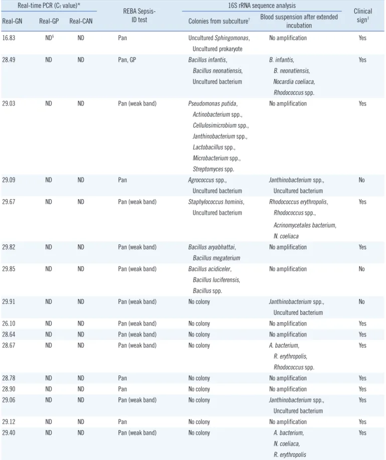
3. Negative blood culture samples
Among the NBC samples, 97.6% (1,074/1,100) had CT values above 30.0. The remaining 2.4% (26/1,100) NBC samples had CT values below 30.0, including four with values less than 25.0, one between 26.0 and 27.0, and 21 between 28.0 and 30.0. Of these samples, 69.2% (18/26) were Real-GN positive, 19.2%
(5/26) were Real-GP positive, and 11.5% (3/26) were Real-CAN positive by TaqMan real-time PCR assay (Table 3). Most Real- GP, and Real-GN positive cases produced PCR products that weakly hybridized to the pan-bacteria probe. Isolates from two Real-CAN positive cases were identified as containing C. tropi- calis and one was identified as containing C. tropicalis plus C.
parapsilosis by the REBA Sepsis-ID test. Thirteen of 26 NBC samples with positive real-time PCR results were not amplified Table 1. Continued
Bacterial pathogen or resistance gene by conventional methods (N)
Identification with the REBA Sepsis-ID test
Consistent results (N) Discrepant results (N)
Moraxella catarhalis (1) Gram negative (1)
Ochrobactrum anthropi (1) Gram negative (1)
Anaerobe bacteria (1) Gram negative (1)
Candida
Candida albicans (7) Candida albicans (7)
Candida parapsilosis (4) Candida parapsilosis (4)
Candida glabrata (2) Candida glabrata (2)
Candida tropicalis (1) Candida tropicalis (1)
Antimicrobial resistance
Methicillin resistance (92) mecA (90) No detection (2)ll
Vancomycin resistance (1) vanA (1)
*One was not amplified and gave no result by the REBA Sepsis-ID test; †The 16S rRNA sequence analysis reported as uncultured bacterium; ‡REBA Sepsis- ID test results agreed with 16S rRNA sequence analysis; §The PCR product weakly hybridized with the E. coli probe; llThe mecA gene was not identified in two S. saprophyticus isolates by the REBA Sepsis-ID test.
Abbreviation: REBA Sepsis-ID, PCR – based reverse blot hybridization assay.
by sequencing reactions. Eight NBC samples produced colonies that grew on subculture media (Table 3). A significant agree- ment was found between conventional culture method and REBA-Sepsis ID test (kappa coefficient=0.916, P <0.001).
4. Identification of mecA and vanA genes
Ninety of 92 (97.8%) blood culture samples with methicillin-resis- tant Staphylococcus isolates were mecA positive. MecA was not detected in the two methicillin-resistant Staphylococcus sapro- phyticus isolates. No mecA genes were identified by PCR-REBA in the 44 methicillin-susceptible Staphylococcus spp. The vanA gene was detected in one blood culture sample, from which van- comycin-resistant Enterococcus was isolated (Table 1).
DISCUSSION
The clinical treatment of bacterial infections with antibiotics de- pends on the bacterial species, and especially differs among GPB, GNB, and fungal infections. Antibiotic therapies are usu- ally selected empirically until antimicrobial susceptibility test re- sults are completed. Therefore, rapidly identifying pathogens and their resistance genes is important for treating septic pa- tients. Diagnostic methods that can reduce the time to identify a BSI pathogen, and its antimicrobial susceptibility have great po- tential to improve patient care. Recently, a molecular diagnostic approach was proposed to be advantageous [20, 21]. The ad- vantage of molecular approaches is notable when the infectious agent is fastidious or fungal, when blood culture fails to identify the causative agent, or when a quick diagnosis is needed.
The agreement rates between the conventional culture method
and the REBA Sepsis-ID test in identifying GPB, GNB, fungi, and the mecA gene in 300 PBC samples were 94.5%, 97.3%, 100%, and 97.8%, respectively. Steindor et al. [22] reported cor- rect identification of 96.1% of GPB, 89.9% of GNB, and 92.9%
of mecA using the PCR-based DNA strip assay, GenoType BC.
Results from our PBC samples demonstrated that the overall agreement rate between the culture method and the REBA Sep- sis-ID test was high at 95.3%, which was similar to that indicated in other reports [23, 24].
Two monomicrobial PBC samples with discrepant results be- tween culturing and the REBA Sepsis-ID test were proven to have additional S. aureus isolates. In contrast, five samples con- taining Staphylococcus spp., including S. aureus isolates, were not correctly identified the by the REBA Sepsis-ID test. It is diffi- cult to accurately identify more than one organism in a sample by using the culture method, as additional blind subculturing of PBC samples with positive flagging is not a common practice in clinical laboratories. PCR inhibitors have been an obstacle to obtaining accurate results. Achieving accurate PCR results from blood culture samples is difficult owing to PCR inhibitors in the blood such as sodium polyanetholsulfonate, heme, hematin, hemoglobin, lactoferrin, and IgG [25-29]. An optimal PCR sample preparation procedure should efficiently lyse resistant bacterial cell walls, including those in GPB, without being too harsh on the DNA released from the cells [30]. Differentiating S. pneu- moniae from mitis group streptococci is difficult because of their close genetic relationship [31]. However, the REBA Sepsis-ID test discriminated between S. pneumoniae and other strepto- cocci, including viridians group streptococci.
Poor accuracy in the identification of more than one organism Table 2. Comparison of results from conventional culture methods and the REBA Sepsis-ID test for 12 polymicrobial blood culture samples
Bacterial pathogen by conventional methods (N) Identification with the REBA Sepsis-ID test (N) Staphylococcus aureus and Enterococcus faecalis (2) Staphylococcus aureus and Enterococcus spp. (2) Staphylococcus epidermidis and Staphylococcus warneri (1) Staphylococcus spp. (1)
Staphylococcus haemolyticus and Candida utilis (1) Staphylococcus spp. and fungus (1) Enterococcus durans and Staphylococcus epidermidis (1) Enterococcus spp. and Staphylococcus spp. (1) Enterococcus faecium and Candida albicans (1) Enterococcus spp. and Candida albicans (1) Enterococcus avium and Staphylococcus epidermidis (1) Enterococcus spp. and Staphylococcus spp. (1) Escherichia coli and Enterococcus gallinarum (1) Escherichia coli and Enterococcus spp. (1) Escherichia coli and Streptococcus anginosus (1) Escherichia coli (1)
Klebsiella pneumoniae and Enterococcus casseliflavus (1) Klebsiella pneumoniae and Enterococcus spp. (1) Klebsiella pneumoniae and Enterobacter cloacae (1) Klebsiella pneumoniae and Gram negative (1) Proteus mirabilis and Enterococcus faecalis (1) Gram negative and Enterococcus spp. (1) Abbreviation: REBA Sepsis-ID, PCR-based reverse blot hybridization assay.
Table 3. Comparison of results from 16S rRNA sequence analysis, real-time PCR-REBA, and subculture Real-time PCR (CT value)*
REBA Sepsis- ID test
16S rRNA sequence analysis
Clinical sign‡ Real-GN Real-GP Real-CAN Colonies from subculture† Blood suspension after extended
incubation
16.83 ND§ ND Pan Uncultured Sphingomonas, No amplification Yes
Uncultured prokaryote
28.49 ND ND Pan, GP Bacillus infantis, B. infantis, Yes
Bacillus neonatiensis, B. neonatiensis, Uncultured bacterium Nocardia coeliaca,
Rhodococcus spp.
29.03 ND ND Pan (weak band) Pseudomonas putida, No amplification Yes
Actinobacterium spp., Cellulosimicrobium spp., Janthinobacterium spp., Lactobacillus spp., Microbacterium spp., Streptomyces spp.
29.09 ND ND Pan Agrococcus spp., Janthinobacterium spp., No
Uncultured bacterium Uncultured bacterium
29.67 ND ND Pan (weak band) Staphylococcus hominis, Rhodococcus erythropolis, Yes
Uncultured bacterium Rhodococcus spp., Acrinomycetales bacterium, N. coeliaca
29.82 ND ND Pan (weak band) Bacillus aryabhattai, No amplification Yes
Bacillus megaterium
29.85 ND ND Pan (weak band) Bacillus acidiceler, No amplification No
Bacillus luciferensis, Bacillus spp.
29.91 ND ND Pan (weak band) No colony Janthinobacterium spp., No
Uncultured bacterium
26.10 ND ND Pan (weak band) No colony No amplification Yes
28.64 ND ND Pan (weak band) No colony No amplification Yes
28.67 ND ND Pan (weak band) No colony A. bacterium, Yes
R. erythropolis, Rhodococcus spp.
28.78 ND ND Pan No colony No amplification Yes
28.90 ND ND Pan No colony No amplification Yes
29.06 ND ND Pan (weak band) No colony Janthinobacterium spp., Yes
Uncultured bacterium
29.12 ND ND Pan No colony No amplification Yes
29.40 ND ND Pan (weak band) No colony A. bacterium, Yes
N. coeliaca, R. erythropolis
(Continued to the next page)
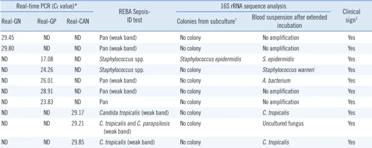
Table 3. Continued
Real-time PCR (CT value)*
REBA Sepsis- ID test
16S rRNA sequence analysis
Clinical sign‡ Real-GN Real-GP Real-CAN Colonies from subculture† Blood suspension after extended
incubation
29.45 ND ND Pan (weak band) No colony No amplification Yes
29.80 ND ND Pan (weak band) No colony No amplification Yes
ND 17.08 ND Staphylococcus spp. Staphylococcus epidermidis S. epidermidis Yes
ND 24.26 ND Staphylococcus spp. No colony Staphylococcus warneri Yes
ND 26.01 ND Pan (weak band) No colony A. bacterium Yes
ND 28.91 ND Pan (weak band) No colony No amplification Yes
ND 23.83 ND Pan No colony No amplification Yes
ND ND 29.17 Candida tropicalis (weak band) No colony C. tropicalis Yes
ND ND 29.21 C. tropicalis and C. parapsilosis
(weak band) No colony Uncultured fungus Yes
ND ND 29.85 C. tropicalis (weak band) No colony C. tropicalis Yes
*The cycle threshold value for real-time PCR was 30.0 in this study; †Samples were subcultured on sheep blood, MacConkey, chocolate, R2A, and plating agar; ‡Clinical signs follow the systemic inflammatory response syndrome definition; §Undetermined result or cycle threshold value was over 30.0 by real- time PCR.
Abbreviations: REBA Sepsis-ID, PCR-based reverse blot hybridization assay; CT, cycle threshold; Real-GN, 16S rRNA gram-negative primers; Real-GP, 16S rRNA gram-positive primers; Real-CAN, 16S rRNA gram-positive primers; Pan, broad-range bacterial 16S rRNA probe.
in a sample is a well-known drawback of PCR-based tests. Mo- lecular tests that detect a limited number of targets may report only one species, which can be misleading. Buchan et al. [32]
reported that the agreement rate between a reference culture and a microarray-based nucleic acid test for polymicrobial cul- tures was only 72%. However, the REBA assay in the present study had an agreement rate of 91.7%. REBA may not detect multiple types of bacteria in a blood culture sample owing to technical limitations, such as interference between multiple probes and undetectable concentrations of minor bacterial con- stituents. The five PBC samples that had a single bacterial spe- cies by blood culture contained two isolates as determined by the REBA Sepsis-ID test, which was confirmed by 16S rRNA sequence analysis. The reason that the REBA Sepsis-ID test de- tected more isolates than the blood culture method is not clear.
However, C. freundii was detected by the blood culture method on the day following its detection by using the REBA Sepsis-ID test. This result indicates that the REBA Sepsis-ID test may be able to detect nonviable bacteria or those at a low concentration that is not detectable by CMBCS. Therefore, the REBA Sepsis- ID test may have practical benefits in the clinical setting, partic- ularly for patients on empirical antibiotic treatment before cul- ture results are obtained.
The REBA Sepsis-ID test did not identify two (2.2%) methicil- lin-resistant S. saprophyticus isolates, which were identified by the conventional method. The two mecA-negative S. saprophyti-
cus isolates were likely methicillin-susceptible organisms. The Clinical and Laboratory Standards Institute interpretive criteria for detecting mecA-mediated resistance in S. saprophyticus may overestimate resistance [33].
Real-time PCR was used to evaluate NBC samples because it reduces the time required to obtain results, decreases contami- nation during the PCR procedure, and increases PCR sensitiv- ity. Among the 26 NBC samples with real-time PCR positive re- sults in this study, 13 cases were considered false positive re- sults because they were not amplified by sequencing reactions.
Matsuda et al. [34] reported that the positivity rate of PCR-hy- bridization using 500 μL of blood from NBC samples was 10.5%
(11/105) and that the 11 culture negative, PCR-hybridization positive samples contained nine CoNS and two GPB species.
Steindor et al. [22] evaluated a PCR-based DNA strip assay, GenoType BC, which detects bacteria from PBC samples and emphasized that the assay required abundant bacteria in the blood. Kocoglu et al. [35] reported that of 904 NBC samples, 2.6% were positive by reculture but not by PCR-based meth- ods. They concluded that subculture was valuable in diagnosis using NBC samples, especially when only one set of blood cul- tures was taken. In this study, using a real-time PCR CT of 30 resulted in a very low positivity rate in NBC samples. Colonies isolated from subculture media may also have been contami- nated as the sequence analysis results were not concordant be- tween the blood suspension and colonies. These results suggest
that it is not necessary for a clinical laboratory to perform sup- plemental subculture as a routine work after 5 days of incuba- tion on CMBCS.
Although the REBA Sepsis-ID test will not absolutely replace the conventional culture method, it is likely to rapidly discrimi- nate between PBC and NBC samples and provide clinical infor- mation relevant to patients by detecting important pathogens and antimicrobial resistance genes.
Authors’ Disclosures of Potential Conflicts of Interest
No potential conflicts of interest relevant to this article were re- ported.
Acknowledgements
This study was supported by a grant from the Korea Health Technology R&D Project, Ministry of Health & Welfare, Republic of Korea (A121030, H.L.).
REFERENCES
1. Angus DC, Linde-Zwirble WT, Lidicker J, Clermont G, Carcillo J, Pinsky MR. Epidemiology of severe sepsis in the United States: analysis of inci- dence, outcome, and associated costs of care. Crit Care Med 2001;
29:1303-10.
2. Barnato AE, Alexander SL, Linde-Zwirble WT, Angus DC. Racial varia- tion in the incidence, care, and outcomes of severe sepsis: analysis of population, patient, and hospital characteristics. Am J Respir Crit Care Med 2008;177:279-84.
3. Dombroskiy VY, Martin AA, Sunderram J, Paz HL. Rapid increase in hospitalization and mortality rates for severe sepsis in the United States:
a trend analysis from 1993 to 2003. Crit Care Med 2007;35:1244-50.
4. Esper AM, Moss M, Lewis CA, Nisbet R, Mannino DM, Martin GS. The role of infection and comorbidity: factors that influence disparities in sepsis. Crit Care Med 2006;34:2576-82.
5. Kung HC, Hoyert DL, Xu J, Murphy SL. Deaths: final data for 2005. Natl Vital Stat Rep 2008;56:1-120.
6. Lever A and Mackenzie I. Sepsis: definition, epidemiology, and diagno- sis. BMJ 2007;335:879-83.
7. Weinstein MP, Towns ML, Quartey SM, Mirrett S, Reimer LG, Parmigiani G, et al. The clinical significance of positive blood cultures in the 1990s:
a prospective comprehensive evaluation of the microbiology, epidemiol- ogy, and outcome of bacteremia and fungemia in adults. Clin Infect Dis 1997;24:584-602.
8. Wilson ML. Outpatient blood cultures: progress and unanswered ques- tions. Eur J Clin Microbiol Infect Dis 2004;23:879-80.
9. Kumar A, Roberts D, Wood KE, Light B, Parrillo JE, Sharma S, et al.
Duration of hypotension before initiation of effective antimicrobial thera- py is the critical determinant of survival in human septic shock. Crit Care Med 2006;34:1589-96.
10. Forrest GN, Roghmann MC, Toombs LS, Johnson JK, Weekes E, Linca-
lis DP, et al. Peptide nucleic acid fluorescence in situ hybridization for hospital-acquired enterococcal bacteremia: delivering earlier effective antimicrobial therapy. Antimicrob Agents Chemother 2008;52:3558-63.
11. Wellinghausen N, Wirths B, Essig A, Wassill L. Evaluation of the Hyplex Blood Screen multiplex PCR-enzyme-linked immunosorbent assay sys- tem for direct identification of gram positive cocci and gram-negative bacilli from positive blood cultures. J Clin Microbiol 2004;42:3147-52.
12. Hansen WL, Beuving J, Bruggeman CA, Wolffs PF. Molecular probes for diagnosis of 260 clinically relevant bacterial infections in blood cul- tures. J Clin Microbiol 2010;48:4432-8.
13. Mühl H, KochemAJ, Disqué C, Sakka SG. Activity and DNA contamina- tion of commercial polymerase chain reaction reagents for the universal 16S rDNA real-time polymerase chain reaction detection of bacterial pathogens in blood. Diagn Microbiol Infec Dis 2010;66:41-9.
14. Lehmann LE, Hunfeld KP, Emrich T, Haberhausen G, Wissing H, Hoeft A, et al. A multiplex real-time PCR assay for rapid detection ad differen- tiation of 25 bacterial and fungal pathogens from whole blood samples.
Med Microbiol Immunol 2008;197:313-24.
15. Choi Y, Wang HY, Lee G, Park SD, Jeon BY, Uh Y, et al. PCR-reverse blot hybridization assay for the screening and identification of patho- gens in sepsis. J Clin Microbiol 2013;51:1451-7.
16. Seok Y, Choi JR, Kim J, Kim YK, Lee J, Song J, et al. Delta neutrophil index: a promising diagnostic and prognostic marker for sepsis. Shock 2012;37:242-6.
17. Atlas RM. Handbook of microbiological media. 3rd ed. London: CRC Press, 2004;1390.
18. Reasoner DJ, BlannonJC, Geldreich EE. Rapid seven–hour fecal coli- form test. Appl Environ Microbiol 1979;38:229-36.
19. Bertani G. Studies on lysogenesis. I. The mode of phase liberation by lysogenic Escherichia coli. J Bacteriol 1951;62:293-300.
20. Mallon PW, Millar BC, Moore JE, Murphy PG, McClurg RB, Chew EW, et al. Molecular identification of Acinetobacter sp. In a patient with cul- ture-nagative endocarditis. Clin Microbiol Infect 2000;6:277-8.
21. Millar B, Moore J, Mallon P, Xu J, Crowe M, McClurg R, et al. Molecular diagnosis of infective endocarditis–a new Duke’s criterion. Scand J In- fect Dis 2001;33:673-80.
22. Steindor M, Weizengger M, Harrison N, Hirschl AM, Schweickert B, Göbel UB, et al. Use of a commercial PCR-based line blot method for identification of bacterial pathogens and the mecA and van genes from BacT Alert blood culture bottles. J Clin Microbiol 2012;50:157-9.
23. Jordan JA and Durso MB. Real-time polymerase chain reaction for de- tecting bacterial DNA directly from blood of neonates being evaluated for sepsis. J Mol Diagn 2005;7:575-81.
24. Liu Y, Han JX, Huang HY, Zhu B. Development and evaluation of 16S rDNA microarray for detecting bacterial pathogens in cerebrospinal flu- id. Exp Biol Med (Maywood) 2005;230:587-91.
25. Akane A, Matsubara K, Nakamura H, Takahashi S, Kimura K. Identifi- cation of the heme compound copurified with deoxyribonucleic acid (DNA) from bloodstains, a major inhibitor of polymerase chain reaction (PCR) amplification. J Forensic Sci 1994;39:362-72.
26. Kreader CA. Relief of amplification inhibition in PCR with bovine serum albumin or T4 gene 32 protein. Appl Environ Microbiol 1996;62:1102-6.
27. Jung R, Lübcke C, Wagener C, Neumaier M. Reversal of RT-PCR inhi- bition observed in heparinized clinical specimens. Biotechniques 1997;23:24, 26, 28.
28. Al-Soud WA, Jönsson LJ, Râdström P. Identification and characteriza- tion of immunoglobulin G in blood as a major inhibitor of diagnostic PCR. J Clin Microbiol 2000;38:345-50.
29. Lam NY, Rainer TH, Chiu RW, Lo YM. EDTA is a better anticoagulant than heparin or citrate for delayed blood processing for plasma DNA
analysis. Clin Chem 2004;50:256-7.
30. Rantakokko-Jalava K and Jalava J. Optimal DNA isolation method for detection of bacteria in specimens by broad-range PCR. J Clin Microbi- ol 2002;40:4211-7.
31. Kawamura Y, Hou XG, Sultana F, Miura H, Ezaki T. Determination of 16S rRNA sequences of Streptococcus mitis and Streptococcus gordo- nii and phylogenetic relationships among members of the genus Strep- tococcus. Int J Syst Bacteriol 1995;45:406-8.
32. Buchan BW, Ginocchio CC, Manii R, Cavagnolo R, Pancholi P, Swyers L, et al. Multiplex identification of gram-positive bacteria and resistance determinants directly from positive blood culture broths: evaluation of
an automated microarray-based nucleic acid test. PLoS Med 2013;
10:e1001478.
33. Higashide M, Kuroda M, Ohkawa S, Ohta T. Evaluation of a cefoxitin disk diffusion test for the detection of mecA-positive methicillin resistant Staphylococcus saprophyticus. Int J Antimicrob Agents 2006;27:500-4.
34. Matsuda K, Iwaki KK, Garcia-Gomez J, Hoffman J, Inderlied CB, Mason WH, et al. Bacterial identification by 16SrRNA gene PCR-hybridization as a supplement to negative culture results. J Clin Microbiol 2011;49:
2031-4.
35. Kocoglu ME, Bayram A, Balci I. Evaluation of negative results of BacT/
Alert 3D automated blood culture system. J Microbiol 2005;43:257-9.