ISSN 2234-3806 • eISSN 2234-3814
http://dx.doi.org/10.3343/alm.2012.32.5.331
Clinical Usefulness of Cell-based Indirect
Immunofluorescence Assay for the Detection of Aquaporin-4 Antibodies in Neuromyelitis Optica Spectrum Disorder
Eun-suk Kang, M.D.1, Ju-Hong Min, M.D.2, Kwang Ho Lee, M.D.2, and Byoung Joon Kim, M.D.2
Departments of Laboratory Medicine and Genetics1, Neurology2, Samsung Medical Center, Sungkyunkwan University School of Medicine, Seoul, Korea Background: The presence of antibodies to aquaporin-4 (AQP4) has been identified as a
key characteristic of neuromyelitis optica spectrum disorder (NMOSD), an autoimmune in- flammatory demyelinating central nervous system (CNS) disorder. We evaluated the perfor- mance of a cell-based indirect immunofluorescence assay (CIIFA) for detecting AQP4 anti- bodies using antigen prepared with a recombinant AQP4 peptide transfection technique and assessed the usefulness of CIIFA for diagnosis of NMOSD in routine clinical practice.
Methods: Forty-six serum samples from 36 patients as a comparison set and another 101 patients enrolled consecutively from a neurology clinic were included. CIIFA and fluores- cence immunoprecipitation assays (FIPA) were performed. CIIFA was performed at 2 dif- ferent institutions for comparison purposes.
Results: CIIFA and FIPA sensitivity in the comparison set was 86% and 79% in neuromyeli- tis optica (NMO) patients and 55% and 36% in high-risk NMO patients, respectively. The semiquantitative titer measured by CIIFA correlated well with the arbitrary unit (fluorescence units [FU]) derived from FIPA (r=0.66). Titers measured by CIIFA and FIPA were elevated in NMO patients compared to high-risk NMO patients (1:240 vs. 1:180 and 8,390 vs. 4,059 FU, respectively). The frequency of AQP4 antibody detection by CIIFA in 101 consecutively enrolled patients was 100% in NMO and 23% in high-risk NMO patients, while only 4.6% in control patients, including those with multiple sclerosis.
Conclusions: Detection of AQP4 antibodies by CIIFA provides sensitive and highly specific diagnostic information for NMO and high-risk NMO patients, which can be used to differ- entiate these conditions from other demyelinating CNS diseases.
Key Words: Neuromyelitis optica, Aquaporin 4, Indirect immunofluorescence assay, Im- munoprecipitation assay
Received: January 5, 2012 Revision received: March 30, 2012 Accepted: June 26, 2012
Corresponding author: Byoung Joon Kim Department of Neurology, Samsung Medical Center, Sungkyunkwan University School of Medicine, 50 Irwon-dong, Gangnam-gu, Seoul 135-710, Korea Tel: +82-2-3410-3594
Fax: +82-2-3410-0052 E-mail: bjkim@skku.edu
© The Korean Society for Laboratory Medicine.
This is an Open Access article distributed under the terms of the Creative Commons Attribution Non-Commercial License (http://creativecom- mons.org/licenses/by-nc/3.0) which permits unrestricted non-commercial use, distribution, and reproduction in any medium, provided the original work is properly cited.
INTRODUCTION
Neuromyelitis optica (NMO; also known as Devic syndrome) is a chronic inflammatory demyelinating disorder of the central nervous system (CNS), which was first described in the late 19th century by E. Devic and others [1]. It preferentially affects the optic nerves and the spinal cord, thus frequently manifests
as recurrent optic neuritis (RON) and longitudinally extensive transverse myelitis (LETM) [2, 3]. The condition which is consid- ered to be a type of neuromyelitis optica spectrum disorder (NMOSD) includes NMO and an array of high-risk NMO disor- ders such as Asian optic-spinal multiple sclerosis (OSMS), re- current myelitis associated with longitudinal extensive spinal cord lesions, recurrent isolated or simultaneous bilateral optic
neuritis (RON/BON), and optic neuritis (ON) or myelitis/LETM in the context of certain organ-specific and non-organ-specific au- toimmune diseases or with brain lesions typically observed in cases of NMO [4, 5]. Due to its relapsing course with rare spon- taneous remission, the accumulation of irreversible deficits and rapid progression of NMO often renders patients severely dis- abled. These characteristics highlight the need to distinguish NMO from other demyelinating CNS conditions as early as pos- sible; however, it is often difficult to differentiate inflammatory demyelinating CNS disorders that have differing etiologies but similar clinical presentations and cerebrospinal fluid (CSF) and magnetic resonance imaging (MRI) findings. In particular, both NMO and multiple sclerosis (MS) have a relapsing-remitting course in the majority of cases, thus NMO has been typically considered to be a localized form of MS due to the lack of an NMO-specific laboratory test [3, 5, 6].
However, recent reports suggest that NMO is a distinct dis- ease entity with a fundamentally different pathogenic mecha- nism than that of MS or other demyelinating diseases. An NMO- specific IgG, designated NMO-IgG, and antibodies to aquapo- rin-4 (AQP4) that serve as relevant antigens of NMO-IgG, are detectable in 60-90% of patients with NMO but are virtually ab- sent in patients with MS and other inflammatory and non-in- flammatory neurological diseases [7-10]. This evidence of auto- antibody-mediated NMO pathogenesis enabled the develop- ment of therapeutic strategies targeted to the humoral arm of the immune system. Treatments based on B cell- and antibody- depleting strategies such as rituximab administration and plas- mapheresis have been effective in combination with conven- tional immunosuppressive treatment, in contrast to the ineffi- cacy of these approaches for treating MS and other diseases associated with inflammatory lesions [11-13].
In 2004, Lennon et al. demonstrated that NMO-IgG was capa- ble of specifically binding to CNS microvessels, pia mater, sub- pia mater, and Virchow-Robin spaces in rodent brain tissue by using an indirect immunofluorescence method [7]. The target antigen of this autoantibody was quickly identified to be AQP4, the most abundant water channel in the CNS [8, 14]; subse- quently, the immunopathogenic role of AQP4 antibodies in NMO was reported [14]. The presence of AQP4 antibodies has been verified by various methods, including visualization of AQP4-anti- body immunoprecipitates by western blotting, fluorescence im- munoprecipitation assay (FIPA), ELISA, and cell-based indirect immunofluorescence assay (CIIFA) [14-18]. The sensitivity and specificity of these tests were variable in approximately 60-90% and 90-100% of cases, respectively, depending on the method
used. With the full native conformation of AQP4 antigen, the an- tibody is known to enhance the sensitivity and specificity of its binding [15]. Therefore, FIPA and CIIFA studies using human embryonic kidney (HEK) cells transfected with AQP4 as the source of antigen resulted in improved performance relative to other assays for the diagnosis of NMO and related disorders, thus differentiating them from other demyelinating disorders [15].
Recently, commercially available CIIFA was introduced by EUROIMMUN AG (Lubeck, Germany), which utilizes fixed, AQP4-transfected HEK cells on slides as an antigenic substrate.
The aim of this study was to evaluate the diagnostic perfor- mance of CIIFA compared to FIPA for the detection of AQP4 an- tibodies and to assess the usefulness of CIIFA for the diagnosis of NMO and high-risk NMO in routine clinical practice using a commercially available CIIFA kit.
METHODS
1. PatientsWe included 36 patients for the initial comparison of different assay methods and another 101 patients for whom AQP4 testing was requested at the neurology clinic from June 2010 through March 2011 to investigate the performance of the CIIFA-based AQP4 antibody test. For the purpose of diagnosis, a systemic work up including demographic features, neurologic manifesta- tions, serologic findings, and brain and spinal cord MRI scans were performed in all patients; patients were initially evaluated clinically without consideration of their NMO-IgG or AQP4 assay status. Clinical diagnosis of NMO, ON, and myelitis with spinal cord lesions extending over 3 or more vertebral segments by MRI, was made according to the revised diagnostic criteria pro- posed by Wingerchuk et al. (2006), with the exception of NMO- IgG or AQP4 assay status [19]. Diagnosis of high-risk NMO in- cluded Asian OSMS, LETM (monophasic or relapsing), BON/
RON, and ON or myelitis/LETM associated with systemic auto- immune disease or with brain lesions typical of NMO. In addi- tion, various neurological disorders were included when differ- ential diagnoses were necessary from NMOSDs, for example MS, acute disseminated encephalomyelitis (ADEM), and clini- cally isolated syndromes (CIS) such as brain stem syndrome (BS), acute myelitis, or ON that did not meet the diagnostic cri- teria of high-risk NMO.
In our comparison study, 14 patients with NMO, 11 patients with NMOSD (6 LETM, 3 RON, and 1 OSMS), 7 patients with MS, and 4 patients with other neurologic diseases (OND) were included. Forty-six serum samples from 36 patients were col-
lected at the time of initial diagnosis and/or during treatment.
The samples were aliquoted and stored at -70°C for the com- parison study. One aliquot from each patient was sent to Dr. An- gela Vincent’s laboratory in the Department of Clinical Neurol- ogy, University of Oxford, Oxford, UK, to be tested by FIPA and CIIFA; another aliquot was used for CIIFA at Samsung Medical Center. The serum samples of 101 patients who consecutively enrolled for the prospective investigation were analyzed for the presence of AQP4 antibodies by CIIFA at Samsung Medical Center. The diagnoses of the 101 patients included 6 NMO, 24 LETM, 11 BON/RON, 31 CIS (17 BS, 10 myelitis, 4 monophasic ON), 7 MS, 6 ADEM, 2 autoimmune neurologic diseases, and 14 miscellaneous OND. This study was approved by the institu- tional review board of Samsung Medical Center.
2. CIIFA for AQP4 antibody detection
CIIFA was performed using the commercially available kit from EUROIMMUN AG according to the manufacturer’s instructions at Samsung Medical Center (CIIFA A); the results from our as- say were compared with those measured in Dr. Vincent’s labo- ratory at the University of Oxford (CIIFA B), according to a proto- col developed in-house [20]. The principles of both tests are ba- sically identical. Briefly, human M23 AQP4 complementary DNA was cloned into a plasmid to yield enhanced green fluorescent protein (EGFP)-tagged AQP4. Then, HEK 293 cells were trans- fected with EGFP-AQP4 using the standard polyethyleneimine transfection method. The transfected cells were subsequently fixed with 0.5% formaldehyde on glass slides and were used as an antigen substrate. The cells were incubated with either pa- tient or control serum samples diluted with phosphate buffered saline (PBS) containing 1% bovine serum albumin or 0.002% Tween 20 in either 1:10 dilutions for commercial CIIFA or 1:20 dilutions for the in-house CIIFA protocol for 30 min to 1 hr at room temperature (RT), washed 3 times with buffer, and then incubated with goat anti-human IgG fluorescein isothiocyanate (FITC)-conjugated secondary antibody. After antibody labeling,
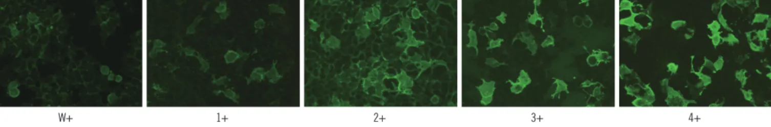
cells were washed 4 times in PBS and observed under a fluo- rescence microscope. Since the EGFP-AQP4 protein is synthe- sized in the cytoplasm and expressed on the cell membrane, a flat, smooth, fine granular green fluorescence signal is detected both in cytoplasm and at the cell surface membrane, but not in the nucleus. The BIOCHIP slide is composed of combinations of transfected and non-transfected cellular substrate for each patient’s test, and the negative and positive control sera are provided to ensure quality control of the entire procedure. The fluorescence was scored from weak positive to 4 positive (w+ to 4+), according to the intensity scale of the conventional fluores- cence anti-nuclear antibody test (FANA; Fig. 1). The presence of AQP4 was confirmed if the intensity was 1+ or stronger. For the semi-quantitative evaluation, the titer was defined as the sample dilution factor for which specific fluorescence was iden- tifiable. The dilution series was 1:10 to 1:640, by a factor of 2.
3. FIPA for AQP4 antibody detection
FIPA was performed according to the in-house protocol de- scribed in detail elsewhere [20]. Cloning of AQP4 and transfec- tion of human HEK 293 cells were performed in a same manner as that of CIIFA. EGFP-AQP4 extract was acquired from lysis and high-speed centrifugation of EGFP-AQP4-transfected HEK 293 cells and was used as an antigen substrate. Individual se- rum samples were incubated with cellular extract at 4°C over- night. Then, the IgG-AQP4 complexes were precipitated using Protein A sepharose beads, washed thoroughly, resuspended in extract buffer, and transferred to a 96-well black PCR plate. The amount of EGFP-AQP4 bound by antibody was detected by counting the green fluorescence at 512 nm (excitation 472 nm;
cut-off 495 nm) on a fluorescence plate reader (SpectraMAX Gemini XS; Molecular Devices, Sunnyvale, CA, USA). Results were given as arbitrary quantitative fluorescence unit (FU) and the mean +3SD from healthy control samples was used as a cut-off value. In this study, the cut-off value was derived from 14 healthy controls and was 575 FU.
Fig. 1. Cell-based indirect immunofluorescence assay (CIIFA) for aquaporin-4 (AQP4) antibody detection with AQP4-transfected HEK 293 cell line as a substrate. Representative samples showing an intensity of w+ to 4+ are provided above. W+ was assigned when the intensity of fluorescence was less than 1+. Of note, fluorescence was also observed in round cells, which might not reflect healthy, viable HEK cells during slide preparation.
W+ 1+ 2+ 3+ 4+
4. Statistical analysis
The sensitivity and specificity of autoantibody detection by each assay, individually and in combination, was determined by refer- ence to the clinical diagnoses. Antibody positivity and levels be- tween groups were analyzed by Fisher’s exact test and the Mann-Whitney test, respectively. The McNemar test was used to analyze the agreement between the 2 CIIFA results and be- tween CIIFA and FIPA, and also to compare the significance of differences in sensitivity and specificity between CIIFA and FIPA. P <0.05 was considered significant. Analyses were per- formed using MedCalc® version 12.0.4.0 (MedCalc Software, Mariakerke, Belgium).
RESULTS
1. Comparison of FIPA and CIIFA for the detection of AQP4 antibodies
We performed the AQP4 antibody detection assay in 46 serum samples of 36 patients using FIPA and 2 different CIIFA meth- ods, the first was commercially available (CIIFA A) and the other was developed in-house (CIIFA B). The concordance rate be- tween the 2 CIIFA methods was 76% (Kappa coefficient (κ=
0.6522, 95% confidence interval [CI]=0.4331-0.8713, P =0.2891) and between CIIFA A and FIPA B was 89% (κ=0.781, 95% CI=
0.5997-0.9623, P =0.3750). There were 11 discordant results be- tween CIIFA A and CIIFA B, where only 3 were true discrepan- cies and the other 8 resulted in discrepancies mainly due to in- ability of the CIIFA B to provide data regarding the conclusive
fluorescence pattern, as the in-house cellular substrate prepara- tion was of variable quality. The results of each test are summa- rized in Table 1.
The sensitivity of CIIFA using the commercial kit (data from CIIFA A) and FIPA in this comparison set was 86% and 79% in NMO patients and 55% and 36% in high-risk NMO patients, re- spectively. The combination of CIIFA and FIPA did not increase the sensitivity of detection of the presence of AQP4. Neither the sensitivity nor the specificity differed significantly in the 2 assays (McNemar test; Tables 1, 2).
The semiquantitative titer of CIIFA was well correlated with the arbitrary quantity (FU) of FIPA (r=0.66; Fig. 2). The AQP4 titers measured by CIIFA and FIPA were elevated in NMO pa- tients compared to those in high-risk NMO patients (1:240 vs.
1:180 and 8,390 FU vs. 4,059 FU, respectively).
2. Frequency of AQP4 antibodies detected by CIIFA in patient populations
For the routine detection of AQP4 antibodies as a clinical labora- tory practice, the commercially available CIIFA was used. AQP4 antibodies were detected in 17 of 101 consecutive patients in whom AQP4 testing was requested at the neurology clinic during a 10-month period. The clinical characteristics and presence of other autoantibodies are summarized in Table 3. NMO patients who were AQP4 antibody-positive were predominantly female (males :females =1:5) and were associated with a longer dis- ease duration than other patients. Autoantibodies such as anti- nuclear antibodies (ANA), anti-Ro antibodies (SSA), and anti-La antibodies (SSB) were found in 1 (17%) NMO patient. AQP4 an- tibodies were present in 6/6 (100%) patients with NMO, in 7/24 (29%) LETM patients, and 1/11 (9%) RON/BON patients, but only 3/65 (4.6%) in control patients (2/31 in CIS patients, 1/2 in Table 1. Results of aquaporin-4 (AQP4) antibody assays in 46 serum
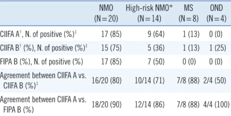
samples
NMO
(N=20) High-risk NMO*
(N=14) MS
(N=8) OND (N=4) CIIFA A†, N. of positive (%)‡ 17 (85) 9 (64) 1 (13) 0 (0) CIIFA B† (%), N. of positive (%)‡ 15 (75) 5 (36) 1 (13) 1 (25) FIPA B (%), N. of positive (%) 17 (85) 7 (50) 0 (0) 0 (0) Agreement between CIIFA A vs.
CIIFA B (%)‡ 16/20 (80) 10/14 (71) 7/8 (88) 2/4 (50) Agreement between CIIFA A vs.
FIPA B (%) 18/20 (90) 12/14 (86) 7/8 (88) 4/4 (100)
*High risk NMO includes bilateral or recurrent optic neuritis and longitudi- nal extensive transverse myelitis; †A and B represent the 2 institutions where each of the tests were performed; ‡Among 11 discordant results between CIIFA A and CIIFA B, 8 of the results performed at institution B using the in- house CIIFA could not provide a conclusive fluorescence pattern due to poor quality of the preparation of cellular substrate.
Abbreviations: NMO, neuromyelitis optica; MS, multiple sclerosis; OND, oth- er neurological diseases; CIIFA, cell-based indirect immunofluorescence as- say; FIPA, fluorescence immunoprecipitation assay.
Table 2. Sensitivity and specificity of CIIFA and FIPA for the diagno- ses of NMO and high-risk NMO
Clinical diagnosis
Estimated sensitivity,
% (95% CI) P value, CIIFA vs.
FIPA
Estimated specificity,
% (95% CI) P value, CIIFA vs.
CIIFA FIPA CIIFA+ FIPA
FIPA* CIIFA FIPA CIIFA+
FIPA*
NMO
(N=14) 86 79 86 1.0 91 100 100 1.0
High-risk NMO
(N=11) 55 36 55 0.5 91 100 100 1.0
*CIIFA+FIPA includes patients who were positive for either CIIFA A or FIPA.
Abbreviations: CIIFA, cell-based indirect immunofluorescence assay; FIPA, fluorescence immunoprecipitation assay; NMO, neuromyelitis optica; CI, confidence interval.
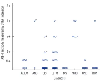
patients with autoimmune neurological diseases (AND), and none in 51 other demyelinating neurological diseases such as MS, ADEM, and OND (Chi-square test, P <0.0001; Fig. 3). These results correspond to a sensitivity of 100% and a specificity of 95% for the test in NMO, and to 39% and 94%, respectively, if NMO and high-risk NMO patients such as LETM and RON/BON patients are considered together. The fluorescence intensities of the CIIFA in NMO patients were significantly higher than those in the other disease groups (Mann-Whitney test, NMO vs. NMOSD, P =0.0026; NMO vs. OND, P <0.0001).
DISCUSSION
In recent years, tests such as CIIFA, radioimmunoprecipitation assay (RIPA), FIPA and ELISA have been developed for the de- tection of the presence of specific AQP4 antibodies. The cell- based assay (CBA), which utilizes AQP4 expressed on the cell surface in a naturally folded state as an antigen, showed advan- tages in sensitivity and specificity over NMO-IgG using mouse brain tissue or ELISA using synthesized recombinant peptide or protein [15, 18, 20, 21]. Subsequently, in-house CBAs based on Table 3. Patient characteristics and seroprevalence of aquaporin-4 (AQP4) antibodies
NMO spectrum disorders (N=36) Controls (N=65)
NMO RON/BON LETM CIS ADEM MS AND OND
Number of patients 6 11 24 31 6 7 2 14
Males:Females 1:5 6:5 11:12 17:14 5:1 1:5 0:2 6:8
Age (range) 36 (27-55) 30 (24-67) 46 (17-71) 36 (19-70) 40 (21-70) 31 (22-44) 54 (48-60) 53 (33-62) Disease duration at sampling (range) 9 (8-11) 2 (1-8) 1 (0.5-11) 1 (1-2) 2 (0.5-4) 4 (2-6) 4 (1-6) - Spinal cord lesion, N. of segments
(range) 6 (2-8) - 4 (3-10) 2 (1-2.5) - 1 (1) - -
N. of attacks, (range) 2 (1-4) 2 (1-5) 2 (1-4) 1 (1-4) - 3 (1-6) 1 (1-2)
ANA/SSA/SSB (%) 1 (17) 1 (9) 10 (42) 1 (3) 0 (0) 1 (14) 0 (0) 1 (7)
Frequency of AQP4 antibodies (%)* 6 (100) 1 (9) 7 (29) 2 (7) 0 (0) 0 (0) 1 (50) 0 (0)
Values are described as medians unless otherwise specified.
*Only included the result that had an intensity greater than 1+ by CIIFA.
Abbreviations: NMO, neuromyelitis optica; RON/BON, recurrent/bilateral optic neuritis; LETM, longitudinal extensive transverse myelitis; CIS, clinical isolated syndrome; ADEM, acute disseminating encephalomyelitis; MS, multiple sclerosis; AND, autoimmune neurologic diseases; OND, other neurological diseases;
ANA, antinuclear antibodies; SSA, anti-Ro antibodies; SSB, anti-La antibodies.
Fig. 2. Distribution of AQP4 antibody values in 46 comparison samples. (A) Correlation of the results between the semiquantitative titer of the cell-based indirect immunofluorescence assay (CIIFA) and the quantitative value derived from the fluorescence immunoprecipitation assay (FIPA) based on arbitrary fluorescence units (FU). (B) Titers of AQP4 antibody measured by CIIFA in sera of patients with various dis- eases.
Abbreviations: MS, multiple sclerosis; NMO, neuromyelitis optica; OND, other neurological diseases.
CIIFA (titer)
800 700 600 500 400 300 200 100
00 5,000 10,000 15,000 20,000 25,000 30,000 FIPA (FU)
R2=0.6565
A
AQP4 antibody measured by CIIFA (titer)
700 600 500 400 300 200 100 0
MS NMO High risk NMO OND
Diagnosis B
different detection principles such as indirect immunofluores- cence [20, 22], flow cytometry [23, 24], or cytotoxicity [25] were developed. All of these methods utilize HEK cell lines trans- fected with full-length recombinant human AQP4 for antigenic preparations, thus providing the naturally folded AQP4 protein as an antigenic target.
Recently, EUROIMMUN AG has introduced the commercially available CIIFA [16]. In our comparison set between the in- house CIIFA and the commercially available CIIFA, we had sev- eral discordant results due to an inconclusive fluorescence pat- tern observed in the in-house CIIFA, which was derived primar- ily from the poor quality of cellular substrate preparation. In contrast, by using the commercially available kit we completely avoided inconclusive results in 147 samples, including 46 com- parison samples and 101 consecutively enrolled patient sam- ples. Nevertheless, the preparation of transfected cells as an antigenic source in the CBA might have caused the variation of results. Test kits manufactured under standardized conditions may improve the consistency of in-house test preparation. In our prospective cohort in which we evaluated the clinical useful- ness of CIIFA, AQP4 antibodies were present in 100% (6/6) of patients with NMO and 23% (8/35) of high-risk NMO such as LETM and BON/RON patients, but was virtually absent in pa- tients with MS and other inflammatory and non-inflammatory
neurological diseases with the exception of 1 patient with an au- toimmune disease and 2 CIS patients. Considering the differ- ences in diagnostic criteria, the study design (i.e., whether pa- tients and sera were acquired consecutively on a clinical basis rather than selected from a pool of known cases and controls), and the test method, direct comparisons of diagnostic values of AQP4 antibody tests reported in other studies may not be appli- cable. However, the sensitivity of the CIIFA used in this study was superior compared to those of other studies, such as 91% sensitivity reported by Takahashi et al. [18, 22] and 73% sensi- tivity reported by McKeon et al. [26]. More importantly, the specificity of CIIFA for detection of AQP4 antibodies was excel- lent (94%), thus the clinical relevance of this test in the discrimi- nation of NMO from MS and other demyelinating diseases was significant.
NMOSDs have been reported in patients with systemic con- nective-tissue diseases such as systemic lupus erythematosus (SLE) or Sjögren’s syndrome [27, 28]; of note, autoantibody markers of SLE or Sjögren’s syndrome are found in almost 40% of patients with NMO and high-risk NMO [14]. However, AQP4 antibodies are not present in patients with systemic connective- tissue diseases in the absence of CNS involvement or with CNS involvement other than NMO [29]. In our study, 4 Sjögren’s syn- drome patients were diagnosed with NMO and LETM (1 patient) and high-risk NMO (3 patients), and 3 of these patients had AQP4 antibodies. Min et al. reported that 75% of Sjögren’s syn- drome patients with recurrent brain lesions had AQP4 antibod- ies and met the criteria for NMOSDs such as NMO in Korean patients with Sjögren’s syndrome [30].
FIPA has been developed by using the principle of an immu- noprecipitation assay for routine use and quantitative measure- ment of AQP4 antibodies [20]. Detection of AQP4 antibodies us- ing FIPA was reported to have comparable sensitivity and speci- ficity and correlate highly with the results of CBAs [15, 20]. The concordance between FIPA and CIIFA performed at our hospital was 86-100%, depending on the diagnosis. The AQP4 antibody level detected by FIPA and titers detected by CIIFA derived from the dilution factor of the patient sera were proportional and had a tendency to correlate well with each other. Since the in vivo pathogenic role of AQP4 antibodies (which are predominantly of the IgG1 subclass and activate complement after binding to ex- tracellular epitopes) is well described [20, 21], the quantitative measurement of AQP4 antibodies may provide insight into the clinical course and treatment response of AQP4 antibody-re- lated diseases. Serial measurements of the AQP4 antibody level by FIPA to monitor the treatment response or relapse during the
AQP4 antibody measured by CIIFA (intensity
) 4+
3+
2+
1+
W+
ADEM AND CIS LETM MS NMO OND RON
Diagnosis
*
*
*
Fig. 3. The fluorescence intensity of AQP4 antibodies measured by the cell-based indirect immunofluorescence assay (CIIFA) in sera of 101 patients with various diseases. There were only 3 AQP4 anti- body-positive patients (*) among non-NMO and non-high-risk NMO patients. Weak positive intensity was not considered to be a signifi- cant positive result.
Abbreviations: ADEM, acute disseminating encephalomyelitis; AND, autoim- mune neurologic diseases; CIS, clinical isolated syndrome; LETM, longitudi- nal extensive transverse myelitis; MS, multiple sclerosis; NMO, neuromyelitis optica; OND, other neurological diseases; RON, recurrent optic neuritis.
clinical course have been reported [18, 29, 31]. Takahashi et al.
[18] observed that the AQP4 antibody titer was related to spinal cord lesion length and Jarius et al. [31] noted that antibody lev- els were higher if serum samples were obtained during a re- lapse and before commencement of immunosuppression. How- ever, in spite of these potential applications, the establishment of in-house FIPA is quite problematic since there are several steps that can cause variability during the test procedure. In particular, the preparation of antigenic material in each batch of test includes multiple procedures such as maintenance of HEK cell lines, preparation of the transfecting vector and DNA, trans- fection, and cell lysate processing. Moreover, establishment of a cut-off point is arbitrary in each laboratory, thus the transferabil- ity of quantitative data is limited, and there is no standardized control material to validate the quantitative value generated from each test. In this respect, CIIFA, a CBA using indirect immuno- fluorescence principles has several advantages over FIPA. First, the antigenic material prepared on slides can be manufactured on a large scale and stored for a relatively long duration of time.
Second, the test procedure is conventional IIFA, which is widely performed in clinical laboratories. Third, the interpretation of fluorescence intensity is a standardized concept among clinical pathologists.
In this study, we demonstrated that the commercially avail- able CIIFA was well correlated with FIPA for the detection and quantitation of AQP4 antibodies, and exhibited a high sensitivity and excellent specificity for the diagnosis of NMO and high-risk NMO diseases. Nevertheless, the usefulness of titration of CIIFA for the prediction of the extent of spinal cord lesions and moni- toring of disease progression or treatment response needs to be actively investigated in a prospective study on a larger scale.
Authors’ Disclosures of Potential Conflicts of Interest
No potential conflicts of interest relevant to this article were re- ported.
Acknowledgements
This work was supported by a grant of the Korea Healthcare technology R&D Project by the Ministry of Health, Welfare, and Family Affairs in the Republic of Korea (Grant No. A080588).
We thank Dr. Angela Vincent, MBBS, MSc, FRCPath of the Neuroscience Group at the Weatherall Institute of Molecular Medicine and Department of Clinical Neurology at the University
of Oxford, for performing the in-house CIIFA and FIPA for our comparison study.
REFERENCES
1. Devic E. Subacute myelitis complicated by optic neuritis. Bull Med 1894; 8:1033-4.
2. Wingerchuk DM, Hogancamp WF, O’Brien PC, Weinshenker BG. The clinical course of neuromyelitis optica (Devic’s syndrome). Neurology 1999;53:1107-14.
3. de Seze J, Stojkovic T, Ferriby D, Gauvrit JY, Montagne C, Mounier-Ve- hier F, et al. Devic’s neuromyelitis optica: clinical, laboratory, MRI and outcome profile. J Neurol Sci 2002;197:57-61.
4. Wingerchuk DM, Lennon VA, Lucchinetti CF, Pittock SJ, Weinshenker BG. The spectrum of neuromyelitis optica. Lancet Neurol 2007;6:805- 15.
5. Kira J. Multiple sclerosis in the Japanese population. Lancet Neurol 2003;2:117-27.
6. Matsushita T, Isobe N, Matsuoka T, Shi N, Kawano Y, Wu XM, et al.
Aquaporin-4 autoimmune syndrome and anti-aquaporin-4 antibody- negative opticospinal multiple sclerosis in Japanese. Mult Scler 2009;15: 834-47.
7. Lennon V, Wingerchuk D, Kryzer T, Pittock S, Lucchinetti C, Fujihara K, et al. A serum autoantibody marker of neuromyelitis optica: distinction from multiple sclerosis. Lancet 2004;364:2106-12.
8. Lennon VA. IgG marker of optic-spinal multiple sclerosis binds to the aquaporin-4 water channel. J Exp Med 2005;202:473-7.
9. Hiroaki Y, Tani K, Kamegawa A, Gyobu N, Nishikawa K, Suzuki H, et al.
Implications of the aquaporin-4 structure on array formation and cell adhesion. J Mol Biol 2006;355:628-39.
10. Paul F, Jarius S, Aktas O, Bluthner M, Bauer O, Appelhans H, et al. An- tibody to aquaporin 4 in the diagnosis of neuromyelitis optica. PLoS Med 2007;4:e133.
11. Papeix C, Vidal JS, de Seze J, Pierrot-Deseilligny C, Tourbah A, Stankoff B, et al. Immunosuppressive therapy is more effective than interferon in neuromyelitis optica. Mult Scler 2007;13:256-9.
12. Cree BA, Lamb S, Morgan K, Chen A, Waubant E, Genain C. An open label study of the effects of rituximab in neuromyelitis optica. Neurology 2005;64:1270-2.
13. Keegan M, Pineda AA, McClelland RL, Darby CH, Rodriguez M, Wein- shenker BG. Plasma exchange for severe attacks of CNS demyelination:
predictors of response. Neurology 2002;58:143-6.
14. Jarius S and Wildemann B. AQP4 antibodies in neuromyelitis optica: di- agnostic and pathogenetic relevance. Nat Rev Neurol 2010;6:383-92. 15. Waters P and Vincent A. Detection of anti-aquaporin-4 antibodies in
neuromyelitis optica: current status of the assays. Int MS J 2008;15:99- 105.
16. Jarius S, Probst C, Borowski K, Franciotta D, Wildemann B, Stoecker W, et al. Standardized methods for the detection of antibodies to aquapo- rin-4 based on a highly sensitive immunofluorescence assay employing recombinant target antigen. J Neurol Sci 2010;291:52-6.
17. Hayakawa S, Mori M, Okuta A, Kamegawa A, Fujiyoshi Y, Yoshiyama Y, et al. Neuromyelitis optica and anti-aquaporin-4 antibodies measured by an enzyme-linked immunosorbent assay. J Neuroimmunol 2008;196: 181-7.
18. Takahashi T, Fujihara K, Nakashima I, Misu T, Miyazawa I, Nakamura M, et al. Anti-aquaporin-4 antibody is involved in the pathogenesis of NMO:
a study on antibody titre. Brain 2007;130:1235-43.
19. Wingerchuk DM, Lennon VA, Pittock SJ, Lucchinetti CF, Weinshenker BG. Revised diagnostic criteria for neuromyelitis optica. Neurology 2006;
66:1485-9.
20. Waters P, Jarius S, Littleton E, Leite MI, Jacob S, Gray B, et al. Aquapo- rin-4 antibodies in neuromyelitis optica and longitudinally extensive transverse myelitis. Arch Neurol 2008;65:913-9.
21. Lucchinetti CF, Mandler RN, McGavern D, Bruck W, Gleich G, Ranso- hoff RM, et al. A role for humoral mechanisms in the pathogenesis of Devic’s neuromyelitis optica. Brain 2002;125:1450-61.
22. Takahashi T, Fujihara K, Nakashima I, Misu T, Miyazawa I, Nakamura M, et al. Establishment of a new sensitive assay for anti-human aquaporin-4 antibody in neuromyelitis optica. Tohoku J Exp Med 2006;210:307-13. 23. Fazio R, Malosio M, Lampasona V, De Feo D, Privitera D, Marnetto F, et
al. Antiacquaporin 4 antibodies detection by different techniques in neuromyelitis optica patients. Mult Scler 2009;15:1153-63.
24. De Vidi I, Boursier G, Delouche N, Portalès P, Cadars E, Bouthier M, et al. Strategy for anti-aquaporin-4 auto-antibody identification and quanti- fication using a new cell-based assay. Clin Immunol 2011;138:239-46. 25. Hinson SR, McKeon A, Fryer JP, Apiwattanakul M, Lennon VA, Pittock
SJ. Prediction of neuromyelitis optica attack severity by quantitation of complement-mediated injury to aquaporin-4-expressing cells. Arch
Neurol 2009;66:1164-7.
26. McKeon A, Fryer JP, Apiwattanakul M, Lennon VA, Hinson SR, Kryzer TJ, et al. Diagnosis of neuromyelitis spectrum disorders: comparative sensitivities and specificities of immunohistochemical and immunopre- cipitation assays. Arch Neurol 2009;66:1134-8.
27. Pittock SJ, Lennon VA, de Seze J, Vermersch P, Homburger HA, Wing- erchuk DM, et al. Neuromyelitis optica and non organ-specific autoim- munity. Arch Neurol 2008;65:78-83.
28. Jarius S, Jacobi C, de Seze J, Zephir H, Paul F, Franciotta D, et al. Fre- quency and syndrome specificity of antibodies to aquaporin-4 in neuro- logical patients with rheumatic disorders. Mult Scler 2011;17:1067-73. 29. Hinson SR, McKeon A, Lennon VA. Neurological autoimmunity target-
ing aquaporin-4. Neuroscience 2010;168:1009-18.
30. Min JH, Kim HJ, Kim BJ, Lee KW, Sunwoo IN, Kim SM, et al. Brain ab- normalities in Sjogren syndrome with recurrent CNS manifestations: as- sociation with neuromyelitis optica. Mult Scler 2009;15:1069-76. 31. Jarius S, Aboul-Enein F, Waters P, Kuenz B, Hauser A, Berger T, et al.
Antibody to aquaporin-4 in the long-term course of neuromyelitis optica.
Brain 2008;131:3072-80.