http://dx.doi.org/10.4174/astr.2014.86.5.227 Annals of Surgical Treatment and Research
Cervical bronchogenic cysts mimic metastatic lymph nodes during thyroid cancer surgery
Hak Hoon Jun, Seok Mo Kim1, Yong Sang Lee1, Soon Won Hong2, Hang-Seok Chang1, Cheong Soo Park1
Department of Surgery, CHA Bundang Medical Center, CHA University, Seongnam, 1Department of Surgery, Thyroid Cancer Center, Gangnam Severance Hospital, Yonsei University College of Medicine, Seoul, 2Department of Pathology, Gangnam Severance Hospital, Yonsei University College of Medicine, Seoul, Korea
INTRODUCTION
Bronchogenic cysts are benign congenital anomalies related to the abnormal budding of the tracheobronchial tree during embryological development [1,2]. Most bronchogenic cysts occur in the mediastinum or within the pulmonary parenchyma, whereas cysts in the thyroid or perithyroidal area are rare [3].
Since many adults with bronchogenic cysts do not experience any symptoms, the cysts are often detected incidentally during
the diagnosis of other diseases [4]. Although symptomatic bronchogenic cysts should be excised, the treatment of asymptomatic cervical bronchogenic cysts remains unclear [5]. Cystic lesions in the neck include branchial cleft cysts, thyroglossal duct cysts, bronchogenic cysts, thymic or thyroid cysts, and metastatic lymph nodes [6]. Cystic degeneration is also common in metastatic lymph nodes associated with thyroid cancer [7,8]. Metastatic lymph nodes affect the extent of lymph node dissection, especially in the lateral neck region.
Purpose: Although congenital bronchogenic cysts in the cervical region, especially in the thyroid or perithyroidal area, are rare, distinguishing them from other cervical cystic lesions (e.g., thyroglossal duct and branchial cleft cysts) and metastatic cervical lymph nodes is difficult preoperatively. Additionally, cystic degeneration of metastatic lymph nodes is common in patients with thyroid cancer. We investigated the clinical characteristics and proper treatment for individuals with cervical bronchogenic cysts.
Methods: Of the 18,900 patients treated for thyroid cancer, 18 patients with pathologically confirmed bronchogenic cysts were retrospectively reviewed. Bilateral total thyroidectomy or less than total thyroidectomy with central compartment node dissection, including cystic mass excision was done and cystic mass was confirmed by postoperative pathologic examination.
Results: All cervical bronchogenic cysts were asymptomatic. Their mean size was 1.2 cm (range, 0.3 to 3 cm). Of these 18 patients, 15 did not have any abnormal radiological findings, except for lymphadenopathy during preoperative evaluations.
Most bronchogenic cysts were detected around the thyroid and paratracheal areas. On preoperative imaging and intraoperatively, most were indistinguishable from metastatic cervical lymph nodes or other cystic lesions.
Conclusion: Although cervical bronchogenic cysts are rare and benign, they should be distinguished from other cystic cervical masses, especially metastatic cervical lymph nodes associated with thyroid cancer. Possible cervical bronchogenic cysts found during thyroid cancer evaluation or surgery should be surgically excised.
[Ann Surg Treat Res 2014;86(5):227-231]
Key Words: Bronchogenic cyst, Thyroid neoplasms
Received October 1, 2013, Revised January 3, 2014, Accepted January 3, 2014
Corresponding Author: Hang-Seok Chang
Department of Surgery, Thyroid Cancer Center, Gangnam Severance Hospital, Yonsei University College of Medicine, 211 Eonju-ro, Gangnam- gu, Seoul 135-720, Korea
Tel: +82-2-2019-3370, Fax: +82-2-3462-5994 E-mail: surghsc@yuhs.ac
Copyright ⓒ 2014, the Korean Surgical Society
cc Annals of Surgical Treatment and Research is an Open Access Journal. All articles are distributed under the terms of the Creative Commons Attribution Non- Commercial License (http://creativecommons.org/licenses/by-nc/3.0/) which permits unrestricted non-commercial use, distribution, and reproduction in any medium, provided the original work is properly cited.
Annals of Surgical Treatment and Research 2014;86(5):227-231
Therefore, correctly diagnosing cystic lesions in the neck is important for thyroid cancer surgery. However, benign bronchogenic cysts appear similar to metastatic lymph nodes, both by preoperative imaging and intraoperative field findings.
The aim of this study was to review the clinical characteristics and proper management for cervical bronchogenic cysts that mimic metastatic lymph nodes during thyroid cancer surgery.
METHODS
Of the 18,900 patients treated for thyroid cancer at the Thyroid Cancer Center, Gangnam Severance Hospital, Younsei University College of Medicine, between January 2007 and April 2012, 18 patients (3 males, 15 females; mean age, 52.6 years) with bronchogenic cysts confirmed by postoperative pathologic examination were retrospectively reviewed in this study. Preoperative neck ultrasound and CT were performed for thyroid evaluation, and thyroid cancer was confirmed by fine needle aspiration cytology. Extent of thyroidectomy and lymph node dissection was determined using American Thyroid Association guidelines [9]. This retrospective study was approved by the Institutional Review Board of Gangnam Severance Hospital, Younsei University College of Medicine, Seoul, Korea.
RESULTS
Of the 18,900 patients who underwent thyroid cancer surgery, 18 (0.1%) were confirmed as having cervical bronchogenic cysts;
their clinical characteristics are shown in Table 1. All cervical bronchogenic cysts were asymptomatic, and cystic lesions were found during either preoperative diagnostic evaluation or thyroid cancer surgery. The mean size of the cervical bronchogenic cysts was 1.2 cm (range, 0.3 to 3 cm). Eleven cysts were located in the right paratracheal area, 3 in the left paratracheal area, and 1 each in the left thyroid cartilage, left inferior thyroid, right level III region, and anterior mediastinum.
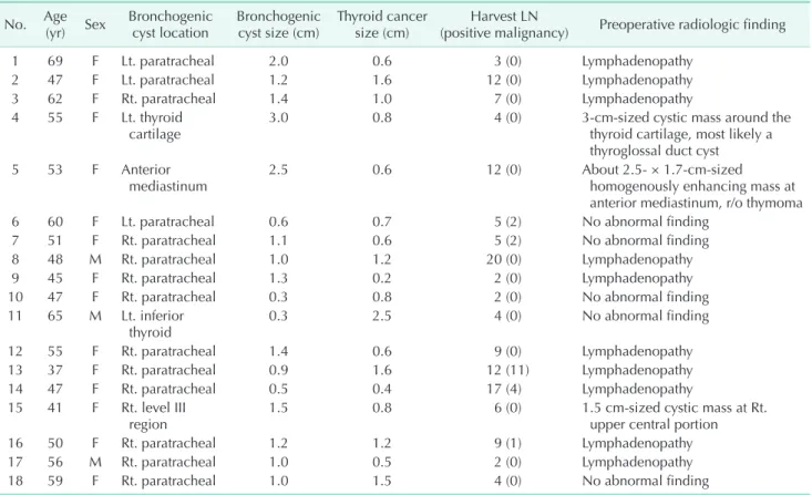
Fifteen patients underwent bilateral total thyroidectomy, whereas three underwent less than total thyroidectomy. All patients underwent central compartment node dissection, including cystic mass removal. None of these patients had any postoperative complications. Metastatic lymph nodes associated with thyroid cancer were found in five patients. Patient 4 was a 55-year-old female with an asymptomatic soft mass in the neck (Table 1). CT showed a homogeneous cystic mass that measured 3 cm × 1.5 cm around the thyroid cartilage (Fig. 1A).
Although it was initially diagnosed as either a thyroglossal duct cyst or a metastatic lymph node with cystic degeneration, postsurgical analysis determined that it was a cyst lined with
Table 1. Clinical characteristics of the 18 included patients No. Age
(yr) Sex Bronchogenic
cyst location Bronchogenic
cyst size (cm) Thyroid cancer
size (cm) Harvest LN
(positive malignancy) Preoperative radiologic finding
1 69 F Lt. paratracheal 2.0 0.6 3 (0) Lymphadenopathy
2 47 F Lt. paratracheal 1.2 1.6 12 (0) Lymphadenopathy
3 62 F Rt. paratracheal 1.4 1.0 7 (0) Lymphadenopathy
4 55 F Lt. thyroid
cartilage 3.0 0.8 4 (0) 3-cm-sized cystic mass around the
thyroid cartilage, most likely a thyroglossal duct cyst
5 53 F Anterior
mediastinum 2.5 0.6 12 (0) About 2.5- × 1.7-cm-sized
homogenously enhancing mass at anterior mediastinum, r/o thymoma
6 60 F Lt. paratracheal 0.6 0.7 5 (2) No abnormal finding
7 51 F Rt. paratracheal 1.1 0.6 5 (2) No abnormal finding
8 48 M Rt. paratracheal 1.0 1.2 20 (0) Lymphadenopathy
9 45 F Rt. paratracheal 1.3 0.2 2 (0) Lymphadenopathy
10 47 F Rt. paratracheal 0.3 0.8 2 (0) No abnormal finding
11 65 M Lt. inferior
thyroid 0.3 2.5 4 (0) No abnormal finding
12 55 F Rt. paratracheal 1.4 0.6 9 (0) Lymphadenopathy
13 37 F Rt. paratracheal 0.9 1.6 12 (11) Lymphadenopathy
14 47 F Rt. paratracheal 0.5 0.4 17 (4) Lymphadenopathy
15 41 F Rt. level III
region 1.5 0.8 6 (0) 1.5 cm-sized cystic mass at Rt.
upper central portion
16 50 F Rt. paratracheal 1.2 1.2 9 (1) Lymphadenopathy
17 56 M Rt. paratracheal 1.0 0.5 2 (0) Lymphadenopathy
18 59 F Rt. paratracheal 1.0 1.5 4 (0) No abnormal finding
LN, lymph node; r/o, rule out.
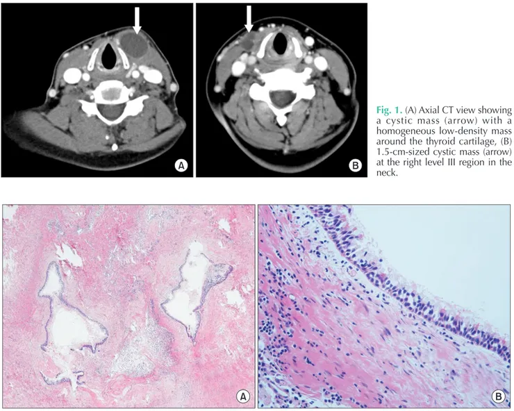
pseudostratified, ciliated, columnar epithelium containing underlying seromucinous glands. CT of patient 15 showed a 1.5-cm-sized cystic mass at the right level III region in the neck (Fig. 1B). The cystic mass was excised during thyroidectomy and was identified as a bronchogenic cyst (Fig. 2). Of these 18 patients, 15 did not have any abnormal radiological findings, except for lymphadenopathy during preoperative evaluations.
Cervical bronchogenic cysts were less than 1.5 cm in size and were detected around the paratracheal area. Although these cysts resembled central lymph nodes in operative field analyses, final pathologic examinations identified them as bronchogenic cysts.
DISCUSSION
The tracheobronchial tree, which consists of the ventral
trachea and the dorsal esophagus, is formed during the fifth week of embryogenesis. Bronchogenic cysts are often caused by an abnormal budding of the primitive foregut’s tracheobronchial tree. When the connection with the tracheobronchial tree is lost, the bronchial buds may migrate to an aberrant position [1,10]. Bronchogenic cysts have been classified according to their site of origin: paratracheal, carinal, hilar, paraesophageal, and atypical (such as diaphragmatic, abdominal, intracutaneous, or subcutaneous, or in the supraclavicular neck area) [11].
Abnormal budding during tracheal development can cause bronchogenic cysts to form on the midline of the upper neck. However, cysts may also develop in the lower and lateral portions of the neck, if abnormal budding occurs during bronchial system development [12]. The thyroid and paratracheal regions are more frequently affected than the supraclavicular region and suprasternal notch, and the majority Fig. 1. (A) Axial CT view showing a cystic mass (arrow) with a homogeneous low-density mass around the thyroid cartilage, (B) 1.5-cm-sized cystic mass (arrow) at the right level III region in the neck.
Fig. 2. The pathologic findings of bronchogenic cyst. (A) The lesion comprises variable sized cysts and fibrotic tissue with inflammation (H&E, ×40). (B) The cysts are lined by respiratory type epithelium, which is characterized by pseudostratified columnar cells and cilia (H&E, ×400).
Annals of Surgical Treatment and Research 2014;86(5):227-231
1. Shimizu J, Kawaura Y, Tatsuzawa Y, Maeda K, Suzuki S. Cervical bronchogenic cyst that presented as a thyroid cyst. Eur J Surg 2000;166:659-61.
2. Teissier N, Elmaleh-Berges M, Ferkdadji L, François M, Van den Abbeele T. Cervical
bronchogenic cysts: usual and unusual clinical presentations. Arch Otolaryngol Head Neck Surg 2008;134:1165-9.
3. Min KW, Jang SH, Song YS, Cho SH, Chon SH, Paik SS. An unusual case of bron- chogenic cyst mimicking thyroid cystic
tumor. Otolaryngol Head Neck Surg 2007;137:520-1.
4. Bocciolini C, Dall'olio D, Cunsolo E, Latini G, Gradoni P, Laudadio P. Cervical bronchogenic cyst: asymptomatic neck mass in an adult male. Acta Otolaryngol
REFERENCES
of cysts are midline [13]. In our study, 16 of the 18 cysts were detected around the thyroid and paratracheal areas, with the right paratracheal area more frequently affected, but we could not find any connection with the trachea.
Bronchogenic cysts in the cervical area are usually asympto- matic and are often detected incidentally through diagnostic evaluation of other diseases. Large cysts may cause dyspnea, respiratory distress, cough, and dysphagia in some individuals.
Occasionally, secondary infection may occur, resulting in sinus tract formation, external drainage of purulent material if the cyst is superficial, or abscess formation if the cyst is deep [14]. The majority of intrathoracic bronchogenic cysts can be detected by chest x-rays, but these have little diagnostic value for cysts located in the neck [15]. CT provides a more accurate localization of these lesions, showing that typically they are round, fluid-filled, well-circumscribed masses of varying densities. Barium studies have also demonstrated abnormal communication of the respiratory tract with the esophagus or stomach [4]. However, definitive identification of these cysts can only be achieved by histopathological examination.
Bronchogenic cysts are characterized by a pseudostratified, ciliated, columnar epithelial lining with underlying seromu- cinous glands [16]. Cervical bronchogenic cysts should be examined to differentiate them from branchial cleft cysts, thyroglossal duct cysts, thymic and thyroid cysts, dermoids and lymphangiomas, cystic hygromas, teratomas, and cystic neuromas [6]. Thyroglossal duct cysts and branchial cleft cysts are more common congenital abnormalities initially seen in the upper triangles of the neck [17]. Branchial cleft cysts are usually located high and lateral, whereas thyroglossal duct cysts are typically located midline on the anterior portion close to the hyoid bone. Bronchogenic cysts should also be distinguished from metastatic lymph nodes associated with thyroid cancer.
Cystic degeneration is a common feature of metastatic lymph nodes associated with thyroid cancer [7,8]. Furthermore, the presence of metastatic lymph nodes affects the extent of lymph node dissection, especially in the lateral neck, and postoperative radioactive iodine therapy. In our study, we could not differentiate them from lymph node metastasis or other cystic lesions in preoperative imaging study or intraoperative
features. Of these 18 patients, 15 did not have any abnormal radiological findings, except for lymphadenopathy and these cysts resembled central lymph nodes in operative field analyses.
After the patients underwent thyroidectomy and central compartment node dissections, which included cystic mass removal, the bronchogenic cysts were identified via pathologic examination. If we had measured level of thyroglobulin (Tg) in fine-needle aspirate wash-out (FNA wash-out Tg) for the cystic lesion before surgery, that could be helpful for differential diagnosis. However unfortunately, we had not measured FNA wash-out Tg.
Management of asymptomatic cysts in adults remains contro- versial. Although continued observation has been suggested [18], recent studies recommend surgical excision to prevent infection, rupture, compressive symptoms, and malignant degeneration [2,12,13,19]. Our results showed that many bron- chogenic cysts resemble metastatic lymph nodes and therefore required removal, dissection, and examination to be identified correctly. Furthermore, several case studies have indicated that carcinomas can develop from bronchogenic cysts, emphasizing the importance of complete surgical excision. Mucoepidermoid carcinomas arising from bronchogenic cysts have been reported in the thymus of a 59-year-old Japanese woman [20] and in the thyroid of a 44-year-old woman [21]. In addition, a recurrent malignant melanoma arising from a cutaneous bronchogenic cyst in the left scapular area has been reported in a 46-year-old Japanese man [22], and a poorly differentiated adenocarcinoma arising from a bronchogenic cyst in the cervical region was recently described [23]. Although cervical bronchogenic cysts are rare and benign, they should be distinguished from other cystic cervical masses, especially metastatic cervical lymph nodes associated with thyroid cancer. Furthermore, possible cervical bronchogenic cysts found during thyroid cancer evaluation or surgery should be surgically excised.
CONFLICTS OF INTEREST
No potential conflict of interest relevant to this article was reported.
2006;126:553-6.
5. Patel SR, Meeker DP, Biscotti CV, Kirby TJ, Rice TW. Presentation and management of bronchogenic cysts in the adult. Chest 1994;106:79-85.
6. Ibanez Aguirre J, Marti Cabane J, Bordas Rivas JM, Valenti Ponsa C, Erro Azcarate JM, De Simone P. A lump in the neck:
cervical bronchogenic cyst mimicking a thyroid nodule. Minerva Chir 2006;61:71- 2.
7. Choi JS, Kim J, Kwak JY, Kim MJ, Chang HS, Kim EK. Preoperative staging of papillary thyroid carcinoma: comparison of ultrasound imaging and CT. AJR Am J Roentgenol 2009;193:871-8.
8. Kim E, Park JS, Son KR, Kim JH, Jeon SJ, Na DG. Preoperative diagnosis of cervical metastatic lymph nodes in papillary thyroid carcinoma: comparison of ultra- sound, computed tomography, and com- bined ultrasound with computed tomo- graphy. Thyroid 2008;18:411-8.
9. American Thyroid Association (ATA) Guidelines Taskforce on Thyroid Nodules and Differentiated Thyroid Cancer, Coo- per DS, Doherty GM, Haugen BR, Kloos RT, Lee SL, et al. Revised American Thy- roid Association management guide lines for patients with thyroid nodules and differentiated thyroid cancer. Thyroid
2009;19:1167-214.
10. Hadjihannas E, Ray J, Rhys-Williams S.
A cervical bronchogenic cyst in an adult.
Eur Arch Otorhinolaryngol 2003;260:216- 8.
11. Maier HC. Bronchiogenic cysts of the mediastinum. Ann Surg 1948;127:476-502.
12. Ustundag E, Iseri M, Keskin G, Yayla B, Muezzinoglu B. Cervical bronchogenic cysts in head and neck region. J Laryngol Otol 2005;119:419-23.
13. Moz U, Gamba P, Pignatelli U, D'Addazio G, Zorzi F, Fiaccavento S, et al. Bronchogenic cysts of the neck: a rare localization and review of the literature. Acta Otorhi nola- ryngol Ital 2009;29:36-40.
14. McManus K, Holt GR, Aufdemorte TM, Trinkle JK. Bronchogenic cyst presenting as deep neck abscess. Otolaryngol Head Neck Surg 1984;92:109-14.
15. Yerman HM, Holinger LD. Bronchogenic cyst with tracheal involvement. Ann Otol Rhinol Laryngol 1990;99(2 Pt 1):89-93.
16. Mehta RP, Faquin WC, Cunningham MJ.
Cervical bronchogenic cysts: a consi de- ration in the differential diagnosis of pediatric cervical cystic masses. Int J Pediatr Otorhinolaryngol 2004;68:563-8.
17. Hadi UM, Jammal HN, Hamdan AL, Saad AM, Zaatari GS. Lateral cervical bron- chogenic cyst: an unusual cause of a lump
in the neck. Head Neck 2001;23:590-3.
18. Fontenelle LJ, Armstrong RG, Stanford W, Lindberg EF, Dooley BN. The asympto- matic mediastinal mass. Arch Surg 1971;
102:98-102.
19. Kim HG, Jeong MR, Kang H, Cheong O, Ju JK, Park YK, et al. Intraperitoneal Bronchogenic cyst misidentified as gastric submucosal tumor. J Korean Surg Soc 2010;79:149-51.
20. Tanaka M, Shimokawa R, Matsubara O, Aoki N, Kamiyama R, Kasuga T, et al.
Mucoepidermoid carcinoma of the thymic region. Acta Pathol Jpn 1982;32:703-12.
21. Mizukami Y, Matsubara F, Hashimoto T, Haratake J, Terahata S, Noguchi M, et al.
Primary mucoepidermoid carcinoma in the thyroid gland: a case report including an ultrastructural and biochemical study.
Cancer 1984;53:1741-5.
22. Tanita M, Kikuchi-Numagami K, Ogoshi K, Suzuki T, Tabata N, Kudoh K, et al.
Malignant melanoma arising from cutane- ous bronchogenic cyst of the scapular area. J Am Acad Dermatol 2002;46(2 Suppl Case Reports):S19-21.
23. Calzada AP, Wu W, Salvado AR, Lai CK, Berke GS. Poorly differentiated adeno- carcinoma arising from a cervi cal bron- chial cyst. Laryngoscope 2011;121:1446-8.