ISSN 2234-3806 • eISSN 2234-3814
https://doi.org/10.3343/alm.2017.37.6.536
Identification of the PROM1 Mutation p.R373C in a Korean Patient With Autosomal Dominant Stargardt- like Macular Dystrophy
Jong Min Kim, M.D.1, Chung Lee, B.S.2,3, Ga-In Lee, M.D.1, Nayoung K. D. Kim, Ph.D.2, Chang-Seok Ki, M.D.4, Woong-Yang Park, M.D.2,5, Byoung Joon Kim, M.D.6, and Sang Jin Kim, M.D.1
Department of Ophthalmology1, Samsung Medical Center, Sungkyunkwan University School of Medicine, Seoul; Samsung Genome Institute2, Samsung Medical Center, Sungkyunkwan University School of Medicine, Seoul; Department of Health Sciences and Technology3, SAIHST, Sungkyunkwan University, Seoul; Department of Laboratory Medicine and Genetics4, Samsung Medical Center, Sungkyunkwan University School of Medicine, Seoul; Department of Molecular Cell Biology5, Sungkyunkwan University School of Medicine, Seoul; Department of Neurology6, Samsung Medical Center, Sungkyunkwan University School of Medicine, Seoul, Korea
Stargardt-like macular dystrophy 4 (STGD4) is a rare macular dystrophy characterized by bull’s eye atrophy of the macula and the underlying retinal pigment epithelium. Patients with STGD4 show decreased central vision, which often progresses to severe vision loss.
The PROM1 gene encodes prominin-1, which is a 5-transmembrane glycoprotein also known as CD133 and is involved in photoreceptor disk morphogenesis. PROM1 muta- tions have been identified as genetic causes for STGD4 and other retinal degenerations such as retinitis pigmentosa. We report a case of STGD4 with a PROM1 p.R373C muta- tion in a Korean patient. Ophthalmic examinations of a 38-yr old man complaining of de- creased visual acuity revealed bilateral atrophic macular lesions consistent with STGD4.
Targeted exome sequencing of known inherited retinal degeneration genes revealed a het- erozygous missense mutation c.1117C>T (p.R373C) of PROM1, which was confirmed by Sanger sequencing. To the best of our knowledge, this is the first case of a PROM1 muta- tion causing STGD4 in Koreans.
Key Words: Macular dystrophy, Prominin-1, PROM1, Stargardt-like macular dystrophy
Received: January 31, 2017 Revision received: March 21, 2017 Accepted: July 12, 2017
Corresponding author: Sang Jin Kim Department of Ophthalmology, Samsung Medical Center, Sungkyunkwan University School of Medicine, 81 Irwon-ro, Gangnam-gu, Seoul 06351, Korea Tel: +82-2-3410-6775
Fax: +82-2-3410-0074 E-mail: [email protected]
© Korean Society for Laboratory Medicine This is an Open Access article distributed under the terms of the Creative Commons Attribution Non-Commercial License (http://creativecom- mons.org/licenses/by-nc/4.0) which permits unrestricted non-commercial use, distribution, and reproduction in any medium, provided the original work is properly cited.
Stargardt-like macular dystrophy 4 (STGD4; OMIM No. 603786), an extremely rare autosomal dominant macular dystrophy, is characterized by bilateral bull’s eye atrophy of the macula and the presence of yellow flecks in the posterior pole [1, 2]. Patients with STGD4 show decreased central vision during the first sev- eral decades of life, which often progresses to severe vision loss with visual acuity of 20/200 or less [1-4]. STGD4 is caused by mutations in the PROM1 gene (encoding prominin-1) on chro- mosome 4 [2]. The p.R373C missense mutation in the PROM1 gene causes several types of autosomal dominant retinal degen- eration, including STGD4, bull’s eye macular dystrophy (MCDR2), retinitis pigmentosa, and cone-rod dystrophy [2, 3]. It has been
proposed that prominin-1, a 5-transmembrane glycoprotein also known as CD133, is involved in photoreceptor disk morphogen- esis [2]. The dysfunction of this protein may cause serious vi- sual impairment. We report the first case of a PROM1 p.R373C mutation in a Korean patient with an STGD4 phenotype, using an exome-sequencing panel of known inherited retinal degener- ation genes.
This study was approved by the Institutional Review Board at Samsung Medical Center (IRB No. 2014-09-052), Seoul, Korea, and written informed consent was obtained from the patient. A 38-yr old man visited the Retina Clinic at Samsung Medical Cen- ter complaining of central visual disturbance without photopho-
2017-03-16 https://crossmark-cdn.crossref.org/widget/v2.0/logos/CROSSMARK_Color_square.svg
bia. He has a family history of visual impairment, suggesting an autosomal dominant inheritance pattern (Fig. 1). A review of systems revealed no remarkable findings. His decimal best cor- rected visual acuity was 0.6 in both eyes. Slit lamp biomicro-
scopic examination showed no remarkable findings in anterior segments. Fundus examination showed bilateral atrophic macu- lar lesions with small flecks in the posterior pole (Fig. 2A). Fun- dus autofluorescence showed a bull’s-eye pattern of hypo-auto- fluorescent macular lesions surrounded by hyper-autofluores- cence (Fig. 2B). Spectral-domain optical coherence tomography (Spectralis; Heidelberg Engineering, Heidelberg, Germany) showed retinal pigment epithelium atrophy and photoreceptor layer de- fects (Fig. 2C). An automated visual field test (Humphrey C30- 2) showed bilateral central scotoma (Fig. 2D). Full-field electro- retinogram (ERG, Ganzfeld Q450; Roland Consult, Branden- burg a.d. Havel, Germany) according to the International Society for Clinical Electrophysiology of Vision standards showed slightly reduced amplitudes in both cone and rod responses (Fig. 2E).
Genomic DNA was extracted from peripheral blood of the af- fected proband. Targeted sequencing of 98 candidate genes (Supplemental Table S1) known to be associated with inherited retinal degenerations, including ABCA4, ELOVL4, MCDR2, and PROM1, was performed. We captured genomic DNAs within these 98 target genes using a SureSelect customized kit (Agilent Technologies, Santa Clara, CA, USA) and prepared libraries us- Fig. 1. Pedigree of the Korean family with Stargardt-like macular
dystrophy 4.
Fig. 2. Fundus photographs (A), fundus autofluorescence photographs (B), Spectral-domain optical coherence tomography (C), automat- ed visual field (D), and full-field electroretinogram (ERG) (E) of the affected proband with Stargardt-like macular dystrophy 4.
Dark-adapted 0.01 ERG
250 μV
250 μV
250 μV
250 μV
250 μV 20 ms
20 ms
20 ms
20 ms
20 ms Dark-adapted 3 ERG
D ark-adapted oscillatory potentials
Light-adapted 3 ERG
Light-adapted 30 Hz flicker ERG
R R R R R
L L L L L A
B
C
D
E
ing the SureSelect Target Enrichment System protocol. Targeted exome sequencing was performed on an Illumina HiSeq 2500 platform (Illumina, San Diego, CA, USA). Sequencing reads were aligned to the Genome Reference Consortium human genome (build37) using Burrow-Wheeler Aligner (v0.7.5) [5] with an al- gorithm, which works by seeding alignments with maximal exact matches. Aligned Sequence Alignment Map (SAM)/Binary Align- ment Map (BAM) format files were sorted and indexed by SAM- TOOLS (v1.2) [6], and duplicated reads were removed by Picard tools (v1.124) (https://broadinstitute.github.io/picard/). Second- ary reads alignment was performed following known insertions/
deletions (indels) from gold-standard (The Single Nucleotide Polymorphism database [dbSNP] build 138, Mills, 1000 Ge- nome Project phase 1 indels) by Genome Analysis ToolKit (GATK) (v3.3-1) [7]. Finally, base recalibration was performed and annotated by GATK. Single nucleotide variants (SNVs) and indels were calculated by Unified Genotyper in GATK. Each called variant was functionally annotated by ANNOVAR [8].
Based on annotated information, the variants were filtered ac- cording to the following criteria: i) located in exonic region, ii) mi- nor allele frequency <0.01 of The Exome Aggregation Consor- tium (ExAC) [9], iii) damaged in prediction tools (SIFT [10], Polyphen2 [11], GERP++ [12]), iv) clinical significance (ClinVar [13]). Structural variants and copy number alterations were cal- culated by using a cigar string of reads containing breakpoints
and normalized read depth in target regions by in-house tools, respectively. Depth of coverage and statistical analysis were cal- culated from BAM files by CalculateHsMetrics of Picard Tools.
Targeted exome sequencing data were generated with three million sequencing reads, covering 97.8% of targeted genes with a sequencing coverage of ≥100X as well as 99.3% of ≥50X.
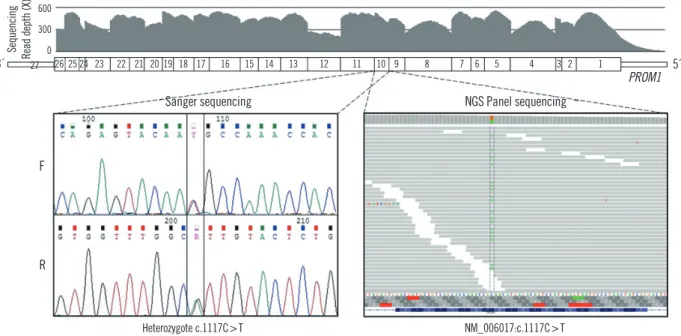
Targeted regions of PROM1 were fully covered, with a mean depth ranging from 168X to 513X. A heterozygous missense variant in PROM1 (NM_006017.1: c.1117C>T [p.R373C]) was detected in the affected proband with a high read depth. Sanger sequencing using newly-designed forward (5’-GGCTGGATGAT- GTTTATCAG-3’) and reverse primers (5’-GACTCTTGATGAC- CATATAATG-3’) confirmed the heterozygous presence of the variant in the proband (Fig. 3). No pathogenic variants were identified in the other 97 genes. On the basis of the characteris- tic retinal phenotype and PROM1 p.R373C mutation, the pa- tient was diagnosed as having STGD4.
Classical Stargardt disease (STGD1; OMIM No. 248200), one of the most common hereditary macular dystrophies, is caused by mutations in the ABCA4 gene on chromosome 1p with auto- somal recessive transmission [14]. In addition to the autosomal recessive pattern, which is more common, some families with autosomal dominant inheritance have also been described, in- cluding STGD3 (OMIM No. 600110), caused by mutations in ELOVL4, and STGD4 (OMIM No. 603786) [1, 2, 15]. In 1999,
Fig. 3. DNA electropherograms of a fragment of the PROM1 gene by Sanger sequencing. Proband DNA sequence shows a heterozygous missense variant (NM_006017.1: c.1117C>T).
Abbreviation: NGS, next-generation sequencing.
Heterozygote c.1117C>T NM_006017:c.1117C>T
F
R
600 300 0
Sanger sequencing
3´ 27 26 25 24 23 22 21 20 19 18 17 16 15 14 13 12 11 10 9 8 7 6 5 4 3 2 1 5´
Sequencing Read depth (X)
NGS Panel sequencing
PROM1
STGD4 was mapped to a locus on chromosome 4p [1]. It had an overlap with bull’s eye macular dystrophy (MCDR2; OMIM Mo. 608051) [16]. In 2008, mutation analysis of genes within the overlapping disease interval revealed a PROM1 p.R373C mutation [2]. PROM1 is normally localized to the photoreceptor outer segments in the retina. The p.R373C mutation showed defective disk morphogenesis in transgenic mice with this mu- tation [2]. Ocular phenotypes of the STGD4 families with PROM1 p.R373C mutations may be similar to the phenotypes of STGD1 and STGD3 as well as MCDR2 and cone-rod dystro- phy [1-4]. Therefore, molecular genetic diagnosis is essential for diagnosing these rare diseases.
Prominin-1 is also known as CD133, which was originally iden- tified as a surface marker of hematopoietic stem cells and endo- thelial progenitor cells [17]. CD133 is also involved in hippocam- pal neurogenesis [18]. Although an extraocular phenotype is not remarkable in patients with PROM1 mutation, one study found that ex vivo endothelial function is abnormal in patients with p.R373C PROM1 mutation; One family has exhibited empty sella turcica on brain magnetic resonance imaging and microhematuria [19].
Although the proband in our study had no systemic symptoms or previous medical history, possible systemic involvement associ- ated with PROM1 mutation needs to be examined when the pa- tient shows certain systemic symptoms or signs.
In summary, we report a Korean patient with STGD4 diag- nosed by targeted exome sequencing. To the best of our knowl- edge, this is the first case of STGD4 in Koreans. Targeted exome sequencing seems to be an efficient approach to find genetic causes of macular dystrophy.
Authors’ Disclosures of Potential Conflicts of Interests
No potential conflicts of interest relevant to this article were re- ported.
Acknowledgments
This work was supported by Collaborative Genome Program for Fostering New Post-Genome industry through the National Re- search Foundation of Korea (NRF) funded by the Ministry of Sci- ence, ICT and Future Planning (NRF-2014M3C9A 2064620).
REFERENCES
1. Kniazeva M, Chiang MF, Morgan B, Anduze AL, Zack DJ, Han M, et al.
A new locus for autosomal dominant stargardt-like disease maps to chromosome 4. Am J Hum Genet 1999;64:1394-9.
2. Yang Z, Chen Y, Lillo C, Chien J, Yu Z, Michaelides M, et al. Mutant prominin 1 found in patients with macular degeneration disrupts photo- receptor disk morphogenesis in mice. J Clin Invest 2008;118:2908-16.
3. Michaelides M, Gaillard MC, Escher P, Tiab L, Bedell M, Borruat FX, et al. The PROM1 mutation p.R373C causes an autosomal dominant bull’s eye maculopathy associated with rod, rod-cone, and macular dys- trophy. Invest Ophthalmol Vis Sci 2010;51:4771-80.
4. Palejwala NV, Gale MJ, Clark RF, Schlechter C, Weleber RG, Pennesi ME. Insights into autosomal dominant stargardt-like macular dystrophy through multimodality diagnostic imaging. Retina 2016;36:119-30.
5. Li H and Durbin R. Fast and accurate short read alignment with Bur- rows-Wheeler transform. Bioinformatics 2009;25:1754-60.
6. Li H, Handsaker B, Wysoker A, Fennell T, Ruan J, Homer N, et al. The Sequence Alignment/Map format and SAMtools. Bioinformatics 2009;
25:2078-9.
7. McKenna A, Hanna M, Banks E, Sivachenko A, Cibulskis K, Kernytsky A, et al. The Genome Analysis Toolkit: a MapReduce framework for ana- lyzing next-generation DNA sequencing data. Genome Res 2010;20:
1297-303.
8. Wang K, Li M, Hakonarson H. ANNOVAR: functional annotation of ge- netic variants from high-throughput sequencing data. Nucleic Acids Res 2010;38:e164.
9. Lek M, Karczewski KJ, Minikel EV, Samocha KE, Banks E, Fennell T, et al. Analysis of protein-coding genetic variation in 60,706 humans. Na- ture 2016;536:285-91.
10. Ng PC and Henikoff S. SIFT: Predicting amino acid changes that affect protein function. Nucleic Acids Res 2003;31:3812-4.
11. Adzhubei I, Jordan DM, Sunyaev SR. Predicting functional effect of hu- man missense mutations using PolyPhen-2. Curr Protoc Hum Genet 2013;Chapter 7:Unit7.20.
12. Davydov EV, Goode DL, Sirota M, Cooper GM, Sidow A, Batzoglou S.
Identifying a high fraction of the human genome to be under selective constraint using GERP++. PLoS Comput Biol 2010;6:e1001025.
13. Landrum MJ, Lee JM, Riley GR, Jang W, Rubinstein WS, Church DM, et al. ClinVar: public archive of relationships among sequence variation and human phenotype. Nucleic Acids Res 2014;42:D980-5.
14. Allikmets R, Singh N, Sun H, Shroyer NF, Hutchinson A, Chidambaram A, et al. A photoreceptor cell-specific ATP-binding transporter gene (ABCR) is mutated in recessive Stargardt macular dystrophy. Nat Genet 1997;15:236-46.
15. Zhang K, Kniazeva M, Han M, Li W, Yu Z, Yang Z, et al. A 5-bp deletion in ELOVL4 is associated with two related forms of autosomal dominant macular dystrophy. Nat Genet 2001;27:89-93.
16. Michaelides M, Johnson S, Poulson A, Bradshaw K, Bellmann C, Hunt DM, et al. An autosomal dominant bull’s-eye macular dystrophy (MCDR2) that maps to the short arm of chromosome 4. Invest Ophthal- mol Vis Sci 2003;44:1657-62.
17. Yin AH, Miraglia S, Zanjani ED, Almeida-Porada G, Ogawa M, Leary AG, et al. AC133, a novel marker for human hematopoietic stem and pro- genitor cells. Blood 1997;90:5002-12.
18. Kempermann G, Chesler EJ, Lu L, Williams RW, Gage FH. Natural vari- ation and genetic covariance in adult hippocampal neurogenesis. Proc Natl Acad Sci U S A 2006;103:780-5.
19. Arrigoni FI, Matarin M, Thompson PJ, Michaelides M, McClements ME, Redmond E, et al. Extended extraocular phenotype of PROM1 mutation in kindreds with known autosomal dominant macular dystrophy. Eur J Hum Genet 2011;19:131-7.
Supplemental Table S1. Inherited retinal degeneration gene panel – gene list
Panel of Inherited Retinal Degenerations (n=98)
Gene DHDDS, RPE65, ABCA4, PRPF3, SEMA4A, PDC, CRB1, NEK2, FLVCR1, USH2A, ZNF513, C2ORF71, EFEMP1, FAM161A, SNRNP200, CNNM4, MERTK, CERKL, SAG, ARL6, IMPG2, RHO, CLRN1, PDE6B, RAB28, PROM1, GPR125, WDR19, CNGA1, LRAT, PDE6A, MAK, TULP1, GUCA1A, GUCA1B, PRPH2, EYS, RIMS1, IMPG1, ELOVL4, KLHL7, RP9, IMPDH1, KIAA1549, RP1L1, ADAM9, RP1, CNGB3, C8ORF37, KCNV2, TOPORS, PRPF4, RBP3, CDHR1, RGR, PDE6C, BEST1, ROM1, MYO7A, C1QTNF5, CACNA2D4, PDE6H, RDH5, MVK, RPGRIP1, NRL, RDH12, SPATA7, TTC8, NR2E3, RLBP1, ARL2BP, CNGB1, PRPF8, AIPL1, PITPNM3, GUCY2D,UNC119, CA4, RGS9, USH1G, PRCD, FSCN2, PDE6G, RAX2, CRX, PRPF31, IDH3B, MKKS, PRPF6, C21ORF2, TIMP3, OFD1, RPGR, RP2, CACNA1F, CHM