ISSN 2234-3806 • eISSN 2234-3814
http://dx.doi.org/10.3343/alm.2015.35.1.62
Evaluation of Matrix-Assisted Laser Desorption
Ionization–Time of Flight Mass Spectrometry-Based VITEK MS System for the Identification of
Acinetobacter Species from Blood Cultures:
Comparison with VITEK 2 and MicroScan Systems
Seung Yeob Lee, M.D., Jong Hee Shin, M.D., Soo Hyun Kim, M.D., Myung Geun Shin, M.D., Soon Pal Suh, M.D., and Dong Wook Ryang, M.D.
Department of Laboratory Medicine, Chonnam National University Medical School, Gwangju, Korea
Background: Acinetobacter species are the leading cause of bloodstream infection (BSI), but their correct identification is challenging. We evaluated the matrix-assisted laser de- sorption ionization-time of flight mass spectrometry (MALDI-TOF MS)-based VITEK MS (bioMérieux, France), and two automated systems, VITEK 2 (bioMérieux) and MicroScan (Siemens, USA) for identification of Acinetobacter BSI isolates.
Methods: A total of 187 BSI isolates recovered at a university hospital in Korea between 2010 and 2012 were analyzed. The identification results obtained using VITEK MS and two automated systems were compared with those of rpoB sequencing.
Results: Of 187 isolates analyzed, 176 were identified to the species level by rpoB se- quencing: the Acinetobacter baumannii group (ABG; 101 A. baumannii, 43 A. nosocomi- alis, 10 A. pittii isolates) was most commonly identified (82.4%), followed by Acineto- bacter genomic species 13BJ/14TU (5.3%), A. ursingii (2.1%), A. soli (2.1%), A. ber- eziniae (1.1%), and A. junii (1.1%). Correct identification rates to the species group (ABG) level or the species level was comparable among the three systems (VITEK MS, 90.3%;
VITEK 2, 89.2%; MicroScan, 86.9%). However, VITEK MS generated fewer misidentifica- tions (0.6%) than VITEK 2 (10.8%) and MicroScan (13.1%) (P <0.001). In addition, VI- TEK MS demonstrated higher specificity (100%) for discrimination between ABG and non-ABG isolates than the other systems (both, 31.8%) (P <0.001).
Conclusions: The VITEK MS system is superior to the VITEK 2 and MicroScan systems for identification of Acinetobacter BSI isolates, with fewer misidentifications and better dis- crimination between the ABG and non-ABG isolates.
Key Words: VITEK MS, VITEK 2, MicroScan, Acinetobacter, Identification
Received: April 9, 2014 Revision received: April 30, 2014 Accepted: October 19, 2014 Corresponding author: Jong Hee Shin Department of Laboratory Medicine, Chonnam National University Medical School, 42 Jebong-ro, Dong-gu, Gwangju 501-757, Korea
Tel: +82-62-220-5342 Fax: +82-62-224-2518 E-mail: shinjh@chonnam.ac.kr
© The Korean Society for Laboratory Medicine This is an Open Access article distributed under the terms of the Creative Commons Attribution Non-Commercial License (http://creativecom- mons.org/licenses/by-nc/3.0) which permits unrestricted non-commercial use, distribution, and reproduction in any medium, provided the original work is properly cited.
INTRODUCTION
Acinetobacter species are one of the leading causative agents of healthcare-associated infections worldwide, including blood-
stream infection (BSI), pneumonia, urinary tract infection, and meningitis [1-3]. To date, more than 32 species of the Acineto- bacter genus have been identified by gene sequencing [4-8].
Members of the Acinetobacter baumannii group (ABG), which
consists of A. baumannii, A. pittii, and A. nosocomialis, share important clinical and epidemiological characteristics that can- not be distinguished by most of the currently available pheno- typic identification (ID) systems [2]. Although the ABG remains the most common Acinetobacter species recovered from clini- cal specimens, the non-ABG species are also often clinically rel- evant [3, 4, 9-11]. Because Acinetobacter species may differ in their pathogenicity, epidemiology, antimicrobial susceptibility, and clinical outcomes [5, 9, 11-13], information on the relative frequency of various Acinetobacter species causing BSI may be useful for establishing protocols for infection control and treat- ment of Acinetobacter BSI. However, to date, data regarding distributions of BSI isolates of Acinetobacter species in hospitals are limited, because the correct ID of Acinetobacter isolates is difficult to determine when using the currently available, com- mon phenotypic methods [2-4, 9, 10, 14].
Recent studies have shown that matrix-assisted laser desorp- tion ionization-time of flight mass spectrometry (MALDI-TOF MS) offers an opportunity for rapid, cost-effective, convenient, and high-throughput bacterial ID in routine diagnostic procedures conducted in clinical microbiology laboratories [15, 16]. How- ever, information regarding the suitability of the MALDI-TOF MS- based VITEK MS system (VITEK MS; bioMérieux, Marcy l’Etoile, France) for the ID of clinical isolates of Acinetobacter species is scarce as compared to that of other commonly used automated ID systems. In this study, we performed molecular ID to investi- gate the rank order of occurrence of the various species of Aci- netobacter isolated from blood cultures obtained from a univer- sity hospital in Korea between 2010 and 2012. We performed a comparative evaluation of MALDI-TOF MS-based VITEK MS sys- tem versus the VITEK 2 (VITEK2 XL; bioMérieux) and MicroScan (MicroScan WalkAway-96 Plus; Siemens, Deerfield, IL, USA) au- tomated systems for the correct ID of Acinetobacter BSI isolates, including evaluation of their discriminative abilities for the ABG versus other (non-ABG) Acinetobacter species.
METHODS
1. Acinetobacter isolation and molecular ID
A total of 187 isolates molecularly identified as belonging to the genus Acinetobacter were analyzed in this study. All isolates were obtained from the blood cultures of 187 patients at the Chonnam National University Hospital (a 1,000-bed tertiary- care hospital in Gwangju, Korea) between January 2010 and December 2012. Duplicate isolates of Acinetobacter species from the same patient were excluded. For molecular ID, DNA
was extracted from the isolates as described previously [17], and a 450-bp sequence (zone 2) of the rpoB gene region of each isolate was sequenced [18]. The primers Ac1055F (5´-GT- GATAARATGGCBGGTCGT-3´) and Ac1598R (5´-CGBGCRTG- CATYTTGTCRT-3´) were used to amplify the rpoB region. All loci were sequenced in both the forward and reverse directions with the same primers as those used for amplification. The amplifi- cation products were purified and sequenced by using an ABI 3730XL sequencer (Applied Biosystems, Foster City, CA, USA).
Sequence data were assembled and compared with previously reported sequences by using the basic local alignment search tool (BLAST) of the national center for biotechnology information (NCBI) database (http://www.ncbi.nlm.nih.gov/blast).
2. ID using the VITEK MS, VITEK 2, and MicroScan systems ID with VITEK MS (in vitro-diagnostic [IVD] mode) was per- formed according to the manufacturer’s instructions by directly smearing an overnight-cultured bacterial specimen onto dispos- able target slides with a 1.0-μL matrix solution (VITEK MS- CHCA). Escherichia coli ATCC 8739 was used as the calibration strain. The advanced spectrum classifier software of the VITEK MS system proposes 3 confidence levels: (a) single choice, with one significant choice (confidence value ≥ 60); (b) low discrim- ination, with more than one significant choice (maximum 4 choices); and (c) unidentified organism, with no significant choice (no class with probability and score higher than the de- fined thresholds) or a number of significant choices greater than the defined threshold of low discrimination.
ID using the VITEK 2 and MicroScan systems was performed by using GN ID Card (bioMérieux) and Gram Negative Break- point Combo Panel Type 42 (Siemens), respectively, according to the manufacturers’ instructions. The ID results from these systems are proposed automatically by the respective accompa- nying software. The tests were repeated only if the initial results indicated “low discrimination” or “no ID”, and the repeat result was used for data analysis.
3. Data analysis
The ID capability of the 3 systems was evaluated by using the Acinetobacter isolates that were identified to the species level by rpoB sequencing. The ID results from the 3 systems were clas- sified into 3 categories by comparison with the results of the ref- erence rpoB sequencing: (a) “correct ID” to the species (identi- cal to sequence-based ID) or species group level; (b) “mis-ID”, when the ID result of the system differed from that of the se- quence-based reference method; and (c) “no ID”, when the
system could not identify the isolate. Because the ID result of the isolates belonging to the ABG (isolates of A. baumannii, A.
pittii, or A. nosocomialis) is assigned by these 3 systems at the species group level only, the ABG isolates that were identified as
“A. baumannii complex” by using VITEK MS IVD and VITEK 2, or those identified as “A. baumannii/haemolyticus” by using the MicroScan system were categorized under the “correct ID” cat- egory. Statistical analysis to compare the ID performance of the 3 systems was performed with the chi-square test, Fisher’s ex- act test, and McNemar test using the IBM SPSS Statistics (ver- sion 21, IBM, Armonk, NY, USA) and GraphPad Prism (version 5, GraphPad Software, San Diego, CA,USA) software packages.
RESULTS
1. Acinetobacter isolation and molecular ID
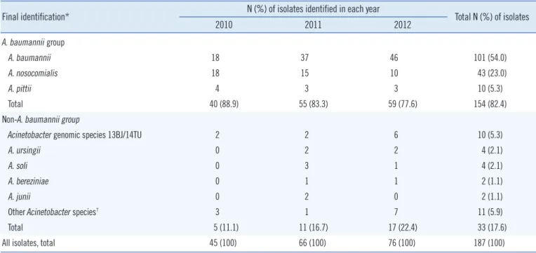
Table 1 presents the species distribution of the 187 Acineto- bacter BSI isolates determined by partial rpoB sequencing. Of the 187 isolates collected during the 3-yr period, 154 (82.4%) isolates belonged to the ABG, which included 101 (54.0% of the total 187 Acinetobacter isolates) A. baumannii, 43 (23.0%) A. nosocomialis, and 10 (5.3%) A. pittii isolates. The remaining 33 (17.6%) isolates belonged to the non-ABG: 10 (5.3%) iso- lates of Acinetobacter genomic species 13BJ/14TU, 4 (2.1%) of A. ursingii, 4 (2.1%) of A. soli, 2 (1.1%) of A. bereziniae, 2
(1.1%) of A. junii, and 11 (5.9%) of other miscellaneous Aci- netobacter species, which were identified as the genus Acineto- bacter but not identified to the species level. Overall, a total of 176 isolates were identified to the species level by partial rpoB sequencing.
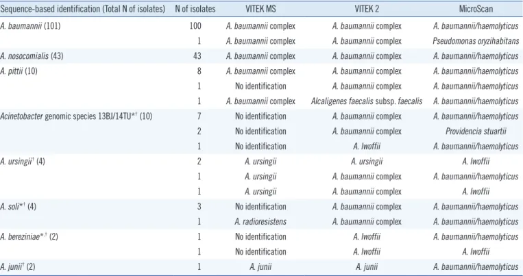
2. ID using the VITEK MS, VITEK 2, and MicroScan systems Details of the ID results from rpoB sequencing and the VITEK MS, VITEK 2, and MicroScan systems are provided in Table 2.
Of the 154 ABG isolates, 153 (99.4%) were correctly identified by all 3 systems. The VITEK MS system correctly identified all 4 A. ursingii isolates and 2 A. junii isolates, and VITEK 2 correctly identified 2 of 4 A. ursingii isolates and 2 A. junii isolates, which were present in their respective databases; by contrast, the Mi- croScan system misidentified each of the 6 isolates absent from its database. Among the 16 isolates of Acinetobacter genomic species 13BJ/14TU, A. soli, and A. bereziniae, which were ab- sent in the databases of the 3 systems, VITEK MS showed 1 (6.3%) mis-ID and 15 (93.8%) no ID results, whereas both VI- TEK 2 and MicroScan showed 16 (100%) mis-ID and 0 (0%) no ID results (P <0.001).
3. ID results for the ABG and non-ABG isolates
The ID results from the 3 tested systems for the 154 ABG and 22 non-ABG isolates are summarized in Table 3. All 3 systems
Table 1. Species distribution of 187 Acinetobacter bloodstream isolates recovered during the 3-yr period Final identification* N (%) of isolates identified in each year
Total N (%) of isolates
2010 2011 2012
A. baumannii group
A. baumannii 18 37 46 101 (54.0)
A. nosocomialis 18 15 10 43 (23.0)
A. pittii 4 3 3 10 (5.3)
Total 40 (88.9) 55 (83.3) 59 (77.6) 154 (82.4)
Non-A. baumannii group
Acinetobacter genomic species 13BJ/14TU 2 2 6 10 (5.3)
A. ursingii 0 2 2 4 (2.1)
A. soli 0 3 1 4 (2.1)
A. bereziniae 0 1 1 2 (1.1)
A. junii 0 2 0 2 (1.1)
Other Acinetobacter species† 3 1 7 11 (5.9)
Total 5 (11.1) 11 (16.7) 17 (22.4) 33 (17.6)
All isolates, total 45 (100) 66 (100) 76 (100) 187 (100)
*The final identification results were obtained by rpoB sequencing; †Isolates were identified as belonging to genus Acinetobacter, but not identified up to the species level by rpoB sequencing.
correctly identified 99.4% (153/154) of the ABG isolates. How- ever, the test results for the 22 non-ABG isolates, including 10 Acinetobacter genomic species 13BJ/14TU, 4 A. ursingii, 4 A.
soli, 2 A. bereziniae, and 2 A. junii isolates, showed correct ID rates of 27.3%, 18.2%, and 0% for the VITEK MS, VITEK 2, and MicroScan systems, respectively (VITEK MS and VITEK 2 versus Table 2. Detailed identification results by rpoB sequencing and the MALDI-TOF MS-based VITEK MS, VITEK 2, and MicroScan systems for 176 Acinetobacter blood isolates
Sequence-based identification (Total N of isolates) N of isolates VITEK MS VITEK 2 MicroScan
A. baumannii (101) 100 A. baumannii complex A. baumannii complex A. baumannii/haemolyticus
1 A. baumannii complex A. baumannii complex Pseudomonas oryzihabitans
A. nosocomialis (43) 43 A. baumannii complex A. baumannii complex A. baumannii/haemolyticus
A. pittii (10) 8 A. baumannii complex A. baumannii complex A. baumannii/haemolyticus
1 No identification A. baumannii complex A. baumannii/haemolyticus 1 A. baumannii complex Alcaligenes faecalis subsp. faecalis A. baumannii/haemolyticus Acinetobacter genomic species 13BJ/14TU*† (10) 7 No identification A. baumannii complex A. baumannii/haemolyticus
2 No identification A. baumannii complex Providencia stuartii
1 No identification A. lwoffii A. baumannii/haemolyticus
A. ursingii† (4) 2 A. ursingii A. ursingii A. lwoffii
1 A. ursingii A. baumannii complex A. baumannii/haemolyticus
1 A. ursingii A. baumannii complex A. lwoffii
A. soli*† (4) 3 No identification A. baumannii complex A. baumannii/haemolyticus
1 A. radioresistens A. baumannii complex A. baumannii/haemolyticus
A. bereziniae*,† (2) 1 No identification A. lwoffii A. baumannii/haemolyticus
1 No identification A. lwoffii A. lwoffii
A. junii† (2) 1 A. junii A. junii A. baumannii/haemolyticus
*Species not included in the databases of VITEK MS and VITEK 2 systems; †Species not included in the database of MicroScan system.
Abbreviation: MALDI-TOF MS, matrix-assisted laser desorption ionization–time of flight mass spectrometry.
Table 3. Identification results of 176 Acinetobacter bloodstream isolates by the MALDI-TOF MS-based VITEK MS, VITEK 2, and MicroScan systems as compared to sequence-based identification
Final identification N of isolates
N (%) of isolates with the indicated ID result*
VITEK MS VITEK 2 MicroScan
Correct ID Mis-ID No ID Correct ID Mis-ID No ID Correct ID Mis-ID No ID A. baumannii group† 154 153 (99.4) 0 (0) 1 (0.6) 153 (99.4) 1 (0.6) 0 (0) 153 (99.4) 1 (0.6) 0 (0)
Acinetobacter genomic species 13BJ/14TUठ10 0 0 10 0 10 0 0 10 0
A. ursingii§ 4 4 0 0 2 2 0 0 4 0
A. soliठ4 0 1 3 0 4 0 0 4 0
A. bereziniaeठ2 0 0 2 0 2 0 0 2 0
A. junii§ 2 2 0 0 2 0 0 0 2 0
Non-A. baumannii group, subtotal 22 6 (27.3) 1 (4.5) 15 (68.2) 4 (18.2) 18 (81.8) 0 (0) 0 (0) 22 (100) 0 (0) All isolates, total 176 159 (90.3) 1 (0.6) 16 (9.1) 157 (89.2) 19 (10.8) 0 (0) 153 (86.9) 23 (13.1) 0 (0)
*Correct ID (identification) included “correct ID” up to the (i) species level; (ii) species group level for the A. baumannii group (e.g., “A. baumannii complex”
by using VITEK MS and VITEK 2 or “A. baumannii/haemolyticus” by using MicroScan); †A. baumannii, A. pittii, and A. nosocomialis were included in the A.
baumannii group; ‡Species not included in the databases of VITEK MS and VITEK 2 systems; §Species not included in the database of the MicroScan system.
Abbreviations: MALDI-TOF MS, matrix-assisted laser desorption ionization–time of flight mass spectrometry; correct ID, identification up to the species or species group level; mis-ID, identification result of the system differed from that of the sequence-based reference method; no ID, the system could not iden- tify the isolate.
MicroScan, P <0.05). The mis-ID rate for the 22 non-ABG iso- lates was 4.5% (1/22) using VITEK MS, which was significantly lower than that obtained using VITEK 2 (81.8%) and MicroScan (100%) (P <0.001). Of the 22 non-ABG isolates, no (0.0%) and 15 (68.2%) isolates were misidentified as A. baumannii com- plex when using VITEK MS and VITEK 2, respectively, and 15 (68.2%) isolates were misidentified as A. baumannii/haemolyti- cus with MicroScan. Therefore, the specificity of VITEK MS for discrimination between the ABG and non-ABG isolates was much higher (100% [22/22]) than that of VITEK 2 and Mi- croScan (both, 31.8% [7/22]; P <0.001), while the sensitivity of the 3 systems was identical at 99.4% (153/154). The false-pos- itive rate of VITEK MS for discrimination between the ABG and non-ABG isolates was 0% (0/22), while that of both VITEK 2 and MicroScan was 68.2% (15/22) (P <0.001).
Overall, for all 176 isolates tested, the correct ID rates obtained with the VITEK MS, VITEK 2, and MicroScan systems were 90.3%, 89.2%, and 86.9%, respectively, which indicate similar rates of correct ID among the 3 systems. However, VITEK MS showed a lower mis-ID rate (0.6%) than VITEK 2 (10.8%) and MicroScan (13.1%) (P <0.001) and a higher no ID rate (9.1%) than VITEK 2 and MicroScan (both, 0%) (P <0.001).
DISCUSSION
To the best of our knowledge, this study reports the first compar- ative evaluation of MALDI-TOF MS-based VITEK MS with both the VITEK 2 and MicroScan systems for the ID of Acinetobacter species from BSI isolates. The current findings demonstrate that the mis-ID rate of VITEK MS was significantly lower (0.6%) than that obtained with the VITEK 2 (10.8%) and MicroScan (13.1%) systems for the ID of Acinetobacter BSI isolates. Moreover, VITEK MS showed the best discrimination between the ABG and non- ABG isolates in this study. Thus, we believe that VITEK MS is a useful system for the rapid and accurate ID of BSI isolates of Acinetobacter species in routine diagnostic procedures con- ducted in clinical microbiology laboratories.
For molecular ID, we analyzed a partial sequence of the rpoB gene region, which has a higher discriminatory power for routine ID of different Acinetobacter species compared to the 16S rRNA gene [18, 19]. Sequence-based analysis of 187 BSI isolates re- covered during the 3-yr period (2010–2012) revealed that the ABG isolates represented 82.4% of all Acinetobacter BSI cases in our hospital. This finding, together with data from other coun- tries, confirmed that the ABG continues to be the leading cause of Acinetobacter BSI worldwide [4, 9, 10]. Although A. bauman-
nii is the most frequent etiological agent of Acinetobacter BSI in our hospital, the prevalence of A. baumannii (54.0%) detected in this study was slightly lower than that reported for the USA (63.4%) [9], but higher than that reported for Norway (8.8%) and Japan (17.9%) in previous studies [4, 10]. Karah et al. [4]
reported that the most prevalent species was A. nosocomialis (46.9%), followed by A. pittii (19.5%) and A. baumannii (8.8%) among the Acinetobacter BSI isolates in Norway. On the other hand, Kishii et al. [10] reported that the most prevalent species was A. pittii (34.1%), followed by A. baumannii (17.9%) and A.
nosocomialis (15.4%) in Japan. Our recent report revealed that Acinetobacter genomic species 13BJ/14TU is innately resistant to colistin, but susceptible to most of the other clinically relevant antimicrobial agents, and that BSI patients infected with this species show excellent clinical outcomes with cleared bactere- mia [11]. In the current study, among the 33 (17.6%) non-ABG isolates, 10 (5.3%) isolates of Acinetobacter genomic species 13BJ/14TU were identified as causative agents of BSI. Overall, our results, together with data from other countries reported pre- viously, demonstrate that the rank order of the occurrence of various Acinetobacter species that cause BSI may differ among hospitals and countries and with respect to time [4, 9, 10].
To date, few evaluations of the VITEK MS system using mo- lecularly identified Acinetobacter clinical isolates have been per- formed, with the exception of one multi-center study for the ID of non-Enterobacteriaceae gram-negative bacilli isolated from various samples [16]. In this previous study, when the VITEK MS ID results of 108 Acinetobacter isolates were compared with those determined by gene sequencing, the rates of correct ID to the species level, mis-ID, and no ID were 83.3% (90/108), 0.9%
(1/108), and 13.0% (14/108), respectively. Similarly, we found that the rates of correct ID, mis-ID, and no ID using VITEK MS for 176 Acinetobacter BSI isolates were 90.3%, 0.6%, and 9.1%, respectively. In addition, the current study demonstrated that the VITEK MS system was better at correctly identifying the ABG isolates (99.4%) than the non-ABG isolates (27.3%). In our study, all isolates of A. ursingii and A. junii, which are in- cluded in the VITEK MS database, were correctly identified. Be- cause the isolates described herein represent almost all of the Acinetobacter BSI isolates encountered in a single hospital over a 3-yr period, our finding that VITEK MS showed a lower correct ID rate for the non-ABG isolates than for the ABG isolates will provide valuable information for the diagnosis and treatment of BSI patients infected with Acinetobacter species.
To date, no comparative evaluation of the VITEK MS, VITEK 2, and MicroScan systems for the ID of BSI isolates of Acineto-
bacter species has been reported. The present study revealed that the correct ID rate of VITEK MS (90.3%) was comparable to that of the VITEK 2 (89.2%) and MicroScan (86.9%) sys- tems; however, VITEK MS showed a lower rate of mis-ID (0.6%) than VITEK 2 (10.8%) and MicroScan (13.1%) for these iso- lates. Instead, VITEK MS yielded more “no ID” results (9.1%) than VITEK 2 and MicroScan (both, 0%). These results suggest that the VITEK MS system can be advantageous in the routine diagnostic procedures conducted in clinical microbiology labo- ratories, because an isolate not identified by the primary testing method can be routinely retested or tested by using other meth- ods [15, 20]. In addition, the low rate of mis-ID obtained using the VITEK MS system offers an important clinical advantage along with offering more rapid and reliable ID of other bacterial isolates [16].
Although the need for species ID of Acinetobacter isolates in routine clinical microbiology laboratories has been questioned, recent studies have highlighted the importance of discriminating the ABG from non-ABG BSI isolates since non-ABG BSI has a more benign clinical course than ABG BSI, and the two groups differ in their antimicrobial susceptibilities [2, 9, 11, 21-23]. Of the 3 systems tested, VITEK MS showed the best discrimination ability between the ABG and non-ABG isolates, with 100%
specificity (0% false-positive rate), while the VITEK 2 and Mi- croScan systems both showed 31.8% specificity (68.2% false- positive rate) for discriminating between these groups. This find- ing highlights the effectiveness of VITEK MS for discriminating the ABG from non-ABG isolates in Acinetobacter BSI cases.
Thus, use of VITEK MS will facilitate prediction of the clinical course and outcome of BSI patients as well as the selection of an appropriate antimicrobial regimen.
The current and previous studies have demonstrated that the limitation of the current VITEK MS system for ID of Acinetobacter species [15, 16]. It cannot differentiate among the ABG species such as A. baumannii, A. pittii, and A. nosocomialis. However, when a cluster analysis was performed on the Acinetobacter isolates tested in this study by using VITEK MS RUO (research- use-only, Saramis database, bioMérieux) mode, the A. bauman- nii, A. pittii, and A. nosocomialis isolates were relatively well-clus- tered (data not shown). In addition, a recent report showed that another MALDI-TOF MS-based system that was unable to reli- ably differentiate between these closely related species has now been improved by using an alternative protocol [24]. Thus, the VITEK MS system shows potential for offering reliable species- level ID of ABG isolates in routine diagnostic procedures con- ducted in clinical microbiology laboratories.
Authors’ Disclosures of Potential Conflicts of Interest
No potential conflicts of interest relevant to this article were re- ported.
REFERENCES
1. Chuang YC, Sheng WH, Li SY, Lin YC, Wang JT, Chen YC, et al. Influ- ence of genospecies of Acinetobacter baumannii complex on clinical outcomes of patients with acinetobacter bacteremia. Clin Infect Dis 2011; 52:352-60.
2. Peleg AY, Seifert H, Paterson DL. Acinetobacter baumannii: emergence of a successful pathogen. Clin Microbiol Rev 2008;21:538-82.
3. Turton JF, Shah J, Ozongwu C, Pike R. Incidence of Acinetobacter spe- cies other than A. baumannii among clinical isolates of Acinetobacter:
evidence for emerging species. J Clin Microbiol 2010;48:1445-9.
4. Karah N, Haldorsen B, Hegstad K, Simonsen GS, Sundsfjord A, Samu- elsen Ø. Species identification and molecular characterization of Aci- netobacter spp. blood culture isolates from Norway. J Antimicrob Che- mother 2011;66:738-44.
5. Dijkshoorn L, Nemec A, Seifert H. An increasing threat in hospitals:
multidrug-resistant Acinetobacter baumannii. Nat Rev Microbiol 2007;
5:939-51.
6. Kim D, Baik KS, Kim MS, Park SC, Kim SS, Rhee MS, et al. Acineto- bacter soli sp. nov., isolated from forest soil. J Microbiol 2008;46:396- 401.
7. Nemec A, Musílek M, Maixnerová M, De Baere T, van der Reijden TJ, Vaneechoutte M, et al. Acinetobacter beijerinckii sp. nov. and Acineto- bacter gyllenbergii sp. nov., haemolytic organisms isolated from hu- mans. Int J Syst Evol Microbiol 2009;59:118-24.
8. Vaneechoutte M, De Baere T, Nemec A, Musílek M, van der Reijden TJ, Dijkshoorn L. Reclassification of Acinetobacter grimontii Carr et al.
2003 as a later synonym of Acinetobacter junii Bouvet and Grimont 1986. Int J Syst Evol Microbiol 2008;58:937-40.
9. Wisplinghoff H, Paulus T, Lugenheim M, Stefanik D, Higgins PG, Ed- mond MB, et al. Nosocomial bloodstream infections due to Acineto- bacter baumannii, Acinetobacter pittii and Acinetobacter nosocomialis in the United States. J Infect 2012;64:282-90.
10. Kishii K, Kikuchi K, Matsuda N, Yoshida A, Okuzumi K, Uetera Y, et al.
Evaluation of matrix-assisted laser desorption ionization-time of flight mass spectrometry for species identification of Acinetobacter strains isolated from blood cultures. Clin Microbiol Infect 2014;20:424-30.
11. Lee SY, Shin JH, Park KH, Kim JH, Shin MG, Suh SP, et al. Identifica- tion, genotypic relation, and clinical features of colistin-resistant isolates of Acinetobacter genomic species 13BJ/14TU from bloodstreams of patients in a university hospital. J Clin Microbiol 2014;52:931-9.
12. Lee YC, Huang YT, Tan CK, Kuo YW, Liao CH, Lee PI, et al. Acineto- bacter baumannii and Acinetobacter genospecies 13TU and 3 bacter- aemia: comparison of clinical features, prognostic factors and outcomes.
J Antimicrob Chemother 2011;66:1839-46.
13. Lee K, Yong D, Jeong SH, Chong Y. Multidrug-resistant Acinetobacter spp.: increasingly problematic nosocomial pathogens. Yonsei Med J 2011;
52:879-91.
14. Bernards AT, van der Toorn J, van Boven CP, Dijkshoorn L. Evaluation of the ability of a commercial system to identify Acinetobacter genomic species. Eur J Clin Microbiol Infect Dis 1996;15:303-8.
15. Dubois D, Grare M, Prere MF, Segonds C, Marty N, Oswald E. Perfor- mances of the Vitek MS matrix-assisted laser desorption ionization-time of flight mass spectrometry system for rapid identification of bacteria in routine clinical microbiology. J Clin Microbiol 2012;50:2568-76.
16. Manji R, Bythrow M, Branda JA, Burnham CA, Ferraro MJ, Garner OB, et al. Multi-center evaluation of the VITEK® MS system for mass spec- trometric identification of non-Enterobacteriaceae Gram-negative bacilli.
Eur J Clin Microbiol Infect Dis 2014;33:337-46.
17. Khamis A, Raoult D, La Scola B. rpoB gene sequencing for identifica- tion of Corynebacterium species. J Clin Microbiol 2004;42:3925-31.
18. La Scola B, Gundi VA, Khamis A, Raoult D. Sequencing of the rpoB gene and flanking spacers for molecular identification of Acinetobacter species. J Clin Microbiol 2006;44:827-32.
19. Gundi VA, Dijkshoorn L, Burignat S, Raoult D, La Scola B. Validation of partial rpoB gene sequence analysis for the identification of clinically im- portant and emerging Acinetobacter species. Microbiology 2009; 155:
2333-41.
20. Won EJ, Shin JH, Lee K, Kim MN, Lee HS, Park YJ, et al. Accuracy of
species-level identification of yeast isolates from blood cultures from 10 university hospitals in South Korea by use of the matrix-assisted laser desorption ionization-time of flight mass spectrometry-based Vitek MS system. J Clin Microbiol 2013;51:3063-5.
21. Gerner-Smidt P, Tjernberg I, Ursing J. Reliability of phenotypic tests for identification of Acinetobacter species. J Clin Microbiol 1991;29:277-82.
22. Seifert H, Strate A, Schulze A, Pulverer G. Bacteremia due to Acineto- bacter species other than Acinetobacter baumannii. Infection 1994;22:
379-85.
23. Wareham DW, Bean DC, Khanna P, Hennessy EM, Krahe D, Ely A, et al.
Bloodstream infection due to Acinetobacter spp: epidemiology, risk fac- tors and impact of multi-drug resistance. Eur J Clin Microbiol Infect Dis 2008;27:607-12.
24. Šedo O, Nemec A, Křížová L, Kačalová M, Zdráhal Z. Improvement of MALDI-TOF MS profiling for the differentiation of species within the Aci- netobacter calcoaceticus-Acinetobacter baumannii complex. Syst Appl Microbiol 2013;36:572-8.