The Value of Ultrasound-Guided
Tattooing Localization of Nonpalpable Breast Lesions
Objective: To investigate the value of ultrasound-guided tattooing localization (US-tattoo) using a charcoal suspension for breast lesions.
Materials and Methods: One hundred sixty-four nonpalpable breast lesions in 134 patients (mean age 47 years; range 30 74 years) were marked with a char- coal suspension under US guidance. The medical records associated with the US-tattoo, the pathology results and the follow-up US results were reviewed.
Results: The average size of the localized lesions was 1.0 cm. The procedure time was < 5 minutes (range, 2 10 minutes) per lesion. The US-tattoo was well tolerated in all cases. The only technical difficulty encountered was a needle tip blockage caused by a large charcoal particle (4.9%). The surgeon easily identi- fied the tattoo with the exception one case. In addition, surgery could be safely delayed from one to 57 days after the making US-tattoo. The pathology result was benign in 108 cases, borderline in five, and malignant in 51. The excised specimen was < 4 cm in 76.6% (82/107) of the benign cases (mean; 2.7 cm). The pathologist could identify the mass around the tattoo and was able to make a spe- cific diagnosis in 81.3% (87/107) of benign lesions. The only complication
encountered was residual charcoal marking along the incision scar (3.6%). All fol- low-up US documented the removal of the lesions.
Conclusion: An US-tattoo for nonpalpable breast lesions is a very simple and accurate method that can help surgeons design and schedule an open biopsy.
he rate of imaging-detected nonpalpable breast lesions has increased with the advent of routine screening for breast cancer (1). A nonsurgical core needle biopsy helps decrease the frequency of unnecessary surgery by obtaining a histological diagnosis of imaging-detected lesions. However, these lesions tend to be an indication for surgery when high-risk lesions such as a papilloma or radial scar are observed by a core needle biopsy, when the patients or surgeons hope for complete removal of the lesions, or when additional lesions are detected in the other quadrant in a breast cancer patient scheduled to undergo conserving surgery.
A preoperative localization is very important for a minimal but accurate excision of a nonpalpable lesion. Several techniques such as needle localization or dye injection such as toludine blue or methylene blue have been used to localize nonpalpable breast lesions (2 6). Needle localization is widely used but the procedure is quite compli- cated. The complications and failure rates has been reported to range from 1 to 10%
due to a lack of communication between the radiologist and surgeon, inadequate wire localization to the dermal calcifications and wire shifting between localization and surgery (3, 7 10).
In Korea, the incidence of US-only detected nonpalpable breast lesions has been Kyungran Ko, MD1
Boo-Kyung Han, MD2 Kyung Mi Jang, MD2 Yeon Hyeon Choe, MD2 Jung Hee Shin MD2 Jung-Hyun Yang, MD3 Suk-Jin Nam, MD3
Index terms :
Breast, nonpalpable lesions Breast, biopsy
Biopsies, technology
Korean J Radiol 2007 ; 8 : 295-301 Received June 21, 2006; accepted after revision February 21, 2007.
1Department of Radiology and Center for Breast Cancer, National Cancer Center, Goyang-si 410-769, Korea; 2Department of Radiology and Center for Imaging Science, Samsung Medical Center, Sungkyunkwan University School of Medicine, Seoul 135-710, Korea;
3Department of General Surgery, Samsung Medical Center, Sungkyunkwan University School of Medicine, Seoul 135- 710, Korea
Address reprint requests to : Boo-Kyung Han, MD, Department of Radiology, Samsung Medical Center, Sungkyunkwan University School of Medicine, 50, Ilwon-dong, Kangnam-gu, Seoul 135-710, Korea.
Tel. (822) 3410-2518 Fax. (822) 3410-2559
e-mail: [email protected]
T
increasing with the increasing use of screening breast US.
A simple and stable localization method is essential for the removal of these lesions, and there are many methods currently available. These include localization with a needle and wire system and the use of a colored dye as a visual marker. Localization with a needle and wire system and dying with toluidine blue or methylene blue should be performed on the day of the operation regardless of the radiologist’s schedule because of the risk of needle displacement and fast dye dispersion (2, 5). However, a charcoal suspension is so stable that surgery can be delayed for days or even weeks.
Svane (6) introduced the use of a charcoal suspension for preoperative marking of nonpalpable breast lesions using stereotactic techniques. However, there are no reports of the use of a charcoal suspension under US guidance. To our knowledge, this is the first study tailored exclusively to US guided cases of charcoal marking. This report
introduces the procedure of US-guided tattooing localiza- tion (US-tattoo) using a charcoal suspension for nonpalpa- ble breast lesions.
MATERIALS AND METHODS
Full informed consent for the procedure was obtained from all patients and institutional review board approval was obtained. Between March 1996 and July 2001, 164
nonpalpable breast lesions detected by US in 134 preoper- ative patients underwent a US-tattoo. The patient’s age was 30 74 years (mean age, 47 years).
Mammograms were available in 115 patients with 136 lesions. The mammographic findings were divided into mass, calcifications, asymmetric density, architectural distortion, or none (Table 1). Mammograms were not performed in 19 patients. The US findings of the 164 targeted lesions were shown as a mass, a non-mass forming lesion, and metastatic lymph nodes (Table 1). The US- tattoo was carried out by one of two board-certified radiol- ogists. The mammographic and sonographic Breast Imaging Reporting and Data System (BI-RADS) catego- rization were also performed (Table 2).
The tattooed lesions had a preoperative pathological result by a core needle biopsy (n = 61) or fine needle aspiration (n = 5) in 66 lesions from 63 patients. The pathology results of the core needle biopsy revealed 25 malignant, 26 borderline, and 10 benign lesions, and the cytology results showed four atypical cells and one papillary neoplasm (Table 3). A surgical excision was performed in 109 lesions. Eleven of these 109 lesions were pathologically verified as benign lesions by a core needle biopsy or fine needle aspiration. These eleven benign lesions and the remaining 98 lesions were surgically removed because they coincided with breast cancer or the imaging findings were typical for surgical indication (e.g.
Table 1. The Imaging Findings of the 164 Tattooed Lesions in 136 Patients
Mammographic Finding Number of Lesions (%)* Ultrasonographic Finding Number of Lesions (%)*
Mass 18 (11.0) Mass 154 (93.9)
Focal asymmetry 34 (20.7) Non-mass 8 (4.9)
Architectural distortion 6 (3.7) Lymph node 2 (1.2)
Calcifications 9 (5.5)
Negative 69 (42.1)
Not done 28 (17.1)
Total 164 (100.0) Total 164 (100.0)
Note. *The numbers in the parentheses are percentages.
Masses include complex or complicated cysts.
Non-mass lesions are hypoechoic lesions without a definite margin.
Table 2. BI-RADS Categories of the 164 Tattooed Lesions in 136 Patients
BI-RADS Category* Number of Lesions (%) in Mammogram Number of Lesions (%) in Ultrasonogram
3 95 (57.9) 95 (57.9)
4 55 (33.6) 53 (32.3)
5 14 (8.5) 16 (9.8)
Total 164 (100.0) 164 (100.0)
Note. *BI-RADS = Breast Imaging Reporting and Data System The numbers in the parentheses are percentages.
suspicious malignancy or intraductal papilloma) or the patient requested the procedure.
A sterilized 3% w/w aqueous suspension of activated charcoal granules (Duksan Pure Chemical, Ansan, Korea) was prepared in the division of the pharmaceutical services at our institution. 0.3 gm of activated charcoal was added to 10 ml of normal saline, and the suspension was filtered through a blue filter with a 0.2 m pore size. The prepara- tion was distributed in 10 ml bottles, and sterilized with 120 steam over a period of 20 minutes.
After identifying the lesions by US, < 1 cc 1% lidocaine of was injected into the skin directly over the lesion selectively in sensitive patients. During the procedure, the assistant technician kept agitating the charcoal bottle to prevent settling. Approximately 2 cc of the charcoal suspension was aspirated using a 10-ml syringe and a needle was substituted with an 18-gauge needle in order to prevent blockage by precipitation. A syringe with an 18- gauge needle was inserted into the lesion vertically (antiparallel to US probe) with freehand US-guidance. The charcoal suspension was injected slowly as the needle was withdrawn to the point of skin entry. Caution was taken to prevent too much charcoal being injected just beneath the skin in order to prevent an unnecessary residual skin tattoo. The injected volume of the charcoal suspension was
< 1 ml.
The procedure time was measured from the time of skin
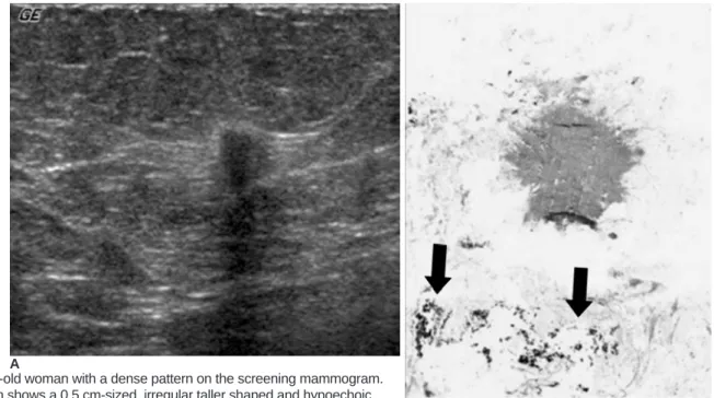
Fig. 1. A 67-year-old woman with a dense pattern on the screening mammogram.
A. Ultrasonogram shows a 0.5 cm-sized, irregular taller shaped and hypoechoic solid nodule.
B. Photomicrograph of the specimen after the US-tattoo shows a small infiltrative ductal cancer. Adjacent to the mass, charcoal markings are observed as black particles (black arrows). (Hematoxylin & Eosin staining; original magnification, 40)
A
B Table 3. The Histology Results of the Preoperative Core
Biopsy or Aspiration in 66 Lesions
Core Biopsy Number of Lesions (%)*
Malignant
Invasive ductal carcinoma 19 (28.8) Invasive lobular carcinoma 1 (1.5) Ductal carcinoma in situ 4 (6.1)
Tubular cancer 1 (1.5)
Total 25 (37.9)
Benign
Fibroadenoma 4 (6.1)
Fibrocystic change 3 (4.5)
Others 3 (4.5)
Total 10 (15.1)
Borderline or high risk
Atypical ductal hyperplasia 10 (15.1)
Mucocele-like lesion 1 (1.5)
Phyllodes tumor 1 (1.5)
Sclerosing Adenosis 1 (1.5)
Papillary lesions 13 (19.7)
Total 26 (39.4)
Aspiration Number of Lesions (%)*
Atypical cell 4
Papillary neoplasm 1
Total 5 (7.6)
Note. *The numbers in the parentheses are percentages.
Benign other lesions include stromal fibrosis and ductal hyperplasia.
Papillary lesions include five papillomas, seven intraductal papillomas and one atypical papilloma.
sterilization using betadine to needle retrieval after infiltra- tion the charcoal suspension.
The US findings, the medical records associated with US- tattoo (technical problem, specimen size, tattoo visualiza- tion, complication) and the pathology results were reviewed. The pathology results were divided into benign lesions (i.e. fibroadenoma, fibrocystic change and others), borderline lesions (i.e. atypical ductal hyperplasia, papilloma and radial scar) and malignant lesions (i.e.
invasive carcinoma, ductal carcinoma in situ and others).
The first follow-up US was carried out within six months after surgery, and an attempt was made to determine if there was some abnormality within or around the tattooing and surgical site.
RESULTS
Of ten benign and 26 borderline lesions by the core biopsy or cytology, five lesions were upgraded by surgery:
two benign lesions to atypical ductal hyperplasia, two atypical ductal hyperplasias to an invasive ductal
carcinoma and one ductal carcinoma in situ and papilloma to a ductal carcinoma in situ (Table 3).
The size of the lesions ranged from 0.4 cm to 3.2 cm, averaging approximately 1.0 cm. Multiple (2 4) lesions were tattooed in 24 patients.
The pathologist could easily identify the targeted lesion around the tattoo in all cases (Figs. 1, 2) except for one.
Although the tumor area was partly masked by the tattoo, there was no significant interference with the pathological diagnosis . The pathology findings were benign lesions (n =
85), borderline lesions (n = 28) and malignant lesions (n = 51) (Table 4). The lesion size was 0.5 3.2 cm (mean size 1.0 cm) and the specimen size was 0.7 4 cm (mean size 2.7 cm).
The procedure took approximately 5 minutes (range, 2 10 minutes) per lesion, and almost all patients tolerated the
Table 4. The Histology Results of Tattooed Lesion Based on the Pathology at Surgery in 164 Cases
Histologic Types Number of Lesions (%)*
Malignant
Invasive ductal carcinoma 31 (18.9)
Metastatic lymph node 2 (1.2)
Ductal carcinoma in situ 10 (6.1)
Tubular cancer 6 (3.6)
Invasive lobular carcinoma 1 (0.6)
Papillary cancer 1 (0.6)
Total 51 (31.1)
Benign
Fibroadenoma 42 (25.6)
Fibrocystic change 23 (14.0)
Others 20 (12.2)
Total 85 (51.8)
Borderline
Papilloma 22 (13.4)
Atypical ductal hyperplasia 5 (3.1)
Radial scar 1 (0.6)
Total 28 (17.1)
Note. *The numbers in the parentheses are percentages.
Other benign lesions include stromal fibrosis, ductal hyperplasia, adenosis, duct ectasia, mucocele-like lesions, and benign phyllodes tumor.
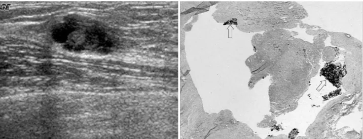
Fig. 2. A 31-year-old woman presented for screening.
A. Ultrasonogram shows a 1.3-cm-sized, complex cystic mass.
B. Photomicrograph of the specimen after the US-tattoo shows a cystically dilated duct, containing charcoal (white arrows) and inflam- matory exudates. (Hematoxylin & Eosin staining; original magnification, 40)
A B
US-tattoo procedure well.
The technical problems encountered include blockage of the needle tip and difficulty in advancing the needle.
Blockage of needle tip by precipitated charcoal particles was an occasional problem (8/164, 4.9%). Strong pressure to the blocked needle relieved the blockage in all eight cases. When resistance was felt within the hard masses, the charcoal solution was injected only superficially into the mass. Sometimes, it was difficult to advance the needle in an extremely dense breast. In these cases, the needle was reinserted through another route adjacent to the first entry site.
Pain and bleeding were minor complications associated with the procedures. These complications were managed by lidocaine infiltration and local compression.
The mean time between the tattooing and surgery was 1.4 days (from immediately before surgery to a maximum of 57 days). Surgery was usually performed on the same day (n = 84) or the next day after the US-tattoo (n = 45) but it was occasionally delayed from three to 57 days after the US-tattoo (n = 5). The surgeon precisely identified the tattoo in 163 out of 164 cases, even in those cases in whom surgery had been delayed. In one case, the on-day tattoo was tinged so scantly in the superficial portion of the lesion that the surgeon could not be guided by the tattoo and an unwanted wide excision was performed. The possible cause of failure in this case might have been the forced injection of a blocked needle tip, which reduced the amount of the diluted tattooed solution remaining in the lesion. The final pathological result was fibrocystic change without a specific diagnosis.
All patients except for two were examined with US within six months after surgery. There were no cases with residual or recurred masses or lesions at tattooing sites.
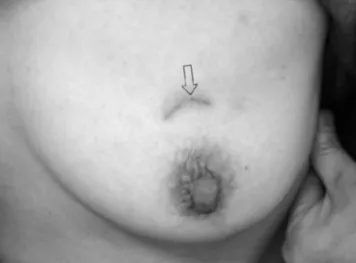
The sole complication was a cosmetic problem, i.e. residual linear tattooing of the skin after surgery in two patients (Fig. 3).
DISCUSSION
The increasing number of nonpalpable breast lesions being detected during US screening of the breast has highlighted the need for the rapid and precise localization.
Among the several techniques, needle localization is widely used. However, there is a high rate of complications and failure (3, 7 10).
Although the optimal time for wire localization was thought to be immediately before surgery, it is difficult in practice. Wire localization is sometimes performed one day before surgery. In this case, the migration or withdrawal of the localized wire might be problematic. Sometimes, the removal of an anchored wire can occur if the operation was suspended due to the poor general condition of the patient, e.g., the heart is not strong enough to endure the procedure and general anesthesia.
In contrast, an US-tattoo is rapid and easy to perform with minimal patient discomfort. The stability of charcoal marking over time is one of its strengths (6, 11, 12).
Because activated charcoal is in particulate form and insoluble in water, it remains within the track and does not disperse to the surrounding tissue, which is in contrast to methylene blue. Therefore, the tracing of the charcoal tattoo during surgery makes it easy to locate the lesion, which means that surgery can be planned over a period of many days. In addition, no particular equipment or instru- ments are needed, and charcoal is biologically inert and has been used in skin tattoos for centuries (13). A histopathology examination of the charcoal specimens revealed only low-grade foreign body reactions. The patients who had received charcoal, including many who were followed up for several years, showed no adverse symptoms (12). Furthermore, the use of charcoal for breast marking has been demonstrated to be safe in other studies (6, 11, 12) that examined stereotactically guided carbon localization in combination with fine-needle aspiration and hookwire localization. The fixation of charcoal particles by phagocytosis subsequently occurs. This permanently marks the track, which means that it can be used before neoadju- vant chemotherapy (14, 15).
Mullen et al. (16) reported that the marking of stereotac- tic biopsy track with a charcoal suspension was safe and effective in place of additional preoperative needle localization (17 20). If the targeted lesion is completely removed with a vacuum-assisted biopsy, the accurate placement of a clip or marker at the biopsy site will facili-
Fig. 3. Photograph of the left breast after breast conserving surgery demonstrates a residual charcoal stain along the scar.
tate accurate placement of the localizing needle in those lesions requiring surgical excision. In these cases, an US- tattoo might be helpful in identifying the previous lesion site (16).
The material needed for an US-tattoo is readily available and cost effective. Moreover the method of preparation is not difficult, as described above.
In our series, the most serious problem during the injection was a blockage of the needle tip by charcoal particles. The pop-up of charcoal particles could be prevented by continuously shaking the bottle. An injection under excessive pressure should be avoided if there is any resistance at the time of the charcoal injection. Another technical complication was that needle insertion was too tight to advance in an extremely dense breast. In this case, another route should be found.
Many complications are possible. Among them, pain and bleeding sometimes occurred, which were controlled by additional local anesthesia and local compression. Residual charcoal marking at the skin after procedure was another complication, which was managed by the additional removal of a skin stripe during surgery.
There are some advantages in US-tattoo compared with stereotactic-charcoal marking. First, breast compression using a fenestrated paddle is essential for performing stereotactic-charcoal marking, which cause pain and discomfort. The second problem that can arise in stereo- tactic-charcoal marking is the loss of the charcoal tract during the decompression of breast after stereotactic guidance. This is not problematic for US-guided charcoal marking because little pressure is applied during US scanning. Furthermore, US-guidance is more comfortable to the patients than stereotactic guidance due to the patient’s supine position. The disadvantage is that the application is limited to a sonographically visible lesion.
This is not possible when the lesions are observed only by mammography. Although the calcifications were also observed by sonography, wire localization is
recommended because charcoal tattooing cannot be identified on a mammogram. Hence, it is impossible to confirm the relationship between a charcoal marked area and a total area of calcification. However, one can substi- tute an US-tattoo for wire localization to localize the lesion on both US and the mammogram. One possible use is in cases of track localization after an US or stereotactic- guided mammotome biopsy.
This study had several limitations. First, a long-term follow up was not performed. Second, the number of skin tattoos could be underestimated because they were based on the patients’ chart. Third, a surgeon unfamiliar with this procedure could confuse the tattoo with the foci of
bleeding or a cauterization effect.
In conclusion, US-tattoo for nonpalpable breast lesions is a very simple, safe and accurate method. After the US- tattoo, the radiologist and surgeon can schedule surgery according to their timetable, and safely delay the surgery time in unavoidable situations.
References
1. Altomare V, Guerriero G, Giacomelli L, Battista C, Carino R, Montesano M, et al. Management of nonpalpable breast lesions in a modern functional breast unit. Breast Cancer Res Treat 2005;93:85-89
2. Czarnecki DJ, Feider HK, Splittgerber GF. Toluidine blue dye as a breast localization marker. AJR Am J Roentgenol 1989;153:261-263
3. Homer MJ, Smith TJ, Safaii H. Prebiopsy needle localization.
Methods, problems, and expected results. Radiol Clin North Am 1992;30:139-153
4. Rosenberg AL, Schwartz GF, Feig SA, Patchefsky AS. Clinically occult breast lesions: localization and significance. Radiology 1987;162:167-170
5. Saarela AO, Kiviniemi HO, Rissanen TJ. Preoperative methyl- ene blue staining of galactographically suspicious breast lesions.
Int Surg 1997;82:403-405
6. Svane G. A stereotaxic technique for preoperative marking of non-palpable breast lesions. Acta Radiol Diagn (Stockh) 1983;24:145-151
7. Bristol JB, Jones PA. Transgression of localizing wire into the pleural cavity prior to mammography. Br J Radiol 1981;54:139- 140
8. Davis PS, Wechsler RJ, Feig SA, March DE. Migration of breast biopsy localization wire. AJR Am J Roentgenol 1988;150:787-788 9. Helvie MA, Ikeda DM, Adler DD. Localization and needle
aspiration of breast lesions: complications in 370 cases. AJR Am J Roentgenol 1991;157:711-714
10. Homer MJ. Transection of the localization hooked wire during breast biopsy. AJR Am J Roentgenol 1983;141:929-930 11. Canavese G, Catturich A, Vecchio C, Tomei D, Estienne M,
Moresco L, et al. Pre-operative localization of non-palpable lesions in breast cancer by charcoal suspension. Eur J Surg Oncol 1995;21:47-49
12. Langlois SL, Carter ML. Carbon localisation of impalpable mammographic abnormalities. Australas Radiol 1991;35:237-241 13. Sperry K. Tattoos and tattooing. Part I: History and methodol-
ogy. Am J Forensic Med Pathol 1991;12:313-319
14. Bonhomme-Faivre L, Mathieu MC, Grossiord JL, Depreatere P, Couarraze G, Orbach-Arbouys S, et al. Formulation of a charcoal suspension for intratumor injection. Part 1: Study of the nature, granulometry, and concentration. Pharm Res 1997;14:218-223
15. Bonhomme-Faivre L, Depraetere P, Savelli MP, Amdidouche D, Bizi E, Seiller M, et al. Charcoal suspension for tumor labelling modifies macrophage activity in mice. Life Sci 2000;66:817-827
16. Mullen DJ, Eisen RN, Newman RD, Perrone PM, Wilsey JC.
The use of carbon marking after stereotactic large-core-needle breast biopsy. Radiology 2001;218:255-260
17. Burbank F, Forcier N. Tissue marking clip for stereotactic breast biopsy: initial placement accuracy, long-term stability, and usefulness as a guide for wire localization. Radiology
1997;205:407-415
18. Fajardo LL, Bird RE, Herman CR, DeAngelis GA. Placement of endovascular embolization microcoils to localize the site of breast lesions removed at stereotactic core biopsy. Radiology 1998;206:275-278
19. Kopans DB. Review of stereotaxic large-core needle biopsy and
surgical biopsy results in nonpalpable breast lesions. Radiology 1993;189:665-666
20. Liberman L, Dershaw DD, Morris EA, Abramson AF, Thornton CM, Rosen PP. Clip placement after stereotactic vacuum- assisted breast biopsy. Radiology 1997;205:417-422