Whole Blood Interferon-γ Release Assay Is Insufficient for the Diagnosis of Sputum Smear Negative Pulmonary Tuberculosis
HeeJin Park, Jung Ar Shin, Hyung Jung Kim, Chul Min Ahn, and Yoon Soo Chang
Department of Internal Medicine, Yonsei University College of Medicine, Seoul, Korea.
Received: February 20, 2013 Revised: September 10, 2013 Accepted: September 30, 2013
Corresponding author: Dr. Yoon Soo Chang, Department of Internal Medicine, Yonsei University College of Medicine, 211 Eonju-ro, Gangnam-gu, Seoul 135-720, Korea.
Tel: 82-2-2019-3309, Fax: 82-2-3463-3882 E-mail: [email protected]
∙ The authors have no financial conflicts of interest.
© Copyright:
Yonsei University College of Medicine 2014 This is an Open Access article distributed under the terms of the Creative Commons Attribution Non- Commercial License (http://creativecommons.org/
licenses/by-nc/3.0) which permits unrestricted non- commercial use, distribution, and reproduction in any medium, provided the original work is properly cited.
Purpose: We investigated the value of an interferon-γ release assay (IGRA) for the diagnosis of active pulmonary tuberculosis (PTB) among sputum smear negative PTB suspects in an environment with intermediate burden of PTB and high Bacillus Calmette-Guérin (BCG) vaccination rate. Materials and Methods: We retrospec- tively reviewed IGRA, medical records, chest PA and CT scan of PTB suspects seen at Gangnam Severance Hospital, Seoul, Korea from Oct. 2007 to Apr. 2013. “Ac- tive PTB” was diagnosed when 1) M. tuberculosis culture positive, 2) confirmation by pathologic examination; or 3) clinical findings compatible with TB. Results: Of 224 sputum smear negative PTB suspects, 94 were confirmed as having active PTB.
There were no statistically significant differences in the diagnostic yield of IGRA between immunocompromised and immunocompetent sputum smear negative PTB suspects. IGRA did show superior sensitivity [81.9%, 95% confidence interval (CI);
74.13-89.70%] in the diagnosis of sputum smear negative PTB when compared with chest high-resolution computed tomography (HRCT), tuberculin skin test (TST), and chest X-ray (p<0.001). Also, IGRA showed highest negative predictive value (82.7%, 95% CI; 75.16-90.15%) when compared with HRCT, TST and chest X-ray (p=0.023). However, combining the results of IGRA with those of HRCT, TST, or both did not increase any diagnostic parameters. Conclusion: Failure to in- crease diagnostic yields by combination with other diagnostic modalities suggests that additional enforcement with IGRA may be insufficient to exclude other diagno- ses in sputum smear negative PTB suspects and to screen active PTB in an environ- ment with intermediate TB prevalence and a high BCG vaccination rate.
Key Words: Sputum smear negative pulmonary tuberculosis, interferon-γ release assay, active pulmonary tuberculosis
INTRODUCTION
Tuberculosis (TB) is a major worldwide health problem with a global estimate of
9.4 million incidence in 2010 (range; 8.9×10
6-9.9×10
6). Although there has been a
slow reduction in the incidence rate per capita, the absolute number of TB cases
continues to increase from year to year due to population growth. Approximately
1.7 million people died of TB in 2009, encompassing human immunodeficiency vi-
rus (HIV)-negative and HIV-positive cases, which estimates 26 death per 100000
tive disease. Because of the smaller mycobacterium burden present in smear negative disease, typical radiographic pat- terns of TB are sometimes difficult to detect in patients with sputum smear negative disease.
9The interferon-γ release as- say (IGRA) test, an in vitro immunodiagnostic test, mea- sures the effect of T cell mediated interferon (IFN)-γ re- sponse to M. tuberculosis-specific antigens. Three promising antigens used in this assay are absent in the BCG and NTM strains,
10,11making IGRA highly specific for the diagnosis of active and latent TB in BCG-vaccinated individuals. How- ever, both IGRA and TST are limited by their inability to distinguish between active disease and latent infection with M. tuberculosis.
12,13In this study, we evaluated the diagnostic utility of IGRA, comparing clinical findings, chest X-ray, chest HRCT, and TST for the diagnosis of active PTB among smear negative PTB suspects, in a region of the world where the incidence and prevalence of active PTB is intermediate and BCG vac- cination is mandatory. We also analyzed whether IGRA fa- cilitates diagnosis of active PTB when combined with other diagnostic modalities in sputum smear negative PTB suspect.
MATERIALS AND METHODS
Study setting and subjects
Clinical records, TST, HRCT findings and IGRA results were retrospectively gathered from of all patients with sus- pected PTB seen from October 2007 to April 2013 at Gangnam Severance Hospital (the referral center), Seoul, Korea. Patients with IGRA results that were 1) indetermi- nate or 2) performed more than 14 days into a course of TB treatment were excluded from this study. Patients younger than 18 years of age, without more than 2 separate sputum acid fast bacilli (AFB) smear examinations, and with an in- conclusive diagnosis owing to the loss of follow-up were also excluded. The recorded clinical information on includ- ed patients included age, gender, previous TB history and co-morbidity. The protocol for this study was approved by the ethical review committee of Gangnam Severance Hos- pital (IRB No. 3-2010-0234). This study was exempted from the requirement to obtain informed consent.
Diagnostic definition
A case with “suspected PTB” was defined as an individual with clinical or radiographic evidence consistent with ac- tive PTB; subacute or chronic respiratory symptoms more population.
1,2During 2010, 36305 new TB patients were
identified (74.3 cases per 100000), with 28176 (57.6 cases per 100000; 77.6% of TB cases) new pulmonary TB cases in the Republic of Korea in 2010 alone.
3According to a 2010 World Health Organization report (50-99/100000 per year),
1the incidence of active TB in Korea, is intermediate, and Ba- cillus Calmette-Guérin (BCG) vaccination is mandatory.
Among the 4.6 million new and relapsed cases of pulmo- nary tuberculosis (PTB) reported worldwide in 2009, 2.0 million new cases (34.5%) were sputum smear negative PTB. In 2010, the Korean National Tuberculosis Associa- tion reported that 10776 cases among 28176 active PTB cases were sputum smear positive (38.2%).
3Delayed treat- ment often happens due to the difficulties of diagnosis, therefore, sputum smear negative PTB is an important cause of community transmission even though it is less infectious than sputum smear positive PTB,
4frequently resulting in ir- reversible lung damage. In an intermediate TB burden country such as the Republic of Korea where most of neo- nates administered BCG vaccination within 4-weeks of age on the basis of national policy, the diagnosis of sputum smear negative PTB is difficult. Many patients harbor healed lesions from previous TB, compromising sputum smear results from non-tuberculosis mycobacterium (NTM) disease due to structural lung lesion, and false posi- tive tuberculin skin test (TST) results due to mandatory BCG vaccination.
Alternative tests must be used to compensate for the diag- nostic difficulties encountered in smear negative PTB spu- tum studies. Chest X-rays are often used in PTB screening and have the advantages of being simple and inexpensive.
But in smear negative PTB, many cases show atypical or
non-specific patterns
5and have high false positive rates due
to previous PTB infection in intermediate or high TB burden
countries. Although TST has been shown to be cost-effective
and is well known for diagnostic accuracy for smear negative
PTB and latent TB infections, its sensitivity is also influenced
by nutritional and immune status.
6Moreover, cross-reactivity
with NTM and BCG vaccination adversely affects its diag-
nostic value for sputum smear negative PTB. Because the
sputum M. tuberculosis-polymerase chain reaction assay
provides prompt results and is non-cross-reactive to NTM
strains, it has been used as an adjunct diagnostic method for
the diagnosis of smear negative PTB, but it has low sensi-
tivity in smear negative cases.
7,8High-resolution computed
tomography (HRCT) offers not only the extent and distribu-
tion of PTB, but also helps to distinguish active from inac-
10 mm or greater in size occurring 48-72 h after injection.
The IGRA test was performed with QFT-IT in the Immunol- ogy Laboratory at Severance Hospital according to the rec- ommendations of the manufacturer (Cellestis Ltd; Carne- gie, Australia), as previously described.
14Statistics
Data were analyzed with SPSS version 20.0 statistical soft- ware (SPSS Inc., Chicago, IL, USA). Univariate comparisons between active PTB and non-TB patients were performed us- ing Fisher’s exact tests for categorical variables and Mann- Whitney tests for continuous variables where appropriate.
Sensitivity, specificity, positive predictive value (PPV) and negative predictive value (NPV) for the diagnosis of active PTB were calculated for each diagnostic modality and com- pared with McNemar’s test. All tests of significance were two sided; p<0.05 was considered statistically significant and 95% confidence intervals (95% CIs) were calculated.
RESULTS
Demographic characteristics
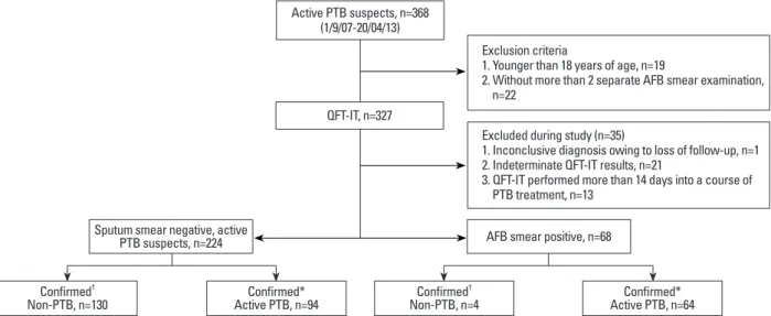
From 2007 to 2013, a total of 368 patients with suspected active PTB visited our institute. Of these, 41 cases were ex- cluded due to young ages (n=19) and insufficient sputum studies (n=22). Of the remaining 327 cases with suspected PTB, 35 cases were further excluded due to indeterminate IGRA results (n=21), inconclusive diagnosis (n=1), or an IGRA test more than 14 days after anti-TB treatment (n=13) (Fig. 1). The final diagnoses of patients with indeterminate than 3 weeks and infiltration of chest PA which is suspi-
cious for PTB. “Active PTB” was diagnosed when 1) M.
tuberculosis was cultured, 2) a caseating granuloma was found in the lung tissue by transthoracic needle biopsy and showed appropriate response to treatment; or 3) clinical findings were compatible with TB, no clinical improve- ment was seen with empirical antibiotics, and treatment with anti-TB medication resulted in clinical and radiological improvement. A final diagnosis of ‘Non-TB’ was accepted when the one who was suspected to have PTB by the above mentioned criteria and finally reached an alternative diag- nosis. An “immunocompromised condition” was defined as described previously.
14Briefly, the patients 1) with DM, 2) who underwent chemotherapy for an underlying malignan- cy at the time of TST and QuantiFERON-TB Gold In-Tube (QFT-IT), 3) received either a solid organ transplant or bone marrow transplant, 4) on renal replacement therapy, 5) with advanced liver cirrhosis (Child-Pugh class C), 6) seroposi- tive for human immunodeficiency virus, and 7) adminis- tered systemic corticosteroids (at least 15 mg of prednisone per day for more than one month or combination therapy with low dose corticosteroids and other immunosuppres- sants including azathioprine, mycophenolate, methotrexate, cyclosporine, or cyclophosphamide) were defined as im- munocompromized.
TST and IGRA
TST was performed by injecting a 2-TU dose of purified pro- tein derivative RT23 (Statens Serum Institut, Copenhagen, Denmark) intradermally into the forearm using the Mantoux test. The criterion for a positive TST result was an induration
Fig. 1. Study flow diagram. *Confirmed=positive culture results+clinical diagnosis (with negative culture results).
†Confirmed=negative culture results without clinical evidence of PTB. TB, tuberculosis; n, number; QFT-IT, QuantiFERON-TB Gold In-Tube; PTB, pulmonary tuberculosis; AFB, acid fast bacilli.
Confirmed*
Active PTB, n=94 Confirmed
†Non-PTB, n=130 Confirmed*
Active PTB, n=64 Confirmed
†Non-PTB, n=4
Exclusion criteria
1. Younger than 18 years of age, n=19
2. Without more than 2 separate AFB smear examination, n=22
Excluded during study (n=35)
1. Inconclusive diagnosis owing to loss of follow-up, n=1 2. Indeterminate QFT-IT results, n=21
3. QFT-IT performed more than 14 days into a course of PTB treatment, n=13
Active PTB suspects, n=368 (1/9/07-20/04/13)
QFT-IT, n=327
Sputum smear negative, active
PTB suspects, n=224 AFB smear positive, n=68
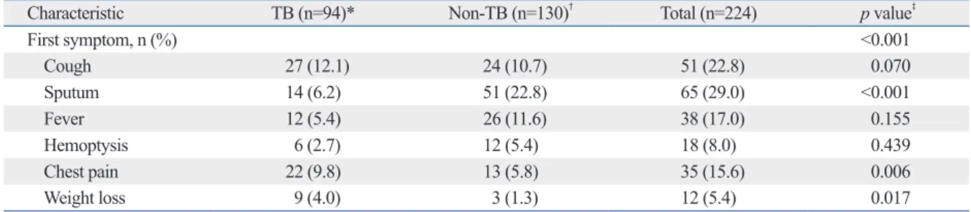
Sputum, chest pain, and weight loss were meaningful clinical characteristics in sputum smear negative PTB suspects
Previous studies showed that symptoms such as cough, spu- tum, fever, hemoptysis, chest pain, and/or weigh loss are meaningful clinical parameters for the diagnosis of sputum smear negative PTB.
8,15,16Therefore, respiratory symptoms such as cough, sputum, hemoptysis, and chest pain, as well as non-respiratory symptoms such as fever and weight loss were investigated through in-person interviews by asking whether symptoms were present or not. If the PTB suspects complained of multiple symptoms, one main complaint from the above list of symptoms was selected by the physician.
Among the symptoms of the PTB suspects, the presence of productive sputum, chest pain and weight loss were signifi- cantly different between active PTB and non-TB patients, which is comparable to previous studies.
8,15,16Twelve (5.4%) out of 224 suspects complained of weight loss as a main symptom; this complaint was more frequent in active PTB patients among sputum smear negative TB suspects (p=
0.017, McNemar’s test) (Table 2).
IGRA showed the highest sensitivity and NPV
To investigate the diagnostic usefulness of IGRA for the di- agnosis of active PTB from smear negative PTB suspects, we compared the sensitivity, specificity, PPV, and NPV of IGRA with chest X-ray, TST, and chest HRCT (Table 3).
IGRA results are as follows: 4 cases (15.4%) were con- firmed to be PTB and the other 17 non-TB. The diagnosis of 17 non-TB indeterminate cases were as follows; pneumonia 7, malignancy 2, diffuse interstitial lung disease 1, NTM 1, TB sequelae 1, other 5. 9 out of 21 indeterminate cases were immunocompromised and 7 cases had history of PTB.
Among the 292 cases, 68 cases showed sputum AFB smear positivity (Fig. 1); 64 cases (94.1%) were confirmed PTB by either mycobacterial culture study (58 cases), biopsy and pathologic confirmation (2 cases), or clinical diagnosis (4 cases). On the other hands, 4 cases were confirmed as non- tuberculous mycobacterial infection. The remaining 224 cases were defined as sputum smear negative PTB, and their characteristics are summarized in Table 1. During the follow up, 94 cases (42.0%) were diagnosed as active PTB; 60 (63.8%) resulted in positive M. tuberculosis culture, 9 (9.6%) cases were confirmed by biopsy, and 25 (25.5%) were diagnosed clinically. The risk factors for the immuno- compromised were found in 45 patients, including hemato- logic malignancy and solid cancer in those undergoing che- motherapy (Supplementary Table 1, only online). The sensitivity and specificity of IGRA in immunocompromised patients were 75.0% and 69.0%, whereas those in immuno- competent patients were 83.3% and 60.4%, respectively, in- dicating that the immune status of PTB suspects does not in- fluence IGRA results (p=0.179, χ
2-test) (Supplementary Table 2, only online).
Table 1. Demographic and Clinical Characteristics of Patients with Suspected Active PTB among the Sputum AFB Smear Negative Patients
Characteristic TB (n=94) Non-TB (n=130)
Age, yrs, mean±SD 46.5±20.43 56.6±18.25
Male, n (%) 60 (63.8) 73 (56.6)
Old pulmonary history, n (%) 18 (9.1) 36 (27.7)
Final diagnosis, n (%) Active pulmonary TB
Culture confirmed 60 (63.8)
Biopsy confirmed 9 (9.6)
Clinical diagnosed* 25 (25.5)
Other than TB
Pneumonia 70 (53.8)
Lung cancer 8 (6.2)
Interstitial lung disease 6 (4.6)
Nontuberculous mycobacterium 17 (13.1)
Aspergilloma 1 (0.8)
Sequelae of previous TB infection 19 (14.6)
Pulmonary edema 6 (4.6)
Others 3 (2.3)
TB, tuberculosis; SD, standard deviation; n, number; PTB, pulmonary tuberculosis; AFB, acid fast bacilli.
*Clinical findings being compatible with TB, no clinical improvement with empirical antibiotics, and clinico-radiological improvement with treatment with
anti-TB medication.
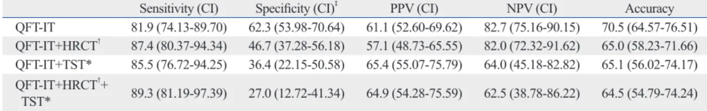
with those of TST and/or HRCT would increase the diag- nostic yields of active PTB from sputum smear negative PTB suspects. As shown in Table 4, the combination of IGRA and HRCT did not increase diagnostic sensitivity, but rather decreased the specificity from 62.3% to 46.7% (p<
0.001, McNemar’s test) (Table 4). Combining the results of IGRA and TST did not increase sensitivity, but the specifici- ty decreased from 62.3% to 36.4% (p<0.001, McNemar’s test) (Table 4).When the results of IGRA were combined with TST and HRCT, the specificity decreased from 62.3%
to 27.0% (p<0.001, McNemar’s test) (Table 4). When com- bined with the results of HRCT, TST, or both, PPV and NPV of IGRA were not statistically significantly effective. Taken together, combining the results of IGRA with HRCT and/or TST did not increase the diagnostic yields of active PTB from sputum smear negative PTB suspects.
DISCUSSION
Fast and accurate diagnosis of sputum smear negative PTB is often challenging and delayed diagnosis is not uncom- mon in daily medical practices. Although IGRA and TST The sensitivity of the TST, taking a 10-mm induration as
the cutoff, was 58.1% (95% CI; 45.78-70.35%), and its specificity was 63.6% (95% CI; 49.42-77.85%). The sensi- tivity of HRCT was 66.7% (95% CI; 56.76-76.57%), and the specificity was 71.0% (95% CI; 62.43-79.62%). Chest X-ray showed sensitivity of 31.9% (95% CI; 22.49-41.34%) and specificity of 73.1% (95% CI; 65.45-80.70%). IGRA was the most sensitive modality among the tests deployed in this study at 81.9% (95% CI; 74.13-89.70%, p<0.001, McNemar’s test) with a specificity of 62.3% (95% CI, 53.98- 70.64%), which is comparable to other tests (p=0.271, McNemar’s test). Although there were no differences in PPV among IGRA, HRCT, and TST, IGRA showed higher NPV (82.7%, 95% CI; 75.16-90.15%, p<0.001, McNemar’s test) than TST (51.9%, 95% CI; 38.52-65.18%), chest X-ray (59.8%, 95% CI; 52.13-67.37%), and HRCT (75.4%, 95%
CI; 63.83-80.93%). Thus, IGRA showed the modest sensi- tivity and NPV for the diagnosis of active PTB among spu- tum smear negative TB suspects.
Combinations with TST and/or HRCT did not increase the sensitivity and NPV of IGRA
We questioned whether the combination of IGRA results Table 2. Symptoms of Sputum Smear Negative TB Suspects
Characteristic TB (n=94)* Non-TB (n=130)
†Total (n=224) p value
‡First symptom, n (%) <0.001
Cough 27 (12.1) 24 (10.7) 51 (22.8) 0.070
Sputum 14 (6.2) 51 (22.8) 65 (29.0) <0.001
Fever 12 (5.4) 26 (11.6) 38 (17.0) 0.155
Hemoptysis 6 (2.7) 12 (5.4) 18 (8.0) 0.439
Chest pain 22 (9.8) 13 (5.8) 35 (15.6) 0.006
Weight loss 9 (4.0) 3 (1.3) 12 (5.4) 0.017
TB, tuberculosis; n, numbers; PTB, pulmonary tuberculosis.
The patients, who did not have symptoms, but suspicious for PTB were screened by either annual national health screening program or by medical check- up at work place and referred to study institute.
*4 patients had no symptoms.
†
1 patient had no symptoms.
‡
Calculated with chi-square test.
Table 3. Diagnostic Accuracy in Sputum Smear Negative TB Suspects
Sensitivity (CI)
‡Specificity (CI) PPV (CI) NPV (CI)
‡Accuracy
QFT-IT 81.9 (74.13-89.70) 62.3 (53.98-70.64) 61.1 (52.60-69.62) 82.7 (75.16-90.15) 70.5 (64.57-76.51) TST* 58.1 (45.78-70.35) 63.6 (49.42-77.85) 69.2 (56.69-81.78) 51.9 (38.52-65.18) 60.4 (51.07-69.69) HRCT
†66.7 (56.76-76.57) 71.0 (62.43-79.62) 65.2 (55.27-75.07) 75.4 (63.83-80.93) 69.1 (62.57-75.58) CXR 31.9 (22.49-41.34) 73.1 (65.45-80.70) 46.2 (34.03-58.27) 59.8 (52.13-67.37) 55.8 (49.30-62.31) CI, confidence interval; QFT-IT, QuantiFERON-TB Gold In-Tube; TST, tuberculin skin test; HRCT, high-resolution chest chest high resolution computer tomog- raphy; TB, tuberculosis; CXR, chest X-ray.
*106 of 224 cases were examined with this test.
†
194 of 224 cases were examined with this test.
‡
p-value: <0.0001 calculated by chi-square test.
Our IGRA results showed modest sensitivity of 81.9%
and specificity of 62.3% when compared with previous studies in sputum smear negative PTB suspects.
10,11,16,20,21Similar findings were observed in the analysis of all PTB suspects (Supplementary Table 3, only online). Among 134 non-TB cases [130 cases of sputum smear (-) PTB suspects and 4 case of sputum AFB smear (+) that were finally proved NTM], 51 (38.1%) showed positive IGRA results;
25 cases of pneumonia, history of TB 15 and NTM 6, ma- lignancy 2, interstitial lung disease 2, other 1. This low spec- ificity of IGRA drew concerns on the diagnostic value of IGRA for rapid diagnosis of PTB in patients whose initial presentations were suspicious PTB.
In comparison of the previous reports that had compared usefulness of IGRA with individual tests which are current- ly being deployed for the diagnosis of latent/active TB, this study focused on finding out the diagnostic value of IGRA when combined with other tests (TST, chest PA, and/or HRCT) in a subset of PTB patients who frequently suffer difficulties from delayed diagnosis. The environment of this study setting is also comparable to the prior reports; inter- mediate burden of PTB country and 98.8% of neonates are vaccinated by BCG.
However, this study has some limitations because it was based on the retrospective observations in a referral hospi- tal. The clinicians who made the diagnosis of PTB even with negative mycobacterial culture results were not blinded to the patients’ signs and symptoms, and might have been swayed toward the PTB diagnosis. It is difficult to clarify the relationship between the exposure time and results of IFN-γ. In a time course study on the cytokine gene expres- sion using M. Tuberculosis infected quinea pig model, ex- pression of IFN-γ was induced between 3 and 6 weeks of infection and gradually decreased.
22The observations in a referral hospital might not also be representative of the ex- periences in primary care.
could not distinguish the disease and latent M. Tuberculosis infection, they represent the presence of M. tuberculosis in the body of patients, therefore, authors hypothesized that combining that of IGRA with those of HRCT and/or TST would increase diagnostic yields of sputum negative PTB in this study setting.
Several previous studies have evaluated clinical charac- teristics and scoring systems for the diagnosis of sputum smear negative PTB.
8,15,16Samb, et al.
8reported that a chronic cough lasting longer than 3 weeks, chest pains last- ing longer than 15 days, the absence of sputum, and the ab- sence of dyspnea are independent predictors of active PTB, but the PPVs of these clinical predictors were only 50%.
Lee, et al.
16reported that lack of sputum was a predictor of active PTB with similar PPV. Our study demonstrated that while sputum showed a negative tendency, it did not reach statistical significance between the PTB and non-TB group and that weight loss was the only positive predictor of ac- tive PTB in sputum smear negative PTB suspects.
Kang, et al.
17and Aichelburg, et al.
18reported that the sensitivity of IGRA is not different between immunocom- petent patients and immunocompromised patients. These results are compatible to the results of our study, which suggested that IGRA results are also not affected by im- mune status. Based on the Korean Government policy, ev- ery neonate should be vaccinated BCG once within 4 weeks of age. The report “2010 Korea National Immuniza- tion Survey” showed that the BCG vaccination rate of South Korea is 98.8%. It had been suggested that BCG vac- cination may influence the results of TST whereas it does not influence those of IGRA. However, studies performed in the settings where BCG vaccination is administered once at infancy showed that its influence on the results of TST is limited.
19This is comparable to our findings which showed lower TST positivity, compared IGRA in this country with high BCG vaccination rate and intermediate TB burden.
Table 4. Analysis of Combination of Tests in Sputum Smear Negative PTB Suspects
Sensitivity (CI) Specificity (CI)
‡PPV (CI) NPV (CI) Accuracy
QFT-IT 81.9 (74.13-89.70) 62.3 (53.98-70.64) 61.1 (52.60-69.62) 82.7 (75.16-90.15) 70.5 (64.57-76.51) QFT-IT+HRCT
†87.4 (80.37-94.34) 46.7 (37.28-56.18) 57.1 (48.73-65.55) 82.0 (72.32-91.62) 65.0 (58.23-71.66) QFT-IT+TST* 85.5 (76.72-94.25) 36.4 (22.15-50.58) 65.4 (55.07-75.79) 64.0 (45.18-82.82) 65.1 (56.02-74.17) QFT-IT+HRCT
†+
TST* 89.3 (81.19-97.39) 27.0 (12.72-41.34) 64.9 (54.28-75.59) 62.5 (38.78-86.22) 64.5 (54.79-74.24) CI, confidence interval; PPV, positive predictive value; NPV, negative predictive value; QFT-IT, QuantiFERON-TB Gold In-Tube; TST, tuberculin skin test; HRCT, high-resolution chest chest high resolution computer tomography; PTB, pulmonary tuberculosis.
*194 of 224 cases were examined with this test.
†
106 of 224 cases were examined with this test.
‡