Assessment of the Arterial Stiffness Index as a Clinical Parameter for Atherosclerotic Coronary Artery Disease
Seong-Mi Park, MD, Hong-Seog Seo, MD, Hong-Euy Lim, MD, Sung-Hee Shin, MD, Chang-Gyu Park, MD, Dong-Joo Oh, MD and Young-Moo Ro, MD
Division of Cardiology, Department of Internal Medicine, Korea University Medical College, Seoul, Korea ABSTRACT
Background:Arterial stiffening increases both the systolic blood and pulse pressures, which is known to be a major contributor to atherosclerosis and the most important cause of cardiovascular disease. The aims of this study were to assess the feasibility of the arterial stiffness index (ASI), using a computerized oscillometric device, by comparison with the pulse wave velocity (PWV), and to investigate its usefulness to patients with clinical coronary artery disease. Methods:60-consecutive patients, who underwent coronary angiography and who’s aorto-femoral PWV were obtained with a Judkins catheter, were the subjects of this study. The ASI was obtained for all patients, using CardioVision®MS-2000 (IMDP, Las Vegas, NV), with cuff pressure on the brachial artery.
The ASI were obtained as follows; 1) at the baseline (ASI-B), 2) after hyperemia induced by compression of the arm with cuff pressure for 5 minutes (ASI-H), 3) after having taken sublingual nitroglycerin (ASI-N). Results: 34-patients had significant coronary artery disease (CAD) from the coronary angiography findings. All the ASI were positively correlated to the PWV, and were also higher in patients with CAD (ASI-B, 85.9±57.8 vs. 48.2±
24.5, p=0.001; ASI-H, 98.1±49.8 vs. 48.1±21.3, p=0.00; ASI-N, 66.7±55.7 vs. 33.2±27.9, p=0.002). The ASI-H was mostly well correlated to the PWV and the severity of CAD (PWV, r=0.49, p=0.00; severity, r=0.52, p=0.00). The ASI was increased after hyperemia in patients with CAD (85.9±57.8 to 98.1±49.8, p=0.01), but was not significantly changed in those without CAD (48.2±24.5 to 48.1±21.3, p=0.68). The ASI-N was de- creased in all patients. Conclusions:The Arterial Stiffness Index, measured non-invasively by computerized oscillometry, was feasible and useful for detection of atherosclerotic coronary disease. Especially, the difference in the ASI between patients with and without CAD was more apparent after hyperemia. These findings suggest that in addition to stiffening of the arterial wall itself, the impairment of flow mediated vasodilation, due to endothelial dysfunction, further increases the arterial stiffness. (Korean Circulation J 2004;34 (7):677-683) KEY WORDS:Compliance;Coronary atherosclerosis;Oscillometry.
Introduction
Stiffening of the arterial tree increase the systolic blood pressure (BP), and simultaneously decreases the diasto- lic BP, resulting in a wide ranging pulse pressure (PP).1)
Many studies have shown that the arterial stiffness, when determined invasively, is the most important cause of cardiovascular complications and a major contributor to atherosclerosis, and thus to strokes, myocardial infarc- tions and renal failure.2-4) However, invasive techniques are of limited value for the screening and risk stratifica- tion of clinical cardiovascular disease. So, considering the increasing emphasis placed on primary prevention of cardiovascular diseases, the development of non-invasive techniques to identify high risk patients and to assess the prognosis of future atherosclerotic burden is very impor- tant. Recently, a new clinical device, the CardioVision® Received:March 23, 2004
Revision Received:April 19, 2004 Accepted:May 18, 2004
Correspondence:Hong-Seog Seo, MD.Division of Cardiology, Department of Internal Medicine, Korea University Medical Col- lege, #97 Gurodong-gil, Guro-gu, Seoul 152-703, Korea Tel:82-2-818-6634, Fax:82-2-864-3062
E-mail:[email protected]
MS-2000 (IMDP, Las Vegas, NV) was developed to si- multaneously measure the arterial stiffness index (ASI), brachial arterial BP and PP. Thus, the aims of this study were to evaluate the feasibility of ASI, using the com- puterized oscillometric device, by comparison with the pulse wave velocity (PWV), via femoral arterial cathe- terization, and investigate the association between clini- cal coronary artery disease and the ASI.
Methods
Study populations
60-consecutive patients (30 men and 30 women; mean age=66±7 years), who underwent coronary angiography and who’s arterial pulse wave was recorded with a right Judkins catheter were the subjects of this study. Patients that had had a myocardial infarction or acute coronary syndrome within the last 3 months, valvular heart disease, arrhythmia, decreased left ventricular function (ejection fraction<50%) or chronic renal failure were excluded. All patients refrained from caffeine containing beverages, alcohol and smoking for at least 4 hours prior to the study. All vasoactive medications were discontinued in all patients for at least 12 hours prior to the study.
Coronary angiography
Routine biplane coronary angiography, using the Jud-
kins technique, was performed in all patients. Signi- ficant coronary artery stenosis was defined as >50%
narrowing in the vessel diameter on the coronary an- giogram. All patients were divided into either those with coronary artery disease (defined as at least one vessel with coronary artery stenosis >50%) or those without coronary artery disease. The severity of the coronary artery disease (CAD) was based on the number of stenotic coronary vessels.
Pulse wave velocity obtainment
After diagnostic coronary angiography in all patients, the pulse waves and electrocardiograms were recorded at the aortic arch and right femoral artery with a Judkins catheter. The time delay between the arrivals of pulse wa- ves at these 2 points was obtained by gating to the peak of the R-wave of the electrocardiogram. The distance tra- veled by the pulse wave was measured with the Judkins catheter length, then the PWV calculated as the distance/
time (m/sec).
Measurement of arterial stiffness index
Assessment of the arterial stiffness was performed non-invasively with the CardioVision®MS-2000 (IMDP, Las Vegas, NV). This device provides the BP, PP and bra- chial artery stiffness. It utilizes a computerized oscillo- metric method of BP measurement and through a pres-
Decreasing cuff pressure
Elastic artery Stiff artery
BP cuff Volumetric oscillations
Systolic BP
Diastolic BP Mean BP
High ASI ASI
Figure 1. Measurement of the arterial stiffness index (ASI). BP: blood pressure.
Low ASI
sure sensor attached to the BP cuff (Figure 1). In all pa- tients, the ASI was measured as follows; 1) at rest as the baseline (ASI-B), 2) after hyperemia induced by com-
pression of the arm with cuff pressure for 5 minutes (ASI- H), 3) after having taken 0.5 mg nitroglycerin sublin- gually (ASI-N).
Statistics
For descriptive purposes, all data are presented as the mean value±SD and relative frequencies, as indicated.
Differences in the mean values between those patients with and without CAD were analyzed using ‘Student T- tests’. Comparison of the arterial stiffness indices in each group was performed using ‘paired t-tests’. A p-value of
<0.05 was considered statistically significant.
Results
Clinical characteristics
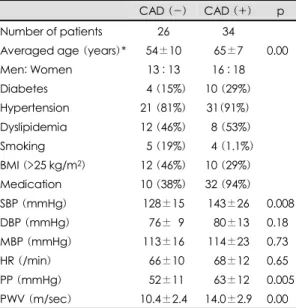
The baseline clinical characteristics of the patients are summarized in Table 1. The patients with CAD were older than those without (65±7 years versus 54±10 years, p=0.00). Most patients had hypertension (86.7%), and the frequencies of hypertension and diabetes were higher in the patients with CAD than in those without.
Table 1. Baseline characteristics of the study patients CAD (-) CAD (+) p Number of patients 26 34
Averaged age (years)* 54±10 65±7 0.000 Men: Women 13 : 13 16 : 18 Diabetes 04 (15%) 10 (29%) Hypertension 21 (81%) 31(91%) Dyslipidemia 12 (46%) 08 (53%) Smoking 05 (19%) 0.4 (1.1%) BMI (>25 kg/m2) 12 (46%) 10 (29%) Medication 10 (38%) 32 (94%) SBP (mmHg) 128±15 143±26 0.008 DBP (mmHg) 076±09 080±13 0.180 MBP (mmHg) 113±16 114±23 0.730 HR (/min) 066±10 068±12 0.650 PP (mmHg) 052±11 063±12 0.005 PWV (m/sec) 10.4±2.4 14.0±2.9 0.000 BMI: body mass index, SBP: systolic blood pressure, DBP:
diastolic blood pressure, HR: heart rate, PP: pulse pres- sure, PWV: pulse wave velocity
Figure 2. Age was one of the greatest factors affecting the arterial stiffness index (ASI). PWV: pulse wave velocity, B:
baseline, H: hyperemia, N: nitroglycerin, p<0.05.
r=0.53 r=0.53 r=0.53 r=0.53
r=0.34 r=0.34 r=0.34 r=0.34 r=0.52
r=0.52
r=0.48 r=0.48 r=0.48 r=0.48
0
0
0
0
0
0
r=0.54 r=0.54
0
0
0
0
0
0 0
0
0
0
0
0
r=0.54 r=0.54
22 20 18 16 14 12 10 8 6
30 40 50 60 70 80 Age
PWV
r=0.53
4.0
3.0
2.0 1.0 0.0
-1.0
30 40 50 60 70 80 Age
Number of stenosis r=0.52
250
200 150 100 50 0
30 40 50 60 70 80 Age
ASI-N
r=0.34 250
200 150 100
50 0
30 40 50 60 70 80 Age
ASI-H
r=0.54 250
200 150 100
50 0
30 40 50 60 70 80 Age
ASI-B
r=0.48
Of note, the difference in the pulse pressure was mediat- ed mainly by a higher systolic BP in the patients with CAD (143±26 mmHg versus 128±15 mmHg, p=
0.008). As known previously, the PWV was higher in patients with CAD than in those without (14.0±2.9 m/
sec versus 10.4±2.4 m/sec, p=0.00) and well correlat- ed to the severity of CAD (r=0.53, p=0.00, Figure 5).
Correlation between pulse wave velocity and arterial stiffness index
Both the PWV and ASI increased with aging (Figure
2). The ASI also increased with the PWV after adjusted for age, but only the ASI-H had a positive correlation with the PWV (r=0.29, p=0.03). The strong factors affecting
Table 3. Comparison of the arterial stiffness index (ASI)
CAD (-) CAD (+) p
ASI-B 48.2±24.5 85.9±57.8 0.001 ASI-B 48.1±21.3 98.1±49.8 0.000 ASI-B 33.2±27.9 36.4±11.3 0.002 Table 2. Correlations with the arterial stiffness index (ASI)
ASI-B ASI-H ASI-N
SBP 0.76 0.68 0.69 DBP 0.39 0.33 0.47
PP 0.81 0.65 0.56
Age 0.48 0.54 0.34 p<0.05 in all cases. B,H,N: baseline, hyperemia, nitrogly-
cerin, SBP (mmHg): systolic blood pressure, DBP (mmHg):
diastolic blood pressure, PP (mmHg): pulse pressure
r=0.48 r=0.48
r=0.31 r=0.31 r=0.31 r=0.52
r=0.52 r=0.52 r=0.53 r=0.53 r=0.53
6 8 10 12 14 16 18 20 22 PWV
4.0 3.0 2.0 1.0 0.0 -1.0
Number of stenosis
r=0.53
0 50 100 150 200 250 ASI-B
4.0 3.0 2.0 1.0 0.0 -1.0
Number of stenosis
r=0.48
0 50 100 150 200 250 ASI-H
4.0 3.0 2.0 1.0 0.0 -1.0
Number of stenosis
r=0.52
0 50 100 150 200 250 ASI-N
4.0 3.0 2.0 1.0 0.0 -1.0
Number of stenosis
r=0.48
Figure 5. Correlation between the arterial stiffness index (ASI) and the severity of coronary artery disease. PWV: pulse wave velocity, B: baseline, H: hyperemia, N: nitroglycerin.
ASI-B ASI-H ASI-N 120
100 80 60 40 20 0
*
†
Figure 3. Changes in the arterial stiffness index (ASI) in patients without coronary artery disease. B: baseline, H:
hyperemia, N: nitroglycerin. *: p=0.68, †: p=0.00
Figure 4. Changes in the arterial stiffness index (ASI) in patients with coronary artery disease. B: baseline, H: hy- peremia, N: nitroglycerin. *: p=0.01, †: p=0.00
ASI-B ASI-H ASI-N 300
250 200 150 100 50 0
* †
the ASI were the systolic BP and PP (Table 2).
Arterial stiffness index and coronary artery disease All the ASI were higher in patients with CAD than in those without (Table 3). In patients without CAD, there were no differences between the ASI-B and ASI-H, and the ASI decreased after having taken nitroglycerin (Fig- ure 3). Compared with the patients without CAD, there were some differences in the response to hyperemia and nitroglycerin in patients with CAD. After hyperemia, the ASI increased (85.9±57.8 to 98.1±49.8, p=0.01) and after having taken nitroglycerin, the ASI decreased (85.9
±57.8 to 66.7±55.7, p=0.00, Figure 4). All the ASI were positively correlated to the severity of CAD, and the ASI-H was especially strongly correlated with the PWV (r=0.52, p=0.00, Figure 5). Comparing the dia- gnostic accuracy between the ASI-B, ASI-H and ASI-N, the ASI-H with a cut-off value of 45 was most valuable;
the sensitivity, specificity and diagnostic accuracy were 74.4, 88.2 and 78.3%, respectively, which were not in- ferior to those of treadmill test.
Discussion
Even when atherosclerotic vascular disease is advan- ced, it remains clinically silent if the blood flow is main- tained. It is very useful to detect imminent clinical athe- rosclerotic coronary artery, with non-invasive methods, for the prevention of its clinical manifestations and com- plications. In the present study, the feasibility of the ASI was evaluated as a parameter of clinical atherosclerotic coronary artery disease. Furthermore, the severity of athe- rosclerosis in the coronary bed was found to correlate positively with the ASI on the brachial artery, which ag- reed well with previous stduies.5)6) Recently, a new clini- cal testing device, the CardioVision®MS-2000 (IMDP, Las Vegas, NV), was developed to simultaneously mea- sure the arterial BP, PP and arterial stiffness index. The changes in the stiffness of the large arteries, such as the aorta and its major braches, largely account for the chan-
ges in the systolic and diastolic BP and PP. In the com- puterized oscillometric method, the cuff is filled with air, and the cuff volume change can be measured as a change in the inner pressure. Because of the arterial pressure/
volume properties, when the cuff pressure equals the mean BP, the elastic modulus of the brachial artery is at a mi- nimum (i.e. the expendability of the artery is at its grea- test.). Therefore, when the cuff pressure decreases to the mean BP, the artery increases in elasticity and the arte- rial volume change caused by the PP of the blood vessel increases. Thus, this device determines the BP and arterial stiffness according to the arterial volume pattern changes caused by the steady decreasing cuff pressure.7)8) The arterial distensibility, defined as the ratio of change of volume to the change in pressure (or the slope of the pres- sure volume curve), has been described as a marker for predicting vascular disease in studies.9-11) The distensi- bility of both the large and small arterial systems serves as a cushion to buffer the pulsatile pressure and flow. A reduced buffering capacity leads to amplification of the reflected pressure waves and a more rapid PWV. The CardioVision®MS-2000 device was based on these me- chanisms, and the ASI was found to correlate well to the PWV in our study. Atherosclerosis refers to a concentric hyaline thickening of the arterial and arteriolar wall, toge- ther with endothelial dysfunction, smooth muscle cell proliferation, deposition of lipid, and accumulation of collagen, elastin and proteoglycans. Enothelial dysfunc- tion, with a release of nitric oxide, produces vasocons- triction, which reduces the compliance. As atherosclerosis progresses, the tunica media thickens and the tunica intimas becomes rigid, thus reducing the arterial elasti- city.12-14) Reduced arterial distensibility has been shown to be associated with atherosclerotic events.15-17) Ste- fanadis et al18) found that coronary ischemic disease was substantially associated with increased aortic stiffness.
When arteries are stiffer and the PWV higher, the re- flected waves arrive earlier and augment the central sys- tolic BP, rather than the diastolic BP, which increases the left ventricular workload and compromises the coronary
blood flow.19)20) With increasing age, the function-al and structural changes in the arterial wall influence the reac- tivity of the arteries and accelerate the atherogenic pro- cess, with an average age for myocardial infarction of 62 years.21) In this study, in addition to the traditional me- thod, the measurement of the ASI after and with nitro- glycerin, to some extent, indicated a relationship between the arterial endothelial and smooth muscle functions.
Our study agreed towel with previous studies, in which the arterial stiffness was inversely correlated to age.22) It was our assumption that all patients had some impair- ment in their flow-mediated vasodilation because they were all older and had other atherosclerotic risk factors.
Therefore, the patients with CAD also had more signi- ficant endothelial dysfunction. The ASI was rather inc- reased after hyperemia. Hypertension has also been well known as another cardiovascular risk factor, which may influence the arterial stiffness.23) Diabetes has been re- ported to accelerate arterial stiffness,24)25) whereas the roles of dyslipidemia and tobacco smoking are unclear.26) In our study patients, the frequencies of hypertension and diabetes were higher in patients with CAD, but these were not significantly different.
Study limitation
The limitations of this study were the relatively small study population and the patients with CAD were older than those without, but by controlling for age, the patients with CAD had higher ASI than those without. Thirdly, the patients were not controlled for comorbid diseases in the analysis, as most patients had hypertension and a few had other diseases.
Clinical implication
In this study, the ASI was found to be a feasible method for the assumption of clinical coronary artery disease.
After hyperemia, induced by compression with cuff pres- sure, its diagnostic accuracy was increased. Thus, the use of the ASI is suggested as an easy, non-invasive screen- ing and risk stratification method for the assessment of clinical atherosclerotic coronary artery disease.
Conclusion
In conclusion, the ASI, as measured by non-invasive computerized oscillometry, provides a sensitive technique, which serves as a parameter for assessing the atheroscle- rotic changes in coronary vessels. The difference in the ASI, especially between patients with and without CAD, was more apparent after hyperemia. These findings sug- gest that in addition to stiffening of the arterial wall itself, the impairment of flow-mediated vasodilation, due to endothelial dysfunction, further increases the arterial stiffness.
REFERENCES
1) ondon GM, Guerin AP. Influence of arterial pulse and re- flected waves on blood pressure and cardiac function. Am Heart J 1999;138:220-4.
2) Hirai T, Sasayama S, Kawasaki T, Yagi S. Stiffness of sys- temic arteries in patients with myocardial infarction: a no- ninvasive method to predict severity of coronary atheros- clerosis. Circulation 1989;80:78-86.
3) Gatzka CD, Cameron JD, Kingwell BA, Dart AM. Relation between coronary artery disease, aortic stiffness, and left ventricular structure in a population sample. Hypertension 1998;32:575-8.
4) Blacher J, Pannier B, Guerin AP, Marchais SJ, Safar ME, London GM. Carotid arterial stiffness as a predictor of car- diovascular and all-cause mortality in end-stage renal di- sease. Hypertension 1998;32:570-4.
5) Kaku B, Mizuno S, Ohsato K, Murakami T, Moriuchi I, Arai Y, et al. The correlation between coronary stenosis index and flow mediated dilation of the brachial artery. Jpn Cir J 1998;62:425-30.
6) Nigam A, Mitchell GF, Lambert J, Tardif JC. Relation be- tween conduit vessel stiffness (assessed by tonometry) and endothelial function (assessed by flow mediated dilation) in patients with and without coronary heart disease. Am J Car- diol 2003;92:395-9.
7) Sorenson KE, Kristensen IB, Celermajer DS. Atherosclero- sis in the human brachial artery. J Am Coll Cardiol 1997;
29:318-22.
8) Shimazu H, Kawarada A, Ito H, Yamakoshi K. Electric im- pedence cuff for the indirect measurement of blood pressure and volume elastic modulus in human limb and finger arte- ries. Med Biol Eng Comput 1989;27:477-83.
9) Cohn JN, Finkelstein S, McVeigh G, Morgan D, LeMay L, Robinson J, et al. Noninvasive pulse wave analysis for the early detection of vascular disease. Hypertension 1995;26:
503-8.
10) Glasser SP, Arnett DK, McVeigh GE, Finkelstein SM, Bank AJ, Morgan DJ, et al. Vascular compliance and cardiovas- cular disease: a risk factor or a marker? Am J Hypertens 1997;10:1175-89.
11) Resnick LM, Militianu D, Cunnings AJ, Pipe JG, Evelhoch
JL, Soulen RL, et al. Pulse waveform analysis of arterial compliance: relation to other techniques, age, and metabo- lic variables. Am J Hypertens 2000;13:1243-9.
12) Nabel EG, Selwyn AP, Ganz P. Large coronary arteries in humans are responsive to changing blood flow: an endothe- lium-dependent mechanism that fails in patients with athe- rosclerosis. J Am Coll Cardiol 1990;16:349-56.
13) Ross R, Glomset JA. The pathogenesis of atherosclerosis. N Engl J Med 1976;295:369-77.
14) Syeda B, Gottsauner-Wolf M, Denk S, Pichler P, Khorsand A, Glogar D. Arterial compliance: diagnostic marker for atherosclerotic plaque burden? Am J Hypertens 2003;16:
356-62.
15) Hirai T, Sasayama S, Kawasaki T, Yagi S. Stiffness of sys- temic arteries in patients with myocardial infarction: a no- ninvasive method to predict severity of coronary atheros- clerosis. Circulation 1989;80:78-86.
16) Stefanadis C, Stratos C, Boudoulas H, Kourouklis C, Tou- touzas P. Distensibility of the ascending aorta: comparison of invasive and non-invasive techniques in healthy men and in men with coronary artery disease. Eur Heart J 1990;11:
990-6.
17) Dart AM, Lacombe F, Yeoh JK, Cameron JD, Jennings GL, Laufer E, et al. Aortic distensibility in patients with isolated hypercholesterolaemia, coronary artery disease, or cardiac transplant. Lancet 1991;338:270-3.
18) Stefanadis C, Wooley CF, Bush CA, Kolibash AJ, Boudo- ulas J. Aortic distensibility abnormalities in coronary artery disease. Am J Cardiol 1987;59:1300-4.
19) Bogren HG, Mohiaddin RH, Klipstein RK, Firmin DN, Un-
derwood RS, Rees SR, et al. The function of the aorta in ischemic heart disease: a magnetic resonance and angiog- raphic study of aortic compliance and blood flow patterns.
Am Heart J 1989;118:234-47.
20) Ohtsuka S, Kakihana M, Watanabe H, Sugishita Y. Chro- nically decreased aortic distensibility causes deterioration of coronary perfusion during increased left ventricular con- traction. J Am Coll Cardiol 1994;24:1406-14.
21) van de Werf F, Topaol EJ, Lee KL, Woodlief LH, Granger CG, Armstrong PW, et al. Variations in patient management and outcomes for acute myocardial infarction in the United States and other countries: global utilization of streptokinase and tissue plasminogen activator for occluded coronary ar- teries. JAMA 1995;273:1586-91.
22) McVeigh GE, Bratteli CW, Morgan DJ, Alinder CM, Glasser SP, Finkelstein SM, et al. Age-related abnormalities in arte- rial compliance identified by pressure pulse contour analysis:
aging and arterial compliance. Hypertension 1999;33:1392-8.
23) Liu ZR, Ting CT, Zhu SX, Yin FC. Aortic compliance in human hypertension. Hypertension 1989;14:129-36.
24) Lehmann ED, Gosling RG, Sonksen PH. Arterial wall com- pliance in diabetes. Diabetic Med 1992;9:114-9.
25) Giannattasio C, Failla M, Piperno A, Grappiolo A, Gamba P, Paleari F, et al. Early impairment of large artery structure and function in type 1 diabetes mellitus. Diabetologia 1999;
42:987-94.
26) Benetos A, Waeber B, Izzo J, Mitchell G, Resnick L, Asmar R, et al. Influence of age, risk factors and cardiovascular and renal disease on arterial stiffness: clinical applications.
Am J Hypertens 2002;15:1101-8.