Oral Biology Research, 2017; September 30, 41(3):187-190 Copyright ⓒ 2017, Oral Biology Research Institute
DOI: 10.21851/obr.41.3.201709.187 ORAL BIOLOGY
RESEARCH
- 187 -
Post-operative maxillary cyst related to LeFort I osteotomy: Case report
Dong-Uk Seo
1, Su-Gwan Kim
1,2*, Ji-Su Oh
1,2, Jae-seek You
1,2, and Bo-Su Shin
11
Deparment of Oral and Maxillofacial Surgery, School of Dentistry, Chosun University, Gwangju 61452, Republic of Korea
2
Oral Biology Research Institute, Chosun University, Gwangju 61452, Republic of Korea (Received Jun 9, 2017; Revised version received Aug 30, 2017; Accepted Aug 31, 2017)
ABSTRACT ···
Post-operative maxillary cysts are locally aggressive lesions that typically develop as delayed complications many years after a Caldwell- Luc operation. This report describes a case of post-operative maxillary cysts following a LeFort I osteotomy performed approximately 7 years ago. The patient complained of stinging pain on her left cheek. Radiographic examination revealed a radiolucency lesion on the left maxillary sinus with a well-defined margin and bone destruction. The cysts were enucleated with the removal of metal plates and screws for pain relief. A histopathological examination confirmed the diagnosis of post-operative maxillary cysts. The patient has remained asymptomatic thus far, and there was no evidence of local recurrence at the 3-year postoperative follow-up.
KEY WORDS: Complication, Jaw cysts, Orthognathic surgery
서 론
술 후 상악 낭종(Postoperative maxillary cyst)은 1927년 Kubo 에 의해 처음 보고되었으며, 외과적 섬모 낭종(surgical ciliated cyst) 또는 부비 낭종(paranasal cyst)라고도 알려져 있다[1]. 이는 중안면골의 절골술, Caldwell-Luc 시술, 상 악동의 기저부를 들어올리는 수술 등 상악동을 포함하는 수술 후 또는 중안면골의 골절이나 총상 등의 외상 후에 지연성 합병증으로 나타날 수 있다[2]. 이 낭종은 국소적 으로 공격적일 수 있으며 단방성이나 다방성으로 나타나 고 임상적으로 협측부위에 부종과 동통을 수반하며 상악 과 상악 치아에 불편감을 나타낸다[3]. 술 후 상악 낭종의 진단은 조직학적 소견뿐 아니라 상악동 관련 수술 또는 외 상의 기왕력 및 방사선학적 소견 임상적 소견을 종합하여 이루어진다.
1990 년에 Sugar 등[4]은 중안면골의 악교정 수술 시행 후 술 후 상악 낭종이 발생한 증례를 보고하였고, 1993년
Hayhurst 등[5]도 악교정 수술 후 발생한 술 후 상악 낭종 에 대해 보고하였다. 이 증례들에서 술 후 상악 낭종은 악 교정 수술 시행 후 3에서 7년 후에 상악이나 경구개 부위 에서 발생하였다.
본 증례는 31세 남환으로 하악골 전돌증 및 안면 비대 칭을 주소로 LeFort I 골절단술과 양측 하악골 상행지 시 상 분할 골절단술을 시행하고 7년이 경과한 후 좌측 상악 동에 술 후 상악 낭종이 발생하였으며, 이를 문헌고찰과 함께 보고하고자 한다.
증례 보고
31세의 환자가 왼쪽 안면부의 부종 및 간헐적으로 발생 하는 통증을 주소로 내원하였다. 내원하기 1년 전에 환자 는 치과의원에서 좌측 제1대구치의 아말감 수복 치료를 받았으며, 지난 6개월 간 반복되는 통증으로 개인치과를 내원하다 조선대학교 치과병원 구강외과로 의뢰되었다.
초진 임상검사 결과 좌측 상악 제1대구치 전정부 점막 의 종창을 보였으며 촉진시 통증을 호소하였다. 전기치수 검사에서 좌측 상악 제1대구치는 양성 반응을 보였으며, 환자는 예통을 호소했다. 파노라마 영상에서는 좌측 상악 견치에서 제1대구치부위까지 경계가 불분명한 방사선투과 Case Report
*Corresponding author: Su-Gwan Kim
Department of Oral and Maxillofacial Surgery, School of Dentistry, Chosun University, 309 Pilmun-daero, Dong-gu, Gwangju 61452, Republic of Korea Tel .: +82-62-220-3815, Fax: +82-62-228-7316
E-mail: [email protected]
Dong-Uk Seo et al.
- 188 - 상이 관찰되었다(Fig. 1). Computed tomography(CT)상에 서는 경계가 뚜렷한 낭성 병소가 관찰되었으며, 주변골의 팽창 및 비박, 천공이 확인되었다(Fig. 2).
7년전에 안면 비대칭 및 하악 전돌증의 개선을 위해 LeFort I 골절술과 양측성 하악골 상행지 시상분리골절술 (BSSRO) 을 시행받은 과거력이 있었으며, 비염이나 축농 증의 진단이나 치료에 대한 기왕력은 없었다. 술후 상악 낭종(postoperative maxillary cyst)으로 잠정적으로 진단하 고 적출술을 계획하였다. 전신마취하에서 좌측 상악 전치
부에서 구치부 후방까지 연장된 수평 점막 절개를 통해서 상악동의 전외측벽을 노출시키기 위해 점막골막피판을 거 상하였다. 병소부위의 피질골 천공이 관찰되었고, 피질골 비박 및 팽창이 확인되었다. 큰 골창을 형성하여 병소에 접근하였다. 두꺼운 벽을 가진 단방성의 낭종이 녹색을 띈 점액과 함께 노출되었고(Fig. 3), 낭종의 벽은 부분적으로
Fig. 1. Ill-defined radiolucency between #23 and #26 was shown in panoramic view.
Fig. 2. Buccal and palatal cortical bone perforation was shown in CT images.
Fig. 3. Photographic view of intra-operation. Revealing a thick unilocular cyst which had displaced the floor of the maxillary sinus superi- orly.
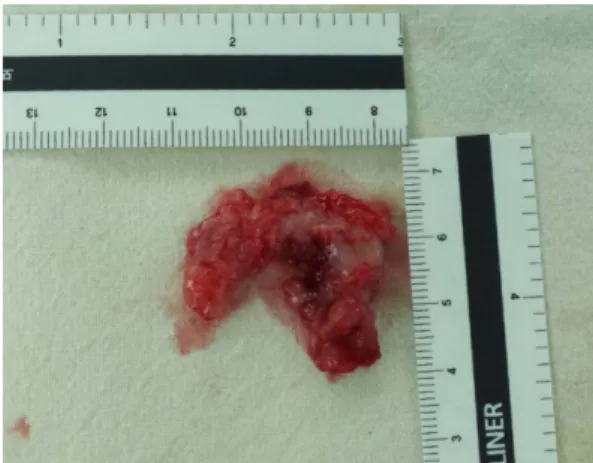
Fig. 4. The lesions measured approximately 30 mm × 30 mm.
POMC related to LeFort I osteotomy
- 189 - 상악동 막에 붙어있었다. 정상조직과 분리 후 낭종은 완전 히 제거되었다(Fig. 4). 조직병리학적 검사상respiratory epithelium 과 squamous epithelium으로 이장되어 있는 술 후 상악 낭종(postoperative maxillary cyst)으로 진단되었다 (Fig. 5). 술 후 3년간 재발 등의 이상 소견 없이 양호한 회 복을 보였다(Fig. 6).
고 찰
술 후 상악 낭종의 발병은 상악동의 외과적인 수술 후 에 상악동 점막의 잔여물이 수술부위로 함입되거나, 상악 동이 재생성 육아조직으로 완전히 채워지기 이전에 상악 동구가 조기 폐쇄 되는 것이 그 기전으로 보고되었다[3].
LeFort I 골절단술 시행 후 발생하는 술 후 상악 낭종은 골 절단술시에 상악동의 점막이나 비강 점막, 혹은 비구개관 의 점막에 손상이 발생하고, 이러한 점막이 골절단술을 시 행한 절단면 사이로 들어가 낭종성 변화가 일어나 발생하
는 것으로 여겨지고 있으나 왜 점막 상피가 낭종성 변화 를 일으키는지는 밝혀지지 않고 있다[4]. Hayhurst 등[5]이 보고한 사례에서도 비점막의 작은 단편이 상악 골절단면 사이에 갇히고, 수년 후에 낭성 변성이 일어난 것으로 추 정하였으며 따라서 비점막을 보존하고 비점막 천공이 발 생했을 경우 이의 처치가 중요하다고 하였다.
악교정 수술 후 발생하는 술 후 상악 낭종을 예방하는 명확한 방법은 정립되지 않았지만, LeFort I 골절단술 시 상악동의 염증성 호흡 점막을 완전히 제거하고, 출혈을 최 소화 하며, 상악동 내에 혈종이 생기지 않도록 하고, 상악 동구의 폐쇄를 예방하고 주위 조직의 자극을 피하는 등의 방법이 필요하다[4,6]. 하지만 술자가 위에서 언급된 요인 들에 대해 주의를 기울이더라도 상악동에서 술 후 상악낭 종의 발생을 완전히 막는 것은 어렵다.
술 후 상악 낭종과 감별을 요하는 질환으로는 종양성 병 소, 재발성 상악동염, 상악동내의 점액 낭종, 치근단 낭종, 비 구개관 낭종 등이 있을 수 있다[2,7]. 이런 질환들과의 감 별을 위해서는 임상적 방사선학적 평가와 기왕력에 대한 평가가 필요하다. 파노라마와 Water’s 방사선 사진이 상악 동 질환들을 발견하는 데 이용될 수 있는데 방사선사진상 술 후 상악 낭종은 단방성 또는 다방성의 경계가 비교적 명확한 소견을 보이지만 경계가 명확하지 않은 경우도 있 다[8]. CT 촬영을 통해 이 병소에 대해 보다 더 정확한 방 사선적 평가를 할 수 있다[9].
술후 상악 낭종의 처치는 일반적으로 본 증례처럼 낭종 의 완전한 적출과 일차 봉합이다[3]. 동시에 상악동 천공 술(nasal antrostomy)역시 추천되며, 얇은 낭종 벽을 가지 고 광범위한 피질골 천공이 동반된 단방성의 낭종에 있어 서는 조대술이 추천된다[6]. 본 증례에서는 술 후 3년간 재 발 소견 없이 양호한 회복을 보였으며, Yoshikawa 등[6]은 부적절한 적출시 재발률을 높일 수 있다고 하였다.
비록 술 후 상악 낭종이 상악의 악교정 수술 이후에 아주 드물게 발생하지만 지연된 진단을 피하기 위해서 각종 상악 동 수술과 외상 뿐 아니라 악교정 수술을 받은 환자에서 증 상을 호소할 시 감별진단에 포함하고 CT 촬영 등을 시행하 여 신속한 진단 및 처치를 해야 할 것으로 생각된다.
Conflict of Interest
The authors declare that they have no competing interests.
ORCID
Dong-Uk Seo 0000-0002-4188-846X Fig. 5. The respiratory epithelium was observed.
Fig. 6. There was no evidence of recurrence 3years after enucle-
ation.
Dong-Uk Seo et al.
- 190 - Su-Gwan Kim 0000-0002-0424-9984
Ji-Su Oh 0000-0002-8369-5025 Jae-seek You 0000-0001-7638-9583 Bo-Su Shin 0000-0001-9140-6345
References