• 검색 결과가 없습니다.

Comparison of Antioxidant Activity and ${\alpha}$-Glucosidase Inhibiting Activity by Extracts of Galla rhois

N/A
N/A
Protected

Academic year: 2021

Share "Comparison of Antioxidant Activity and ${\alpha}$-Glucosidase Inhibiting Activity by Extracts of Galla rhois"

Copied!
6
0
0

로드 중.... (전체 텍스트 보기)

전체 글

(1)

ISSN 2288-0356(Online) http://dx.doi.org/10.14518/crals.2013.31.4.010 Original Article

Comparison of Antioxidant Activity and α-Glucosidase Inhibiting Activity by Extracts of Galla rhois

Seung-Hyun Lee

1

, Sang-Han Lee

1,2*

1

Department of Food Science & Biotechnology, Graduate school, Kyungpook National University, Daegu 702-701, Korea

2

Food & Bio-Industry Research Institute, Daegu 702-701, Korea

Abstract

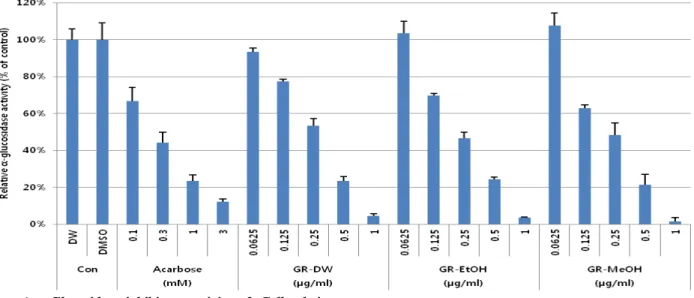
We studied antioxidant activity and inhibitory effect of α-glucosidase from aqueous, ethanolic and methanolic fractions of Galla rhois . In FRAP and ORAC assay for measuring antioxidant activity, we confirmed that Galla rhois extracts had strong antioxidant activity and ethanolic and methanolic extracts were relatively stronger than aqueous extract. We used trolox as a positive control. In order to measure the inhibitory effect of α-glucosidase, we compared acarbose and Galla rhois extracts. As a result of α-glucosidase inhibitory assay, aqueous, ethanolic and methanolic extracts of Galla rhois showed high inhibitory activitity and ethanolic and methanolic extracts were relatively stronger than aqueous extract. The 50% inhibitory concentrations (IC50s) of acarbose, aqueous, ethanolic and methanolic fractions were 0.45 mM, 0.53 μg/mL, 0.415 μg/mL and 0.37 μg/mL, respectively. These results suggest that Galla rhois extracts can be a clinically useful anti-diabetic ingredient, indicating that it needs to be fractionated and isolated and should be further investigated.

Keywords:acarbose, α-glucosidase, enzyme kinetics, FRAP assay, Galla rhois , ORAC assay, trolox,

Introduction 1)

In recent decades, the prevalence of adult diseases is being increased due to modern people's lifetime increase, westernized eating habits, stress and irregular life, etc. Adult diseases include cerebrovascular disease, heart disease and diabetes.

The onset of such adult diseases is mainly caused by oxidative stress. Oxidative stress means unbalance of reactive oxygen species' generation and antioxidant system. Reactive oxygen species (ROS) are chemically reactive molecules containing oxygen and are called harmful oxygen. They can react with other organic matter in cells. ROS in adequate amounts make physiological function such as cell signaling, muscle extension, etc. Inhibition of making ROS or removal of ROS through reaction with already occurred ROS is called antioxidant activity.

People have antioxidant system to maintain adequate amounts of ROS in the body. Excess onset of ROS makes oxidative stress in the body and consequently causes heart disease, cancer and diabetes (Finkel and Holbrook 2000; Heitzer et al. 2001;

Valko et al. 2006). Now butylated hydroxy anisol (BHA) and butylated hydroxy toluene (BHT) are widely used synthetic antioxidants. These are restricted to use because these 50 mg/kg/day or higher dose in the long-term can induce lipid metabolism's imbalances and cancer. We need alternative

functional material for synthetic antioxidant. Not only already proven materials in fork remedies and oriental medicines but also natural substances which being possible to eat and having fewer side-effect are studied actively (Bae et al. 2012).

Diabetes is occurred when glucose homeostasis is broken by insulin metabolism problem. There are two main types of diabetes depending on insulin action. Type 1 diabetes results from the body's failure to produce insulin and Type 2 diabetes results from insulin resistance, a condition in which cells fail to use insulin properly, sometimes combined with an absolute insulin deficiency, reduced insulin receptors. Insulin resistance means that the body normally produces insulin but the cells in the body become resistant to insulin (through changes in their surface receptors) and unable to use it as effectively. It is caused by genetic factors and environmental factors such as aging, obesity, lack of exercise, dose of immunosuppressive drug, stress. Most of the diabetes are type 2 diabetes because most onsets are caused by environmental factors (Lee et al.

2012a, b).

α -Glucosidase inhibitors interrupt carbohydrate digestion such as starch, sugar and they are orally administered anti-diabetic drugs used to treat type 2 diabetes. In digestion steps, carbohydrate is decomposed into monosaccharide, and then Received: December 12, 2013 / Revised: December 20, 2013 / Accepted: December 31, 2013

*

Corresponding Author: Sang-Han Lee, Tel. 82-53-950-7754, Fax. 82-53-950-6772, Email. [email protected]

(2)

absorbed by intestine. α -glucosidase combined with intestinal membrane breaks oligosaccharides, disaccharides and trisaccharides into glucose and monosaccharides. α -Glucosidase inhibitors are competitive inhibitor and they have anti-diabetic activity by interrupting digestion through reaction with α -glucosidase instead of carbohydrate. acarbose (precose), miglitol (glyset), voglibose belong to typical anti-diabetic drugs (Lee et al. 2012b; Yoji et al. 2012).

Galla Rhois is that cocoons which are made by Galla rhois (BELL) BAKER being parasitic on leaf of Rhus chinensis of anacardiaceae is undergone process of drying. Cocoons are collected when their outer walls changed from blue to light brown, and then they were received with boiling or steaming by the time their surface's color is transformed into gray in order to eradicate Galla rhois (BELL) BAKER. Finally they are used after pulverized and dried (Kim et al. 2009). G. Rhois extracts having syringic acid, gallic acid, gallic acid methyl ester, protocatechuic acid and 1,2,3,4,6-penta-O-galloyl-β -D-glucose have strong antioxidant activities and more free radical scavenging ability than BHA (Cha et al. 2000). Their extracts also have strong antimicrobial activity on gram positive and negative bacilli (Choi 2003; Kim et al. 2009).

We performed ferric reducing ability of plasma (FRAP) and oxygen radical absorbance capacity (ORAC) assay for measuring their antioxidant activity. We could expect another effect about G. rhois extracts because of such activity and performed α -glucosidase inhibitory assay in order to measure anti-diabetic activity. 2)

Materials and Methods Samples

Dried Galla rhois was purchased from Yak-Jun Street, Daegu, Korea. These samples were ground using a homogenizer and extracted with distilled water (DW) (1:15 w/v, 60℃), 100%

ethanol (1:10 w/v, 45℃) and 100% methanol (1:10 w/v, 45℃) for 24 hours, respectively. Each obtained solution was filtered using filter paper (No. 4, Whatman, Buckinghamshire, UK) to remove debris, and then their filtrates were pulverized using vacuum freezing dryer. Each yield of aqueous, 100% ethanolic and 100% methanolic extracts was 36.70 g, 42.56 g and 48.14 g per sample 100 g after being freeze-dried at -80℃ under vacuum. Each part of powders was deposited for voucher specimen. The powder obtained from aqueous extract was dissolved in DW and the powders obtained from 100% ethanolic and 100% methanolic extracts were dissolved in dimethyl sulfoxide (DMSO), respectively. All powders were prepared

to be various concentrations using DW and DMSO.

Measurement of antioxidant activity

Antioxidant activity was measured using FRAP (ferric ion reducing antioxidant power) and oxygen radical antioxidant capacity (ORAC) assay (Benzie and Strain 1996; Yang et al.

2011; Zulueta et al. 2009). In FRAP assay, 2 μL of sample or L-ascorbic acid (positive control) was placed in 96-well plate, and then 200 μL of FRAP solution [sodium acetate trihydrate, acetic acid glacial, Iron(Ⅲ) chloride hexahydrate, TPTZ (2,4,6-tripyridyl-s-triazine)] was added. 96-well plate was incubated at room temperature (25℃) for 10 min prior to measurement of the absorbance. The absorbance was measured using spectrophotometer (Wallac Victor3 PerkinElmer, MA, USA) at 595 nm.

In ORAC assay, 2 μL of sample or trolox [positive control, (±)-6-Hydroxy-2,5,7,8-tetramethylchromane-2-carboxylic acid, 238813, Sigma] was placed in 96-well black plate (3603, Corning Inc., NY, USA), and then 400 nM fluorescein (F2456, Sigma) was added. 96-well black plate was incubated at 37°C for 10 min before the addition of AAPH [2,2'-azobis (2-methylpropionamidine) dihydrochloride, 440914, Sigma].

100 μL of AAPH (40 mM) was added into 96-well black plate, and then the fluorescence was measured immediately using spectrophotometer (Wallac Victor3 PerkinElmer, MA, USA) with fluorescence filters for an excitation wavelength of 490 nm and an emission wavelength of 535 nm after the addition.

Measurements were taken every 5 min for 70 min. AUC (the area under the curve) was calculated to compare the sample, blank and trolox, and is the area below the fluorescence decay curve of the sample, blank and trolox, respectively, calculated by applying the following formula in a Microsoft Excel spread sheet (Microsoft, Washington, USA)

AUC = (0.5 + f

5

/ f

0

+ f

10

/ f

0

+...+ f

n+5

/ f

0

)×5

where f

0

+is the initial fluorescence and f

n

is the fluorescence at time n.

Cells and Materials

RAW 264.7 cells were obtained from the American Type Culture

Collection (ATCC No. TIB-71, Manassas, VA) and cultured

in DMEM containing 10% FBS (Lonza, US), 100 U/mL of

penicillin (Lonza, US), and 100 μg/ml of streptomycin (Lonza,

US) at 37℃ in a 5% CO

2

humidified incubator.

(3)

Cell viability assay 3)

Cellular cytotoxic activity of the sample extracts was carried out as previously described by Mosmann (1983) with slight modification. Cell viability was measured by the MTT [3-(4,5-dimethylthiazol-2-yl)-2,5-diphenyltetrazolium bromide, Sigma Chemical Co., US] assay (Klostergaard 1985). RAW 264.7 cells were seeded on 96-well plates at a concentration of 4×10⁴cells/well, and then incubated at 37℃, 90% humidity, 5% CO

2

concentrations for 1 hour. The sample extracts of various concentrations (0.03, 0.1, 0.3, 1, 3, 10, 30 mg/mL) were added, and then cells were incubated at 37℃, 90% humidity, 5% CO

2

concentrations for 18 hours. After 18 hours, cells in each well were incubated with MTT solution (5 μg/μL) at 37℃

for 1 hour. The medium was discarded and formazan crystal adhered to the well floor was solubilized with dimethyl sulfoxide, and then incubated in darkness at 10 min. Cell viability was assessed by measuring absorbance at 595 nm with Spectra MAX (Molecular Devices, US).

Measurement of antidiabetic activity

α -Glucosidase inhibitory activity was determined using a modified version of the method according to Kim (2013). 2 μ L of sample or acarbose (A8980, Sigma) was placed in 96-well plate, and then 100 μL of α -glucosidase (0.2 U/mL) was added.

100 μL of PNPG (p-nitrophenyl α-D-glucopyranoside, N1377,

Figure 1. Antioxidant activities of Galla rhois extracts on FRAP value.

Con: control (not treated sample group), Vit C: ascorbic acid as a positive control, GR: Gallar hois , DW: aqueous fraction, EtOH: ethanolic fraction, MeOH: methanolic fraction. In FRAP assay, 2 μL of sample or L-ascorbic acid (positive control) was placed in 96-well plate, and then 200 μL of FRAP solution was added. When a ferric tripyridyltriazine (Fe

3+

TPTZ) is reduced to the ferrous(Fe

2+

)-form by a reductant (antioxidant), an intense blue color with an absorption maximum at 593 nm develops. After incubation at room temperature for 10 minutes, the absorbance was measured using spectrophotometer at 595 nm.

Sigma) was added into 96-well plate, and then the absorbance was measured immediately using spectrophotometer (Wallac Victor3 Perkin Elmer, MA, USA) with wavelength of 405 nm and measurements were taken every 2 min for 20 times.

Statistical analysis

Data are presented as means ± standard deviation (SD).

Statistical differences between mean values ± SD were determined by the Dunnett's multiple range test using Statistical Analysis System (NC, USA). The significance was set at P<0.05.

Results and Discussion

As a result of FRAP and ORAC assay for measuring antioxidant activity of Galla rhois , all extracts showed dose dependent increase in antioxidant activity. FRAP assay showed more than 100 μg/mL of all extracts has stronger antioxidant activity than 25 μM of ascorbic acid (Figure 1). ORAC assay also showed more than 10 μg/mL of all extracts has stronger antioxidant activity than that of 10 μM of trolox (Figure 2).

The MTT assay was carried out using RAW 264.7 (mouse

macrophage cell) to know cellular cytotoxic activity of G. rhois

extracts. The MTT assay is a colorimetric assay for measuring the

activity of cellular enzymes that reduce the tetrazolium dye, MTT,

to its insoluble form, formazan, giving a purple color. Therefore,

the absorbance means concentration of living cells. Aqueous

(4)

Figure 2. Antioxidant activities of Galla rhois extracts on ORAC value.

Con: control (not treated sample group), Trolox: a positive control, GR: Galla rhois , DW: aqueous fraction, EtOH: ethanolic fraction, MeOH: methanolic fraction.

In ORAC assay, 2 μL of sample or trolox was placed in 96-well black plate, and than 400 nM fluorescein was added. After incubation at 37℃ for 10 minutes, 100 μL of AAPH (40 mM) was added into 96-well black plate, and then the fluorescence was measured immediately using spectrophotometer with fluorescence filters for an excitation wavelength of 490 nm and an emission wavelength of 535 nm after the addition. Measurements were taken every 5 min for 70 minutes.

Fluorescence intensity is decreased by AAPH (oxidant) over time but antioxidants maintain fluorescence decay curve. The potent antioxidant power results in increasing AUC (area under the curve). AUC was calculated to compare the sample, blank and trolox, and is the area below the fluorescence decay curve of the sample, blank and trolox, respectively, calculated by applying the following formula in a Microsoft Excel spread sheet.

4)

Figure 3. Cell viability of mouse macrophage cells (RAW 264.7) against Galla rhois extracts by MTT assay.

Con: control (not treated sample group), GR: Galla rhois , DW: aqueous fraction, EtOH: ethanolic fraction, MeOH: methanolic fraction.

Cell viability was measured by the MTT assay. RAW 264.7 cells were seeded on 96-well plates at a concentration of 4×10⁴cells/well, and then incubated at 37℃,

90% humidity, 5% CO

2

concentrations for 1 hour. The sample extracts of various concentrations were added, and then cells were incubated at 37℃, 90% humidity,

5% CO

2

concentrations for 18 hours. After 18 hours, cells in each well were incubated with MTT solution (5μg/μL) at 37℃ for 1 hour. Succinate dehydrogenase

is capable of reducing the tetrazolium dye MTT to its insoluble formazan, which has a purple color. The medium was discarded and formazan crystal adhered to

the well floor was solubilized with dimethyl sulfoxide, and then incubated in darkness at 10 minutes. Cell viability was assessed by measuring absorbance at 595

nm with Spectra MAX.

(5)

extract showed cell viability was more than 80% until 10 μg/mL of concentration but less than 60% in 300 μg/mL of concentration. Ethanolic and methanolic extracts showed cell viability was more than 80% until 100 μg/mL of concentration but 45 % and 70% in 300 μg/mL of concentration, respectively (Figure 3). 5)

As a result of α-glucosidase inhibitory assay for measuring antidiabetic activity of G. rhois extracts, all extracts showed dose dependent increase in antidiabetic activity. The best straight section was selected from the absorbance growth curve of acarbose. The absorbance increase quantity of its section was obtained about acarbose and samples, and then showed in bar graphs (Figure 3). In bar graphs, 3 mM of acarbose and 0.5 μ g/mL of aqueous extract showed almost the same inhibitory activity and ethanolic and methanolic extracts showed stronger inhibitory activity than 3 mM of acarbose. Ethanolic and methanolic extracts were relatively stronger than aqueous extract. The 50% inhibitory concentrations (IC

50

s) of acarbose, aqueous, ethanolic and methanolic fractions were 0.45 mM, 0.53 μg/mL, 0.415 μg/mL and 0.37 μg/mL, respectively.

By Cha et al. (2000), the methanol extract of G. rhois , indicating strong antioxidant activity, was fractionated and isolated, and then their chemical structures were identified as syringic acid, gallic acid methyl ester, protocatechuic acid, gallic acid, 1,2,3,4,6-penta-O-galloyl-β-D-glucose. Gallic acid methyl ester, protocatechuic acid and 1,2,3,4,6-penta-O-galloyl-β-D-glucose

Figure 4. α -Glucosidase inhibitory activity of Galla rhois extracts.

α -Glucosidase inhibitory activities were calculated according to relative percentage of control group (100%). Con: control (not treated sample group), Acarbose: a positive control, GR: Galla rhois , DW: aqueous fraction, EtOH: ethanolic fraction, MeOH: methanolic fraction.

2 μL of sample or acarbose was placed in 96-well plate, and then 100 μL of α -glucosidase (0.2 U/mL) was added. is a chromogenic α -glucosidase substrate yielding a yellow solution upon cleavage. 100 μL of PNPG was added into 96-well plate, and then the absorbance was measured immediately using spectrophotometer with wavelength of 405 nm and measurements were taken every 2 minutes 20 times.

showed more potent radical scavenging effect than butylated hydroxy anisol (BHA) using DPPH method. BHA and butylated hydroxy toluene (BHT) are synthetic antioxidant which widely used today, but there are side effects when more than 50 mg/kg/day was dosed in the long term. The study of G. rhois which having antioxidant capacity, fewer side effects and being possible to eat, a natural substance is more important.

By Lee et al. (2012), diabetes become a serious problem in Korea and around the world because of the recent rapid increase in the prevalence of it. However, pre-existing diabetic drugs have side effects such as hypoglycemia, weight gain, digestive side effects, renal dysfunction, heart failure, fracture etc. For this reason, the navigation of new functional materials that can replace them is important. In this experiment, G. rhois was found to be sufficient for possibility to replace pre-existing diabetic drugs.

G. rhois has been used as oriental traditional medicine described

in Korean Donguibogam and fork remedy etc and has already

been recognized its functionality from leading papers. This

experiment showed G. rhois extracts have strong antioxidant and

antidiabetic activity. Maybe, there will be highly effective

compound when these are fractionated and isolated. We need to

study the mechanism through identification of these active

substances and also to investigate candidate substances of α

-glucosidase inhibitor. Through this process, new antidiabetic

drugs having almost no side effects will be able to be developed.

(6)

Summary 6)

We measured antioxidant and antidiabetic activity from aqueous, ethanolic and methanolic fractions of Galla rhois . Aqueous, ethanolic and methanolic extracts were dose-dependent increase in antioxidant activity in both FRAP and ORAC assay. FRAP assay showed more than 100 μg/mL of all extracts has stronger antioxidant activity than 25 μM of ascorbic acid. ORAC assay also showed more than 10 μg/mL of all extracts has stronger antioxidant activity than 10 μM of trolox. The MTT assay was carried out for to know cellular cytotoxic activity of G. rhois extracts. aqueous extract showed cell viability was more than 80% until 10 μg/mL of concentration and ethanolic and methanolic extracts showed cell viability was more than 80%

until 100 μg/mL of concentration. As a result of α -glucosidase inhibitory assay, 3 mM of acarbose and 0.5 μg/mL of aqueous extract showed almost the same inhibitory activity and ethanolic and methanolic extracts showed stronger inhibitory activity than 3 mM of acarbose. Ethanolic and methanolic extracts were relatively stronger than aqueous extract. The 50% inhibitory concentrations (IC

50

s) of acarbose, aqueous, ethanolic and methanolic fractions were 0.45 mM, 0.53 μg/mL, 0.415 μg/mL and 0.37 μg/mL, respectively. This study shows G. rhois extracts have strong antioxidant and antidiabetic activity. Maybe, there will be highly effective compound when these are fractionated and isolated.

References

Finkel T, Holbrook NJ (2000) Oxidants, oxidative stress and the biology of ageing. Nature 408:239-247.

Heitzer T, Schlinzig T, Krohn K, Meinertz T, Munzel T (2001) Endothelial dysfunction, oxidative stress, and risk of cardiovascular events in patients with coronary artery disease. J Am Heart Assoc 104:2673-2678.

Valko M, Rhodes CJ, Moncola J, Izakovic M, Mazura M (2006) Free radicals, metals and antioxidants in oxidative stress-induced cancer. Chem Biol Interact 160:1-40.

Bae JS, Lee HS, Lee HY, Yoo BH, Kim TW, Kim YH, Kim TH (2012) Evaluation of acetylcholinesterase inhibitiory and antioxidative activities of Rhus javanica. Korean J Food Preserv 19:751-756.

Lee SH, Lee JK, Kim IH (2012a) Trends and perspectives in the development of antidiabetic drugs for type 2 diabetes mellitus. Korean J Microbiol Biotechnol 40180-185.

Lee SY, Kim IS, Park SL, Lim SI, Lim SI, Choi HS, Choi SY (2012b) Antidiabetic activity and enzymatic activity of commercial Doenjang certified for traditional foods. Korean J Biotechnol Bioeng 27:361-366.

Yoji H, Hiroshi N, Masahiro F, Shinji F, Yusuke S, Jiro N, Yutaka O (2012) α - Glucosidase inhibitor maglitol affects bile acid metabolism and ameliorates obesity and insulim resistance in diabetic mice. Metabolism 62:734-742.

Kim SJ, Yang HY, Lee SG (2009) Antimicrobial effect and cytotoxicity of Chinensis Galla. Korean J Orient Physiol Pathol 23:412-415

Cha BC, LEE SB, Rhim TJ, Lee KH (2000) Constituents of antioxidantive activity and free radical scavenging effect from Galla rhois (Rhus javanica Linne). Korean J Pharmacogn 31:185-189.

Choi I (2003) Antimicrobial activity of Rhus javanica extracts against animal husbandry disease-related bacteria. Korean J Soc Food Sci Nutr 32:1214-1220.

Benzie IF, Strain JJ (1996) The ferric reducing ability of plasma (FRAP) as a measure of "antioxidant power": FRAP assay.

Anal Biochem 230:70-79.

Zulueta A, Esteve MJ, Frigola A (2009) ORAC and TEAC assays comparison to measure the antioxidant capacity of food products. Food Chem 114:310-316.

Yang YJ, Kim HJ, Kang SH, Kang SC (2011) Screening of natural herb resources for anti-oxidative effects in Korea.

Korean J Plant Res 24:1-9.

Mosmann T (1983) Rapid colorimetric assay for cellular growth and survival: application to proliferation and cytotoxicity assay. J Immunol Methods 65:55-63.

Klostergaard J (1985) A rapid extremely sensitive, quantitative micro-assay for cytotoxic cytokines. Lymphokine Res 4:309-317.

Kim SD (2013), α -Glucosidase inhibitor from Buthus martensi

Karsch. Food Chem 136:297-300.

수치

Figure 1. Antioxidant activities of Galla rhois extracts on FRAP value.
Figure 2. Antioxidant activities of Galla rhois extracts on ORAC value.
Figure 4. α -Glucosidase inhibitory activity of Galla rhois extracts.

참조

관련 문서

Chemical antioxidant activities of β-carotene, lycopene, and Trolox were examined by measuring ferric reducing antioxidant power (FRAP) and 2,2’-di- phenyl-1-picrylhydrazyl

Antioxidant activity was evaluated by measuring total phenolic contents, reducing power, 1,1-diphenyl-2-picrylhydrazyl (DPPH) radical scavenging activity,

Antioxidant activity and protective effects of α-TP were evaluated by DPPH radical-scavenging assay, morphological assay, MTT reduction assay, and lactate dehydrogenase

The activities of each extracts were measured by antioxidant tests, such as total phenolic contents, total flavonoid contents, hydroxyl radical activity, DPPH

In additional results by the xanthine oxidase assay, antioxidant activity showed that water extract of chestnut leaves showed the highest xanthine oxidase inhibitory activity

α -Glucosidase inhibitory activity of MC layer and EA layer from Pueraria thunbergiana MeOH extract.. Each value

Abstract This study evaluated in vitro antioxidant activity, antioxidant content, and α-glucosidase and pancreatic lipase inhibitory activities of ethanol extracts of samnamul

Ferric reducing antioxidant power (FRAP) assay of dependent on various cultured regions and concentration from extracts of Wofiporia extensa.. Data were expressed as the means ± SD