배양대뇌신경세포 저산소증모델에서 반하에 의한 유전자표현의 변화
권건록․정현정․신길조․문일수1․이원철2․정승현*
동국대학교 한의과대학 내과학교실, 1동국대학교 의과대학 해부학교실,2부산대학교 한의학전문대학원
Received January 5, 2009 /Accepted May 8, 2009
Microarray Analysis of Gene Expression Affected by Water-extracts ofPinelliae rhizoma in a Hypoxic Model of Cultured Rat Cortical Cells. Gun-Rok Kwon, Hyun-Jung Jung, Gil-Jo Shin, Il-Soo Moon1, Won-Chul Lee2and Seung-Hyun Jung*.Dept. of Oriental Medicine, and1Department of Anatomy, Dongguk University,2Pusan National University School of Oriental Medicine - Pinelliae rhizoma (Pr, 半夏) is a tradi- tional medicine used in the treatment of incipient stroke. We investigated the effects of Pr on gene expression in a hypoxic model using cultured rat cortical cells. Pr (2.5 μg/ml) was added to the cul- ture medium on DIV 12. A hypoxic shock (2% O2/5% CO2, 37oC, 3 hr) was given two days later (on DIV 14), and total mRNAs were isolated at 24 hr post-shock from both Pr-treated samples and un- treated control cultures. Microarray using TwinChipTMRat-5K (Digital Genomics, Seoul) indicated that Pr upregulated genes for cell growth and differentiation (tubb5, tgfα, ptpn11, n-ras, pdgfa) and anti- apoptosis (mcl-1), while downregulating the apoptosis-induced gene (tieg). Therefore, it is interpreted that Pr protects neurons from hypxoic shock by maintaining cell growth and differentiation and by preventing apoptosis.
Key words : Cortical cell culture, hypoxia, microarray, Pinelliae rhizoma
*Corresponding author
*Tel:+82-31-961-9040, Fax:+82-31-961-9000
*E-mail : [email protected]
서 론
반하(半夏,Pinelliae rhizoma :Pr)는 천남성과의 15~30 cm 되는 다년생 초본의 구근으로 조습화담(燥濕化痰), 소비산결 (消痞散結), 강역지구(降逆止嘔)하는 효능이 있다[28]. 반하에 는 phenol류인 homegentisic acid, homegentisic acid gluco- side, 3,4-dihydroxybenzaldehyde와 alkaloid인 ephedrine이 0.002% 정도 함유되어 있다[57]. 반하에 관한 연구로는 기침억 제[57], 구토억제[37], 항암효과[54] 등이 보고되어 있다. 반하 는 반하후박탕(半夏厚朴湯), 반하사심탕(半夏瀉心湯) 등 호흡 기계와 소화기계에 작용하는 처방 뿐만 아니라, 성향정기산 (星香正氣散), 도담탕(導痰湯) 등 중풍(中風) 급성기의 처방(處 方)에도 많이 사용된다[22].
현대 사회는 인구의 노령화로 뇌혈관질환의 발병률이 점차 증가하고 있으며[15], 2006년 사망원인 통계연보에 따르면 뇌 혈관질환은 전체 사망인구의 12.3%로, 악성 신생물(27%) 다음 으로 높은 비중을 차지하고 있다. 뿐만 아니라 뇌혈관질환은 비가역적인 신경손상을 초래하여 심각한 후유증을 동반하게 된다. 신경세포는 세포 생존을 위하여 혈액 순환에 의한 지속 적인 영양과 산소의 공급을 필요로 한다. 임상에서 뇌신경세 포는 뇌허혈 및 심정지 등으로 인한 심박출량 감소, 폐질환 및 이산화탄소 중독 등으로 인한 혈중 산소분압의 저하 등에 의해 저산소증 상태에 빠지게 된다[15]. 결국 신경세포는 산소
공급이 저하되면 신경세포막을 통해 이온들의 이동을 조절하 고, 에너지 생산 저하에 따른 에너지 소모를 줄이며, hypo- xia-inducible factor (HIF)-1과 nuclear factor kappa B (NFκB) 등의 transcription factor를 활성화시켜 특정 유전자들의 발현 을 유도한다[21,40,47]. 그러나 신경세포가 저산소증에 대해 전 혀 방어능력이 없는 것은 아니다[23,38]. 신경세포에는 다수의 산소감지자(O2 sensor)가 있는데 이 가운데 산소에 민감한 Na+이온통로와 K+이온통로, Nicotinamide adenine dinucleo- tide phosphate (NADPH) 등은 직접 O2와 접촉하여 신경세포 의 흥분성을 조절하게 된다[35]. 동물세포는 세포내 산소압의 변화에 반응할 수 있는 유전자 프로그램을 갖고 있다. 즉, 몸에 공급되는 산소가 부족하면 산화적 스트레스에 대응하기 위해 항산화스트레스 유전자들의 발현을 증가시키며, 적혈구 생성 (erythropoiesis)과 혈관신생(angiogenesis)이 촉진된다[17,31].
또한 줄어든 ATP 생성을 만회하기 위하여 해당(glycolysis)과 포도당 수송(glucose transport)에 관여하는 유전자 발현을 증 가시켜 포도당 이용 속도를 증가시킨다[14,46,48-50].
지금까지 의학을 포함한 생명과학 분야의 대부분의 연구는 관심의 대상이 되는 특정 유전자를 먼저 선정하고, 그 유전자 발현의 변화에 초점을 맞춘 연구가 주종을 이루어왔다. 그 방 법으로 mRNA에 대한 실험은 northerm blot이나 in situ hy- bridization이 이용되어 왔고, 단백질 수준에서의 변화는 western blot이나 면역세포화학법(immunocytochemistry)을 이용한 실험이 사용되어 왔다. 그러나 최근 동 시간대에 변화 를 보이는 많은 유전자를 한꺼번에 관찰할 수 있는 기법들이 개발되었다. 여기에는 differential display, subtraction hy-
bridization[32]을 포함해 가장 최근에 개발되어 사용되고 있 는 cDNA microarray 기법 등이 있다[6,45]. 대부분의 조직내 mRNA의 발현 변화는 아주 미약하기 때문에 검사 기법 선택 의 주요관건은 어떤 방법이 이 미약한 발현 변화를 보다 더 민감하게 발견해 낼 수 있는가 하는 점이다. cDNA microarray 는 검사 속도의 측면에서나 반복 검사 시 일관성 있는 결과를 얻을 확률이 가장 높다는 점에서 다른 두 검사법에 비하여 보다 더 관심의 대상이 되고 있다. 이론대로라면 다양한 조건 에서 수백 혹은 수천 개 유전자의 발현 변화를 동시에 관찰할 수 있는 획기적인 실험 기법이다. 최근에는 관련된 유전자끼 리 그룹화한 microarray가 상품화되어 더욱 더 그 검사가 용이 해졌다[8].
반하가 예로부터 뇌졸중의 치료와 예방에 사용되었음에도 불구하고 뇌신경세포의 보호 작용과 그 기전에 대한 연구가 없었으므로, 저산소증을 유발한 흰쥐 대뇌신경세포에 반하를 처리하여 cDNA microarray 기법을 이용하여 유전자 표현 변 화를 조사하였다.
재료 및 방법 동물
본 연구는 임신 18일(Embryonic day 18, E18)의 Sprague- Dawley계 흰쥐(대한동물실험센터)를 사용하였다.
약재 및 추출액 제조
실험에 사용된 반하는 대한약전 및 대한약전 외 한약 규격 주해[25]에 근거하여 동국대학교 부속 한방병원에서 구입하 였다. 반하(2 g)에 20 ml의 증류수를 넣어 균질화하고 4oC에 서 18시간 진탕하였다. 이를 15,000 rpm에서 15분간 원심 분 리하여 상등액을 얻고 여과멸균(0.45 μm)한 후 소량씩 분주하 여 -20oC에 보관하였다. 건조물의 량은 1 ml 추출액 튜브를 이동하여 3 반복으로(triplicate) 동결건조한 후 평균치를 사용 하였다.
신경세포 배양
E18의 Sprague-Dawley계 흰쥐 대뇌피질신경세포를 Brewer 등[2]의 방법에 따라 배양하였다. E18의 흰쥐를 dry ice 가 들어있는 통속에 3~5분간 넣어 마취하고, 자궁을 가른 후 흰쥐 태아를 취하고 뇌를 잘라내었다. 대뇌피질 조직을 37oC 에서 5분간 0.25% trypsin으로 처리하고 1 mM sodium pyr- uvate와 10 mM HEPES (pH 7.4)가 첨가된 HBSS용액(Hank's Balanced Salt Solution, Invitrogen Life Technology) 5 ml로 4~5회 세척하여 반응을 중단시켰다. 조직을 1 ml HBSS용액 으로 옮기고, 끝을 불에 달구어 구멍을 작게 한 pasteur pipette 으로 6~7회 통과시켜 세포를 분산시켰다. 이후 분산된 세포 의 밀도를 측정한 다음 B27을 첨가한 plating neurobasal me-
dia (Invitrogen Life Technology; 100 ml neurobasal, 2 ml B27 supplement, 0.25 ml glutamax I, 0.1 ml 25 mM glutamate, 0.1 ml 25 mM 2-mercaptoethanol)에 약 1,500 cells/mm2가 되도록 접종하여 5% CO2배양조에서 배양하였다. 2~3일 간 격으로 배양액을 feeding neurobasal media (100 ml neuro- basal, 2 ml B27 supplement, 0.25 ml glutamax I)로 1/3씩 교환하였다.
저산소증 유발
Culture plate를 CO2 Water Jacketed Incubator (Forma Scientific Inc.)를 이용하여 2% O2/5% CO2, 37oC 환경에서 3시 간 처리하여 저산소증을 유발하였다. 저산소 처리가 끝나면 배양세포를 정상산소 환경 배양조에 옮기고 계속 배양하였다.
유전자표현 분석(microarray)을 위한 RNA 추출
배양 신경세포의 경우 DIV 14일 경에 가지돌기의 성장은 거의 완성되며, 이 시기에 왕성한 연접생성이 일어난다[29,36].
한편 DIV 7 세포의 경우 glutamate, AMPA, 혹은 NMDA에 의한 흥분독성을 일으키지 않지만, DIV 14 세포는 심한 독성 반응을 일으킨다[29]. 따라서 본 연구에서는 DIV 12에 반하 2.5 μg/ml를 처리하고 DIV 14에 저산소증을 유발하였다. 저산 소 처리가 끝나고 24 시간 후에 배양액을 제거하고 5~10 ml의 ice-cold PBS로 세척하였다. PBS를 제거하고 용액D [4 M gua- nidinium thiocyanate, 25 mM sodium citrate, 0.5%(w/v) so- dium lauryl sarcosinate, 0.1 M β-mercaptoethanol] 1 ml를 각 culture dish에 넣어 세포를 용해시켰다. 용해된 lysate를 microfuge tube에 옮기고 tissue homogenizer로 15~30초간 균질화하였다. 여기에 용액D 1 ml 당 0.1 ml의 2 M sodium acetate (pH 4.0), 1 ml의 phenol (4oC), 0.2 ml의 chloro- form-isoamyl alcohol을 넣고 섞어주었다. Tube를 얼음에 15 분간 넣어둔 후 원심분리(10,000×g, 20분, 4oC)하여 상등액을 취하고 동일 부피의 isopropanol을 넣어 잘 섞어주었다. 이를 -20oC에서 2시간 처리한 후 원심분리(10,000× g, 30분, 4oC)하 여 RNA를 침전시키고 용액D로 녹였다. 이를 isopropanol로 한 번 더 침전시킨 후 75% alcohol로 2회 세척한 후 diethyl pyrocarbonate로 처리한 물에 녹이고 -70oC에 보관하였다.
Microarray
Microarray는 Digital Genomics (서울)에 의뢰하여 분석하 였다. 대조군과 실험군의 저산소증 유발 세포에서 각각 분리 한 20~50 μg의 total RNA로부터 oligo (dT) primer와 역전사 효소를 이용하여 first cDNA strand를 만들고, 대조군은 Cy3 dye (green)로, 실험군은 Cy5 dye (red)로 표지하였다. 두 probe를 1:1 혼합하여 TwinChipTMRat-5K (Digital Genomics) 을 hybridization (3X SSC, 42oC, 16시간)하고 최종적으로 0.1X SSC로 상온에서 1분씩 4회 세척하였다.
결과 및 고찰
저산소증에서 반하에 의한 유전자 표현의 변화
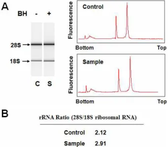
저산소증에서 반하의 신경세포 보호 기전을 알아보기 위하 여 microarray 기법으로 반하가 유전자 표현에 미치는 영향을 조사하였다. 본 연구에 사용한 흰쥐 대뇌신경세포는 저산소증 유발 후 3일 후부터 급격히 사망하기 시작한다. 따라서 저산소 증 유발 후 3일 이전에 많은 유전자의 표현이 달라질 것으로 예상되어 1일째에 total RNA를 분리하였다. 즉, DIV 12의 배 양세포에 반하 2.5 μg/ml를 처리하고 DIV 14에 저산소증을 유발하였으며, DIV 15에 세포로부터 guanidinium thio- cyanate 방법으로 total RNA를 분리하였다. BioAnalyzer를 이 용하여 RNA quality를 조사한 결과 대조군과 실험군 모두 rRNA ratio (28S/18S)가 2.0 이상으로 RNA 분해가 거의 일어 나지 않은 매우 좋은 상태임을 확인할 수 있었다(Fig. 1).
Microarray 전문회사인 Digital Genomics (서울)에 의뢰하 여 TwinChipTMRat-5K microarray chip을 형광사진으로 분석 하였다. 대조군의 cDNA는 Cy3 형광물질(green)로, 실험군은 Cy5 형광물질(red)로 표지하였으며, hybridization 결과 TwinChip의 upper array와 lower array의 형광이미지가 매우 유사하여 재현성이 높음을 알 수 있었다. MA plot에서 보면 [M=log2(R/G), A={log2(R×G)}/2], 대부분의 M 값이 -0.5에서 +0.5 사이로서 40% 정도 이내의 증감을 나타내었다(Fig. 2).
이 가운데 Global M 값이 +0.2 이상 즉, 14% 이상 표현이 증가
Fig. 1. Quality of isolated total RNA. Total RNA was isolated from Pr-treated and Pr-untreated cortical cultures at 24 hrs after hypoxic insult. Small aliquots of RNA prepara- tions were subjected quality analysis by polyacrylamide gel electrophoresis and scanning at A260 (A) using a Bioanalyzer. The RNA quality was assessed by measur- ing 28S rRNA/18S rRNA ratios (B).
Fig. 2. MA plots. The y-axis indicate the R/G ratio [M=log2
(R/G)] and the x-axis signal intensities [A={log2(R×G)}
/2]. Note that the M values of the majority of signals are between -0.5 and +0.5.
된 유전자는 530 여종, -0.2 이하 즉, 14% 이상 표현이 감소된 유전자는 524 여종이었다.
Apoptosis/cell death 관련 유전자 표현의 변화
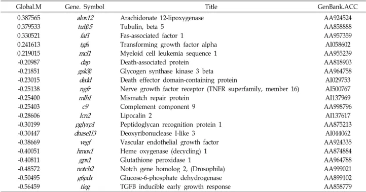
저산소증에서 반하에 의하여 표현이 변하는 apoptosis/cell death 관련 유전자 중 Global M 값이 ±0.2 이상 즉, ±14% 이상 표현이 증가된 유전자는 5종, 감소된 유전자는 15종이었다 (Table 1). Apoptosis 관련 유전자 중 arachidonate 12-lipoxygenase (alox12)는 약 30% (Global M=0.39)로 가장 많이 증가하였다.alox12는 세포분화와 세포사의 중요한 조절 자로서 ROS의 일종인 ONOO-에 의한 신경세포의 자연사[56]
와 glutathione 고갈 및 arachidonic acid에 의한 세포사에 중 요한 역할을 하는 것으로 알려졌다[53]. 본 연구에서 세포사를 촉진하는 것으로 알려진alox12 유전자가 많이 표현된 이유는 알 수 없다. Tubulin β5 (tubβ5)가 약 30% (Global M=0.38)로 두 번째로 많이 증가되었다. α-/β-tubulin heterodimer로 구성 된 미세소관(microtubule)은 진핵세포에서 세포모양의 유지, 신호전달, 염색체 이동, mRNA 국소화, 축삭수송, 소기관의 자리매김, 그리고 소포 및 소기관의 이동 등 다양한 기능을 갖는다[13] 최근 proteosome 연구에 의하면 정상 상피세포에 비하여 식도편평세포암세포에서는tubβ5을 포함한 15종의 단 백질 표현이 증가하였다[41]. 이러한 결과들은tubβ5가 세포의 분화, 성장 및 신호전달에 기여하는 단백질로서 저산소증에서 세포의 활성을 증가시키거나 세포 성장을 촉진하여 세포손상 을 방지하는 것으로 해석된다. 다음은 Fas-associated factor 1 (faf1 gene)로 약 27% (Global M=0.33)의 증가를 보였다.
Mouse Faf1 (mfaf1)을 일시적으로 과표현시키면 L cell에서 Fas에 의한 세포 자연사를 촉진시키며[6], Human Faf1 (hfaf1) 은 CK2에 의하여 serine 289 및 291에 인산화되며, hFAF1- CK2 complex 형성이 세포 자연사에서 증가한다[20,24]. 세포 사를 촉진하는 것으로 알려진faf1이 왜 저산소증에서 반하에 의하여 표현이 증가하는지는 알 수 없다. Transforming
Table 1. Apoptosis/cell death-related genes
Global.M Gene. Symbol Title GenBank.ACC
0.387565 0.379533 0.330521 0.241613 0.219015 -0.20987 -0.21851 -0.23015 -0.25138 -0.25400 -0.25403 -0.28606 -0.30199 -0.30447 -0.38669 -0.40051 -0.40811 -0.48572 -0.50495 -0.56459
alox12 tub
β
5 faf1tgfα
mcl1dap gsk3β
dedd ngfr mlh1c9 pglyrp1lcn2 dnase1l3vegf hmox1 notch2gpx1 g6pdx tieg
Arachidonate 12-lipoxygenase Tubulin, beta 5
Fas-associated factor 1
Transforming growth factor alpha Myeloid cell leukemia sequence 1 Death-associated protein
Glycogen synthase kinase 3 beta Death effector domain-containing protein
Nerve growth factor receptor (TNFR superfamily, member 16) Mismatch repair protein
Complement component 9 Lipocalin 2
Peptidoglycan recognition protein 1 Deoxyribonuclease I-like 3
Vascular endothelial growth factor Heme oxygenase (decycling) 1 Glutathione peroxidase 1
Notch gene homolog 2, (Drosophila) Glucose-6-phosphate dehydrogenase TGFB inducible early growth response
AA924524 AA858888 AA957359 AI058602 AA955239 AA818903 AA964758 AI029753 AI500767 AI137969 AA998796 AI137617 AA875213 AI044062 AA924335 AA874884 AA964788 AA999021 AA899102 AA858779 growth factor alpha 유전자(tgfα)는 약 19% (Global M=0.24)
로, 네 번째로 많은 증가를 보였다. tgfα는 난모세포의 핵과 세포질의 성숙을 자극하고[43], 다양한 중배엽 및 외배엽 조직 에 세포분열 효능이 있으며 강력한 혈관생성 인자인 것으로 알려져 있다[7]. 최근에는tgfα가 nuclear factor-kappaB를 활 성시켜 c-Myc에 유도된 세포 자연사를 저해하는 것으로 밝혀 졌다[3]. 따라서 저산소증에서tgfα의 표현의 증가는 세포사를 억제하는데 도움이 될 것으로 이해된다. 약 17% (Global M=0.21)의 증가를 보인 Myeloid cell leukemia sequence 1 (mcl-1)은 Bcl-2 family의 antiapoptotic member로서[10], 세포 분화나 세포사의 신호전달과정에서 유도되며[19], multiple myeloma cell과[12,16,55] B cell lymphoma cell의[30] 생존에 필수적이며, myeloma cell의 증식을 촉진시킨다[55].mcl-1은 Bim과 같은 proapoptotic protein들과 대항하는 것으로 생각 되어진다[18]. 따라서mcl-1 표현의 증가는 저산소증에서 세포 사를 억제하는데 도움이 될 것으로 이해된다. Apoptosis 유전 자 그룹 가운데 Tgfβ inducible early growth response 유전자 (tieg)는 약 45% (Global M=-0.56)로 가장 크게 발현이 감소하 였다. 이 유전자는 간세포에서 산화적 스트레스 하에서 증가 하여 세포 자연사를 매개하는 것으로 알려져 있고[44], pan- creatic epithelial cells [44], hepatocyte line Hep 3B [44], 그리 고 epithelial Mv1Lu cells [5]에서 세포 자연사를 유도하는 것 으로 보고되고 있다. 이러한 결과들은tieg 단백질들이 항증식 및 세포 자연사를 유도하는 기능을 갖는 유전자 전사인자임을 의미한다. 따라서 반하에 의한tieg 유전자의 표현 감소는 저산 소증에서 세포사를 억제하는데 도움을 줄 것으로 이해된다.
Fig. 3. Proposed machanisms for neuroprotecting effects by Pr.
Excitotoxic stimulation of neurons results in the influx of Ca2+, which damages mitochondrial functions. High production of reactive oxygen species (ROS) by damaged mitochondria. In turn, ROS damages many important cellular molecules leading to cell death. The Pr up-regu- lates many genes that help survival of neurons in hypo- xia such as antiapoptosis (tgf
α
, mcl-1, tieg), and immunity (cxcl10). Genes that have general neurotrophic effects may have alleviated ROS production and mitochondrial membrane potential (MMP) deprivation.Table 2. Cell cycle-related genes
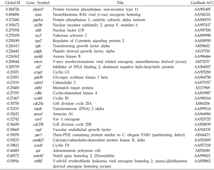
Global.M Gene. Symbol Title GenBank.ACC
0.584726 0.494496 0.372445 0.305672 0.275938 0.275039 0.258189 0.241613 0.226641 0.216562 0.209644 0.205755 -0.21821 -0.21851 -0.23319 -0.25400 -0.27355 -0.27467 -0.30750 -0.32151 -0.32623 -0.32742 -0.36658 -0.38669 -0.39078 -0.39233 -0.39821 -0.44485 -0.48572 -0.50916
ptpn11 ppp1cnras
α
nr2f6nfib tsc2 rgs2tgf
α
pdgfα
aurkb mycnid2 ccng1 gsk3b calm3 mlh1cdk4 ccnb1 cdc25atop2
α
anxa1 vav1 cdc25bvegf camk2δpar3
ccnd1 apc notch2
erbb2
Protein tyrosine phosphatase, non-receptor type 11 Neuroblastoma RAS viral (v-ras) oncogene homolog Protein phosphatase 1, catalytic subunit, alpha isoform Nuclear receptor subfamily 2, group F, member 6 Nuclear factor I/B
Tuberous sclerosis 2
Regulator of G-protein signaling protein 2 Transforming growth factor alpha Platelet derived growth factor, alpha Aurora kinase B
V-myc myelocytomatosis viral related oncogene, neuroblastoma derived (avian) Inhibitor of DNA binding 2, dominant negative helix-loop-helix protein Cyclin G1
Glycogen synthase kinase 3 beta Calmodulin 3
Mismatch repair protein Cyclin-dependent kinase 4 Cyclin B1
Cell division cycle 25A Topoisomerase (DNA) 2 alpha Annexin A1
Vav 1 oncogene Cell division cycle 25B
Vascular endothelial growth factor
Three-PDZ containing protein similar to C. elegans PAR3 (partitioning defect) Calcium/calmodulin-dependent protein kinase II, delta
Cyclin D1
Adenomatosis polyposis coli Notch gene homolog 2, (Drosophila)
V-erb-b2 erythroblastic leukemia viral oncogene homolog 2, neuro/glioblastoma derived oncogene homolog (avian)
AA901405 AA926321 AA900570 AA997437 AA997500 AA899998 AA858950 AI058602 AI137550 AA996466 AI072537 AA964507 AA925280 AA964758 AA875357 AI137969 AA819907 AA998164 AI060206 AA899114 AA964960 AA925725 AA858839 AA924335 AI044473 AA925495 AA957218 AI058280 AA999021 AA858862 세포주기 관련 유전자 표현의 변화
저산소증에서 반하에 의하여 표현이 변하는 세포주기 관련 유전자 중 12종류의 표현이 14% 이상 증가되었으며, 18종류는 감소되었다(Table 2). Protein tyrosine phosphatase, non-re- ceptor type 11 유전자(ptpn11)는 약 47% (Global M=0.58)로 발현이 가장 많이 증가하였다. ptpn11에 의해 만들어 지는 non-receptor protein tyrosine phoshatase Shp2 (Src Homology 2 domain-containing protein tyrosine phoshatase) 는 growth factor, cytokine, 및 extracellular matrix receptor 에 신호를 주는 신호 유인 요소로서 세포의 증식과 분화, 이동에 중요한 역할을 한다. 뿐만 아니라 태아발생과 조혈에 도 중요한 역할을 한다[51]. Neuroblastoma RAS viral (v-ras) oncogene homolog 유전자(n-ras)는 약 40 % (Global M=0.49) 의 증가를 보였다.n-ras 유전자는 thyroid carcinomas와 mye- loid leukemias 등에서 발견되며[30],b-ras 유전자와 n-ras 유 전자는 cutaneous melanoma의 발병과 유지에 중요한 역할을 한다[1]. Platelet derived growth factor alpha 유전자(pdgfα)는
약 19% (Global M=0.23)의 증가를 보였다. pdgfα 유전자들은 중간엽 세포의 이동과 증식에 중요한 역할을 한다. Pdgf re- ceptor/Pdgf system은 배 발생 과정에서 신장, 심혈관 계통, 폐, 뇌와 결합조직의 발생에 중요한 역할을 하며, 어른에서는 상처치유, 염증, 혈관신생에 중요한 역할을 한다.pdgfα 또는 pdgfβ receptor tyrosine kinases의 활성은 골수종에서 보여지 고 pdgfα의 활성은 위장관 종양에서 보여진다[26]. 따라서 ptpn11, nras, pdgfα 단백질들은 세포의 분화, 성장에 기여하는 단백질로 세포사를 방지하는 반하에서 증가하는 것은 저산소 증에서 세포의 분화와 성장을 촉진하여 세포 손상을 방지하는 데 도움을 줄 것으로 이해된다.
면역 관련 유전자 표현의 변화
저산소증에서 반하에 의하여 표현이 변하는 면역 관련 유전 자 중 10종류의 표현이 14% 이상 증가되었으며, 12종류는 감 소되었다(Table 3). 이 가운데cd3d 유전자가 가장 크게 증가되 었다(Global M=0.66).cd3와 cd8은 T-cell의 중요 세포표면단백
Table 3. Immune response-related genes
Global.M Gene. Symbol Title GenBank.ACC
0.6618464 0.4733848 0.3457336 0.3440545 0.2750392 0.2416067 0.2283354 0.2122148 0.2038110 0.2029980 -0.2045649 -0.2341147 -0.2461116 -0.2540251 -0.2939990 -0.3019932 -0.3122543 -0.3173815 -0.3274247 -0.3352131 -0.3409484 -0.3757863
cxcl10cd3d ppargpawr cxcl12tsc2 azgp1 ptprccd5 ptmsstat3 galf2 gbp2c9 pglyrp1
igbp1hrh4 c1qr1vav1
α
hsg β2mCD3 antigen delta polypeptide Chemokine (C-X-C motif) ligand 10 PRKC, apoptosis, WT1, regulator
Peroxisome proliferator activated receptor, gamma Tuberous sclerosis 2
Chemokine (C-X-C motif) ligand 12 Alpha-2-glycoprotein 1, zinc CD5 antigen
Protein tyrosine phosphatase, receptor type, C Parathymosin
Signal transducer and activator of transcription 3 Galanin
Coagulation factor 2 Complement component 9
Guanylate binding protein 2, interferon-inducible Peptidoglycan recognition protein 1
Histamine H4 receptor
Immunoglobulin (CD79A) binding protein 1 Vav 1 oncogene
Lymphocyte antigen 68 Alpha-2-HS-glycoprotein Beta-2 microglobulin
AI137921 AI059907 AA956914
AI111890 AA899998 AA925112 AI059700 AA925584 AA924685 AA899878 AI045179 AA955779
AI071305 AA998796 AA819701 AA875213 AA900657 AA997141 AA925725 AA818744 AA955349 AA817792
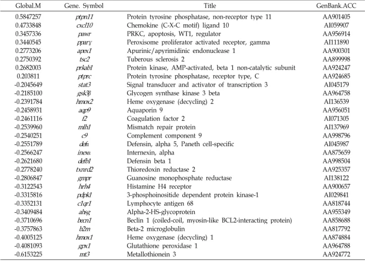
Table 4. ‘Response to stress’-related genes
Global.M Gene. Symbol Title GenBank.ACC
0.5847257 0.4733848 0.3457336 0.3440545 0.2773206 0.2750392 0.2682003 0.203811 -0.2045649 -0.2185100 -0.2391784 -0.2458931 -0.2461116 -0.2539960 -0.2540251 -0.2551789 -0.2566247 -0.2621680 -0.2778240 -0.2806847 -0.3122543 -0.3315816 -0.3352131 -0.3409484 -0.3710696 -0.3757863 -0.4005125 -0.4081093 -0.6153225
ptpn11 cxcl10 pparγpawr apex1 prkab1tsc2 ptprc stat3 gsk3
β
hmox2aqp9f2 mlh1c9 def
α
inexα
defb1 txnrd2gmprhrh4 pdpk1 c1qr1 becn1ahsg hmox1b2m gpx1mt3
Protein tyrosine phosphatase, non-receptor type 11 Chemokine (C-X-C motif) ligand 10
PRKC, apoptosis, WT1, regulator
Peroxisome proliferator activated receptor, gamma Apurinic/apyrimidinic endonuclease 1
Tuberous sclerosis 2
Protein kinase, AMP-activated, beta 1 non-catalytic subunit Protein tyrosine phosphatase, receptor type, C
Signal transducer and activator of transcription 3 Glycogen synthase kinase 3 beta
Heme oxygenase (decycling) 2 Aquaporin 9
Coagulation factor 2 Mismatch repair protein Complement component 9
Defensin, alpha 5, Paneth cell-specific Internexin, alpha
Defensin beta 1 Thioredoxin reductase 2
Guanosine monophosphate reductase Histamine H4 receptor
3-phosphoinositide dependent protein kinase-1 Lymphocyte antigen 68
Alpha-2-HS-glycoprotein
Beclin 1 (coiled-coil, myosin-like BCL2-interacting protein) Beta-2 microglobulin
Heme oxygenase (decycling) 1 Glutathione peroxidase 1 Metallothionein 3
AA901405 AI059907 AA956914
AI111890 AA900301 AA899998 AA924247 AA924685 AI045179 AA964758
AI136539 AA956051
AI071305 AI137969 AA998796
AI045987 AA875659 AA998504 AA925357 AI138122 AA900657
AI029841 AA818744 AA955349 AA858688 AA817792 AA874884 AA964788 AA924772
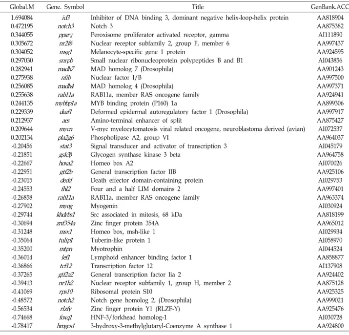
Table 5. Transcription-related genes
Global.M Gene. Symbol Title GenBank.ACC
1.694084 0.472195 0.344055 0.305672 0.304052 0.297030 0.282941 0.275938 0.256085 0.255638 0.244135 0.229339 0.212937 0.209644 0.202134 -0.20456 -0.21851 -0.22667 -0.22951 -0.23015 -0.24553 -0.26858 -0.27902 -0.29744 -0.30694 -0.31248 -0.35064 -0.35200 -0.36014 -0.36866 -0.37265 -0.39413 -0.41069 -0.48572 -0.56534 -0.74668 -0.78417
notch3id3 pparγ nr2f6 msg1 snrpb madh7 madh4nfib rab11a mybbp1a
deaf1 mycnaes pla2g6
stat3 gsk3
β
hoxa2 gtf2b deddfhl2 rab11amyog khdrbs1 znf354a msx1 tulip1 mtpn tcf12lef1 gtf2a2 nr1h2 rps10 notch2
rlzfy foxq1 hmgcs1
Inhibitor of DNA binding 3, dominant negative helix-loop-helix protein Notch 3
Peroxisome proliferator activated receptor, gamma Nuclear receptor subfamily 2, group F, member 6 Melanocyte-specific gene 1 protein
Small nuclear ribonucleoprotein polypeptides B and B1 MAD homolog 7 (Drosophila)
Nuclear factor I/B
MAD homolog 4 (Drosophila)
RAB11a, member RAS oncogene family MYB binding protein (P160) 1a
Deformed epidermal autoregulatory factor 1 (Drosophila) Amino-terminal enhancer of split
V-myc myelocytomatosis viral related oncogene, neuroblastoma derived (avian) Phospholipase A2, group VI
Signal transducer and activator of transcription 3 Glycogen synthase kinase 3 beta
Homeo box A2
General transcription factor IIB
Death effector domain-containing protein Four and a half LIM domains 2 RAB11a, member RAS oncogene family Myogenin
Src associated in mitosis, 68 kDa Zinc finger protein 354A Homeo box, msh-like 1 Tuberin-like protein 1 Myotrophin
Lymphoid enhancer binding factor 1 Transcription factor 12
General transcription factor Iia 2
Nuclear receptor subfamily 1, group H, member 2 Ribosomal protein S10
Notch gene homolog 2, (Drosophila) Zinc finger protein Y1 (RLZF-Y) HNF-3/forkhead homolog-1
3-hydroxy-3-methylglutaryl-Coenzyme A synthase 1
AA818904 AA875382 AI111890 AA997437 AA924595 AI043856 AA901243 AA997500 AA997371 AA924941 AA899306 AA997917 AA875427 AI072537 AA964037 AI045179 AA964758 AI070026 AA925106 AI029753 AA997401 AA963374 AI030924 AA818199 AA965012 AI029934 AI058970 AI044524 AA858877 AI137908 AA924402 AA875128 AA925325 AA999021 AA925476 AI030728 AA924800 질이다. T-cell 활성화 동안, T-cell 수용체(TCR)/cd3 복합체와
결합하고 있는 major histocompatibility complex (MHC) class I 단백질에 cd8이 결합한다[39,52]. cd3 복합체는 4개의 작은 펩티드cd3d, cd3e, cd3g 및 cd3ζ로 구성된다. cd3d는 세포 막 당단백질로서 T-cell의 발생, 갑상선세포(thymocyte)의 발 생에 필수적인 역할을 한다[4,11]. 따라서 반하에 의한 CD3의 표현증가는 세포사를 방지하는데 도움을 줄 것으로 이해된다.
Chemokine (C-X-C motif) ligand 10 유전자(cxcl10)는 약 38 %(Global M=0.47)로 많은 증가를 보였다. Chemokine은 면역세포들이 감염부위로 몰려들도록 조절을 하는데, 그 가운 데 Cxcl10 chemokine은 Cxcr3 수용체를 활성시키는 리간드이 다. 한편 Cxcr3 수용체는 활성 T 임파구와 natural killer cell에
서 표현되며 이들이 감염부위로 이동하는데 관여한다[27,34].
또한cxcl10 유전자는 광견병 virus에 감염시켰을 때 발현이 증가하며[41], mouse hepatitis virus를 뇌 내로 감염시킨 쥐에 서도 발현이 증가한다[33].cxcl10 유전자의 발현 증가는 저산 소증 상태에서 면역과 관련된 반응을 통해 세포 손상을 방지 하는데 도움을 줄 것으로 이해된다.
이 밖에도 스트레스 반응 관련 유전자는 8종이 14%이상 표현이 증가되었으며, 21 종류는 14% 이상 감소되었고(Table 4), 유전자 전사(transcription) 관련 유전자 중 15종류의 표현 이 14% 이상 증가되었으며, 22종류는 14% 이상 감소되었다 (Table 5). 생리대사 관련 유전자는 111 종류의 표현이 14%
이상으로 증가하였다. 이 가운데 Global M 값이 0.4, 즉 32%
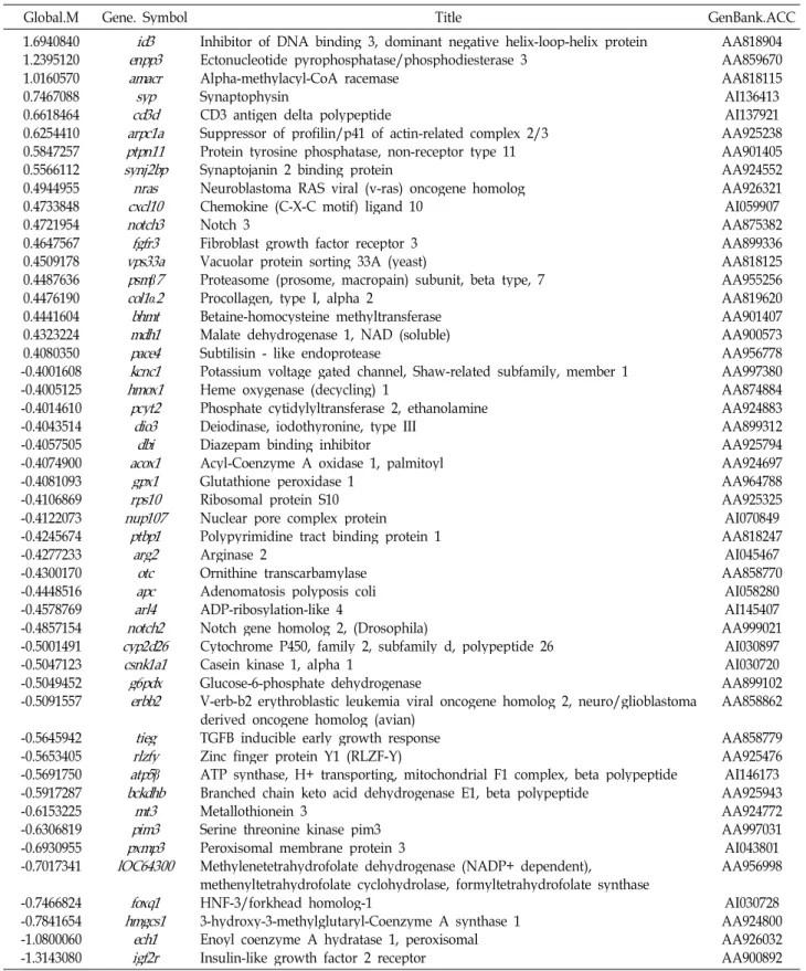
Table 6. Physiological process-related genes
Global.M Gene. Symbol Title GenBank.ACC
1.6940840 1.2395120 1.0160570 0.7467088 0.6618464 0.6254410 0.5847257 0.5566112 0.4944955 0.4733848 0.4721954 0.4647567 0.4509178 0.4487636 0.4476190 0.4441604 0.4323224 0.4080350 -0.4001608 -0.4005125 -0.4014610 -0.4043514 -0.4057505 -0.4074900 -0.4081093 -0.4106869 -0.4122073 -0.4245674 -0.4277233 -0.4300170 -0.4448516 -0.4578769 -0.4857154 -0.5001491 -0.5047123 -0.5049452 -0.5091557 -0.5645942 -0.5653405 -0.5691750 -0.5917287 -0.6153225 -0.6306819 -0.6930955 -0.7017341 -0.7466824 -0.7841654 -1.0800060 -1.3143080
enpp3id3 amacr cd3dsyp arpc1a ptpn11 synj2bp
cxcl10nras notch3 fgfr3 vps33a psm
β
7 col1α
2 mdh1bhmt pace4 kcnc1 hmox1pcyt2 dio3dbi acox1 rps10gpx1 nup107
ptbp1 arg2otc
arl4apc notch2 cyp2d26 csnk1a1 g6pdx
erbb2 rlzfytieg atp5
β
bckdhbpim3mt3 pxmp3 lOC64300
foxq1 hmgcs1
igf2rech1
Inhibitor of DNA binding 3, dominant negative helix-loop-helix protein Ectonucleotide pyrophosphatase/phosphodiesterase 3
Alpha-methylacyl-CoA racemase Synaptophysin
CD3 antigen delta polypeptide
Suppressor of profilin/p41 of actin-related complex 2/3 Protein tyrosine phosphatase, non-receptor type 11 Synaptojanin 2 binding protein
Neuroblastoma RAS viral (v-ras) oncogene homolog Chemokine (C-X-C motif) ligand 10
Notch 3
Fibroblast growth factor receptor 3 Vacuolar protein sorting 33A (yeast)
Proteasome (prosome, macropain) subunit, beta type, 7 Procollagen, type I, alpha 2
Betaine-homocysteine methyltransferase Malate dehydrogenase 1, NAD (soluble) Subtilisin - like endoprotease
Potassium voltage gated channel, Shaw-related subfamily, member 1 Heme oxygenase (decycling) 1
Phosphate cytidylyltransferase 2, ethanolamine Deiodinase, iodothyronine, type III
Diazepam binding inhibitor
Acyl-Coenzyme A oxidase 1, palmitoyl Glutathione peroxidase 1
Ribosomal protein S10 Nuclear pore complex protein Polypyrimidine tract binding protein 1 Arginase 2
Ornithine transcarbamylase Adenomatosis polyposis coli ADP-ribosylation-like 4
Notch gene homolog 2, (Drosophila)
Cytochrome P450, family 2, subfamily d, polypeptide 26 Casein kinase 1, alpha 1
Glucose-6-phosphate dehydrogenase
V-erb-b2 erythroblastic leukemia viral oncogene homolog 2, neuro/glioblastoma derived oncogene homolog (avian)
TGFB inducible early growth response Zinc finger protein Y1 (RLZF-Y)
ATP synthase, H+ transporting, mitochondrial F1 complex, beta polypeptide Branched chain keto acid dehydrogenase E1, beta polypeptide
Metallothionein 3
Serine threonine kinase pim3 Peroxisomal membrane protein 3
Methylenetetrahydrofolate dehydrogenase (NADP+ dependent),
methenyltetrahydrofolate cyclohydrolase, formyltetrahydrofolate synthase HNF-3/forkhead homolog-1
3-hydroxy-3-methylglutaryl-Coenzyme A synthase 1 Enoyl coenzyme A hydratase 1, peroxisomal Insulin-like growth factor 2 receptor
AA818904 AA859670 AA818115 AI136413 AI137921 AA925238 AA901405 AA924552 AA926321 AI059907 AA875382 AA899336 AA818125 AA955256 AA819620 AA901407 AA900573 AA956778 AA997380 AA874884 AA924883 AA899312 AA925794 AA924697 AA964788 AA925325 AI070849 AA818247
AI045467 AA858770
AI058280 AI145407 AA999021
AI030897 AI030720 AA899102 AA858862 AA858779 AA925476 AI146173 AA925943 AA924772 AA997031 AI043801 AA956998
AI030728 AA924800 AA926032 AA900892 이상 증가한 유전자는 18 종이었다. 반면에 14% 이상 감소하
는 유전자는 157 종으로 이 가운데 Global M 값이 -0.4, 즉 32% 이하로 감소하는 유전자는 31 종이었다(Table 6). 신호전
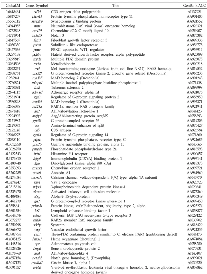
달 관련 유전자는 28종류의 표현이 14% 이상 증가되었으며, 47 종은 감소되었다. 그 가운데 27종류는 Global M 값이 -0.3, 즉 23% 이상 감소되었다(Table 7).
Table 7. Signal transduction-related genes
Global.M Gene. Symbol Title GenBank.ACC
0.6618464 0.5847257 0.5566112 0.4944955 0.4733848 0.4721954 0.4647567 0.4080350 0.3457336 0.3297138 0.3279819 0.3064598 0.3023251 0.2888761 0.282941 0.2760824 0.2750392 0.2674113 0.2581894 0.2560848 0.2556378 0.2471418 0.2294907 0.2171982 0.2129373 0.2122148 0.2046275 0.2038110 -0.3012858 -0.3026250 -0.3122543 -0.3173815 -0.3180748 -0.3253093 -0.3262285 -0.3274084 -0.3274247 -0.3315816 -0.3335970 -0.3409484 -0.3461239 -0.3558641 -0.3601354 -0.3644576 -0.3672277 -0.3750298 -0.3866872 -0.3907754 -0.4005125 -0.4448516 -0.4528926 -0.4578769 -0.4857154 -0.5047123 -0.5091557
ptpn11cd3d synj2bp
cxcl10nras notch3 fgfr3 pace4 pdgfrpawr
α
mpdzmt1a gprk2lmel madh7 minpp1 adrtsc2α
1d madh4rgs2 rab11a argbp2arl1 gpr56cd5aes rgs14 ptprc gn
α
15 ppap2a igbp1hrh4 dgkα
cmkor1anxa1 cacna1
α
pdpk1vav1 alcam ahsggit1 prkar2
α
celsr3lef1 rab26 sdcbp par3vegf hmox1
bmp2apc notch2arl4 csnk1a1
erbb2
CD3 antigen delta polypeptide
Protein tyrosine phosphatase, non-receptor type 11 Synaptojanin 2 binding protein
Neuroblastoma RAS viral (v-ras) oncogene homolog Chemokine (C-X-C motif) ligand 10
Notch 3
Fibroblast growth factor receptor 3 Subtilisin - like endoprotease PRKC, apoptosis, WT1, regulator
Platelet derived growth factor receptor, alpha polypeptide Multiple PDZ domain protein
Metallothionein
Mel transforming oncogene (derived from cell line NK14)- RAB8 homolog G protein-coupled receptor kinase 2, groucho gene related (Drosophila) MAD homolog 7 (Drosophila)
Multiple inositol polyphosphate histidine phosphatase 1 Tuberous sclerosis 2
Adrenergic receptor, alpha 1d
Regulator of G-protein signaling protein 2 MAD homolog 4 (Drosophila)
RAB11a, member RAS oncogene family ADP-ribosylation factor-like 1
Arg/Abl-interacting protein ArgBP2 G protein-coupled receptor 56 Amino-terminal enhancer of split CD5 antigen
Regulator of G-protein signaling 14
Protein tyrosine phosphatase, receptor type, C Guanine nucleotide binding protein, alpha 15 Phosphatidate phosphohydrolase type 2a Histamine H4 receptor
Immunoglobulin (CD79A) binding protein 1 Diacylglycerol kinase, alpha (80 kDa) Chemokine orphan receptor 1 Annexin A1
Calcium channel, voltage-dependent, P/Q type, alpha 1A subunit Vav 1 oncogene
3-phosphoinositide dependent protein kinase-1 Activated leukocyte cell adhesion molecule Alpha-2-HS-glycoprotein
G protein-coupled receptor kinase interactor 1
Protein kinase, cAMP-dependent, regulatory, type 2, alpha Lymphoid enhancer binding factor 1
Cadherin EGF LAG seven-pass G-type receptor 3 RAB26, member RAS oncogene family
Syntenin
Vascular endothelial growth factor
Three-PDZ containing protein similar to C. elegans PAR3 (partitioning defect) Heme oxygenase (decycling) 1
Adenomatosis polyposis coli Bone morphogenetic protein 2 ADP-ribosylation-like 4
Notch gene homolog 2, (Drosophila) Casein kinase 1, alpha 1
V-erb-b2 erythroblastic leukemia viral oncogene homolog 2, neuro/glioblastoma derived oncogene homolog (avian)
AI137921 AA901405 AA924552 AA926321 AI059907 AA875382 AA899336 AA956778 AA956914 AA925099 AA925078 AA900218 AA819532 AA963235 AA901243 AI071438 AA899998 AA924076 AA858950 AA997371 AA924941 AI044423 AI058393 AA819206 AA875427 AA925584 AI071860 AA924685
AI045065 AA818593 AA900657 AA997141 AA818373 AA997721 AA964960 AI045770 AA925725
AI029841 AA875360 AA955349 AA997450 AA925274 AA858877 AI029122 AI030702 AA818429 AA924335 AI044473 AA874884
AI058280 AI070931 AI145407 AA999021
AI030720 AA858862
요 약
본 연구는 저산소증에서 반하가 대뇌신경세포의 유전자 표 현에 미치는 영향을 알아보기 위하여 배양한 E18의 흰쥐 대뇌 신경세포를 반하로 처리하고, 저산소증을 유도한 후 micro- array 기법으로 유전자 표현 변화를 조사하였다. Microarray 결과tubb5, tgfα, ptpn11, n-ras, pdgfa 등 세포의 성장 분화에 관여하는 유전자들의 표현이 증가하였으며, 세포 자연사를 억 제하는mcl-1 유전자의 표현 또한 증가하였다. 한편 세포 자연 사를 유도하는tieg 유전자는 표현이 감소하였다(Fig. 3). 반하 에 의하여 수많은 유전자의 표현이 변화되었고, 세포사를 촉 진하는 유전자의 표현이 크게 증가되는 경우(예,alox12, faf1) 도 있어 본 연구결과만으로 일반적인 결론을 유도하기는 어려 웠다. 그러나 대략적으로 반하는 저산소증에서 주로 세포의 성장과 분화를 유지하고, 세포 자연사를 방지하는 유전자들의 표현을 증가시켜 신경 세포사를 보호하는 것으로 이해된다.
References
1. Akslen, L. A., S. Angelini, O. Straume, I. M. Bachmann, A.
Molven, K. Hemminki, and R. Kumar. 2005. BRAF and NRAS mutations are frequent in nodular melanoma but are not associated with tumor cell proliferation or patient survival. J. Invest. Dermatol. 125, 312-317.
2. Brewer, G. J., J. R. Torricelli, E. K. Evege, and P. J. Price.
1993. Optimized survival of hippocampal neurons in B27-supplemented Neurobasal, a new serum-free medium combination. J. Neurosci. Res. 35, 567-676.
3. Cavin, L. G., F. Wang, V. M. Factor, S. Kaur, M.
Venkatraman, S. S. Thorgeirsson, and M. Arsura. 2005.
Transforming growth factor-alpha inhibits the intrinsic pathway of c-Myc-induced apoptosis through activation of nuclear factor-kappaB in murine hepatocellular carcinomas. Mol. Cancer Res. 3, 403-412.
4. Chalaux, E., T. Lopez-Rovira, J. L. Rosa, G. Pons, L. M.
Boxer, R. Bartrons, and F. Ventura. 1999. A zinc-finger transcription factor induced by TGF-beta promotes apop- totic cell death in epithelial Mv1Lu cells. FEBS Lett. 457, 478-482.
5. Chee, M., R. Yang, E. Hubbell, A. Berno, X. C. Huang, D.
Stern, J. Winkler, D. J. Lockhart, M. S. Morris, and S. P.
Fodor. 1996. Accessing genetic information with high-den- sity DNA arrays. Science274, 610-614.
6. Chu, K., X. Niu, and L. T. Williams. 1995. A Fas-asso- ciated protein factor, FAF1, potentiates Fas-mediated apoptosis. Proc. Natl. Acad. Sci. USA92, 11894-11898.
7. Cirelli, C. and G. Tononi. 1999. Differences in brain gene expression between sleep and waking as revealed by mRNA differential display and cDNA microarray technology. J. Sleep Res. 8, S44-S52.
8. Coffey, R. J., R. Derynck, and J. N. Wilcox. 1987.
Production and autoinduction of transforming growth fac-
tor in human keratinocytes. Nature28, 817-820.
9. Coultas, L. and A. Strasser. 2003. The role of the Bcl-2 protein family in cancer. Semin Cancer Biol. 13, 115-123.
10. Dadi, H. K. A. J. Simon, and C. M. Roifman, 2003. Effect 1of CD3delta deficiency on maturation of alpha/beta and gamma/delta T-cell lineages in severe combined immunodeficiency. N. Engl. J. Med. 349, 1821-1828.
11. Dave, V. P., Z. Cao, C. Browne, B. Alarcon, G. Fernandez- Miguel, J. Lafaille, A. de la Hera, S. Tonegawa, and D. J.
Kappes. 1997. CD3 delta deficiency arrests development of the alpha beta but not the gamma delta T cell lineage, EMBO. 16, 1360-1370.
12. Derenne, S., B. Monia, N. M. Dean, J. K. Taylor, M. J.
Rapp, J. L. Harousseau, R. Bataille, and M. Amiot. 2002.
Antisense strategy shows that Mcl-1 rather than Bcl-2 or Bcl-x(L) is an essential survival protein of human myelo- ma cells. Blood100, 194-199.
13. Dutcher, S. K. 2001. The tubulin fraternity: alpha to eta.
Curr. Opin. Cell Biol.13, 49-54.
14. Ebert, B. L., J. D. Firth, and P. J. Ratcliffe. 1995. Hypoxia and mitochondrial inhibitors regulate expression of glu- cose transporter-1 via distinct Cis-acting sequences.J. Biol.
Chem. 270, 29083-29089.
15. Fuh, Jr., S. J Wang, E. B. Larson, and H. C. Liu. 1996.
Prevalence of stroke in kinmen.Stroke27, 1338-1341.
16. Gojo, I., B. Zhang, and R. G. Fenton. 2002. The cyclin-de- pendent kinase inhibitor flavopiridol induces apoptosis in multiple myeloma cells through transcriptional repression and down-regulation of Mcl1. Clin. Cancer Res. 8, 3527- 3538.
17. Goldberg, M. A., S. P. Dunning, and H. F. Bunn. 1988.
Regulation of the erythropoietin gene: evidence that the oxygen sensor is a heme protein. Science242, 1412-1415.
18. Gomez-Bougie, P., R. Bataille R, and M. Amiot. 2004. The imbalance between Bim and Mcl-1 expression controls the survival of human myeloma cells. Eur. J. Immunol. 34, 3156-3164.
19. Good, L., G. P. Dimri, J. Campisi, and K. Y. Chen. 1996.
Regulation of dihydrofolate reductase gene expression and E2F components in human diploid fibroblasts during growth and senescence.J. Cell Physiol. 168, 580-588.
20. Guerra, B., B. Boldyreff, and O. G. Issinger. 2001. FAS-as- sociated factor 1 interacts with protein kinase CK2 in vivo upon apoptosis induction. Int. J. Oncol.19, 1117-1126.
21. Haddad, J. J. and S. C. Land. 2000. O2-evoked regulation of HIF-1a and NFκB in perinatal lung epithelium requires glutathione biosynthesis. Am. J. Physiol. Lung Cell Mol.
Physiol. 278, L492-L503.
22. Hoe, J. 1999. Dong-Eui-Bo-Gam. pp. 965, 961, 1955, Byuinmoonhwasa, Seoul.
23. Hochachka, P. W. and Lutz P. 2001. Mechanism, origin, and evolution of anoxia tolerance in animals. Comp Biochem. Physiol. B Biochem. Mol. Biol.130, 435-459.
24. Jensen, H. H., M. Hjerrild, B. Guerra, M. R. Larsen, P.
Hojrup, and B. Boldyreff. 2001. Phosphorylation of the Fas associated factor FAF1 by protein kinase CK2 and identi-
fication of serines 289 and 291 as the in vitro phosphor- ylation sites. Int. J. Biochem. Cell Biol. 33, 577-589.
25. Ji, H. J. and S. I. Lee. 2007. Hanyack-Gyugyuk-Juhea. pp.
513, Korean index Co., Seoul.
26. Jones, A. V. and N. C. Cross. 2004. Oncogenic derivatives of platelet-derived growth factor receptors. Cell Mol. Life Sci. 61, 2912-2923.
27. Kim, C. H. and H. E. Broxmeyer. 1999. SLC/exodus2/
6Ckine/TCA4 induces chemotaxis of hematopoietic pro- genitor cells: differential activity of ligands of CCR7, CXCR3, or CXCR4 in chemotaxis vs. suppression of pro- genitor proliferation. J. Leukoc Biol. 66, 455-461.
28. Kim, H. C. 2001. Pharmacology of Korea. pp. 346-347, Jipmoondang, Seoul.
29. King, A. E., R. S. Chung, J. C. Vickers, and T. C. Dickson.
2006. Localization of glutamate receptors in developing cortical neurons in culture and relationship to suscepti- bility to excitotoxicity. J. Comp. Neurol. 498, 277-294.
30. Kroll, T. G. Molecular events in follicular thyroid tumors.
2004. Cancer Treat Res. 122, 85-105.
31. Levy, A. P., N. S. Levy, S. Wegner, and M. A. Goldberg.
1995. Transcriptional regulation of the rat vascular endo- thelial growth factor gene by hypoxia. J. Biol. Chem. 270, 13333-13340.
32. Liang, P. and A. B. Pardee. 1992. Differential display of eukaryotic messenger RNA by means of the polymerase chain reaction. Science257, 967-971.
33. Liu, M. T., H. S. Keirstead, and T. E. Lane. 2001.
Neutralization of the chemokine CXCL10 reduces in- flammatory cell invasion and demyelination and improves neurological function in a viral model of multiple sclerosis. J. Immunol. 167, 4091-4097.
34. Luster, A. 1998. Chemokines-chemotactic cytokines that mediate inflammation. N. Engl. J. Med. 228, 436-445.
35. Michels, J., J. W. O'Neill, C. L. Dallman, A. Mouzakiti, F.
Habens, M. Brimmell, K. Y. Zhang, R. W. Craig, E. G.
Marcusson, P. W. Johnson, and G. Packham. 2004. Mcl-1 is required for Akata6 B-lymphoma cell survival and is converted to a cell death molecule by efficient cas- pase-mediated cleavage. Oncogene23, 4818-4827.
36. Mundy, W. R., B. Robinette, N. M. Radio, and T. M.
Freudenrich. 2008. Protein biomarkers associated with growth and synaptogenesis in a cell culture model of neu- ronal development. Toxicology30, 249(2-3):220-229.
37. Niijima, A, Y. Okui, M. Kubo, M. higuchi, H. Taguchi, H.
Mitsuhashi, and M. Maruno. 1993. Effect of pinellia terna- ta tuber on the efferent activity of gastric vagus nerve in the rat. Brain Res Bull. 32, 103-136.
38. Nilsson, G. E. 2001. Surviving anoxia with the brain turned on. News Physiol. Sci. 16, 217-221.
39. Norment, A. M., R. D. Salter, P. Parham, V. H. Engelhard, and D. R. Littman. 1988. Cell-cell adhesion mediated by CD8 and MHC class I molecules. Nature336, 79-81.
40. Prabhakar, N. R. and J. L. Overholt. 2000. Cellular mecha- nisms of oxygen sensing at the carotid body: heme pro- teins and ion channels. Respir. Physiol. 122, 209-221.
41. Prehaud, C., F. Megret, M. Lafage, and M. Lafon. 2005.
Virus infection switches TLR-3-positive human neurons to become strong producers of beta interferon. J. Virol. 79, 12893-12904.
42. Qi, Y., J. F. Chiu, L. Wang, D. L. Kwong, and Q. Y. He.
2005. Comparative proteomic analysis of esophageal squ- amous cell carcinoma. Proteomics 5, 2960-2971.
43. Reeka, N., F. D. Berg, and C. Brucker. 1998. Presence of transforming growth factor alpha and epidermal growth factor in human ovarian tissue and follicular fluid.Hum.
Reprod.13, 2199-2205.
44. Ribeiro, A., S. F. Bronk, P. J. Roberts, R. Urrutia, and G.
J. Gores. 1999. The transforming growth factor be- ta(1)-inducible transcription factor TIEG1, mediates apop- tosis through oxidative stress. Hepatology30, 1490-1497.
45. Schena, M., D. Shalon, R. Heller, A. Chai, P. O. Brown, and R. W. Davis. 1996. Parallel human genome analysis:
microarray-based expression monitoring of 1000 genes.
Proc. Natl. Acad. Sci. USA93, 10614-10619.
46. Semenza, G. L. 1998. Hypoxia-inducible factor 1: master regulator of O2 homeostasis. Curr. Opin. Genet Dev. 8, 588-594.
47. Semenza, G. L. 1999. Regulation of mammalian O2homeo- stasis by hypoxia-inducible factor 1. Annu. Rev. Cell Dev.
Biol. 15, 551-578.
48. Semenza, G. L., B. H. Jiang, S. W. Leung, R. Passantino, J. P Concordet, P. Maire, and A. Giallongo. 1996. Hypoxia response elements in the aldolase A, enolase 1, and lactate dehydrogenase A gene promoters contain essential bind- ing sites for hypoxia-inducible factor 1.J. Biol Chem.271, 32529-32537.
49. Semenza, G. L, P. H. Roth, H. M. Fang, and G. L Wang.
1994. Transcriptional regulation of genes encoding glyco- lytic enzymes by hypoxia-inducible factor 1.J. Biol. Chem.
269, 23757-23763.
50. Tachibana, I., M. Imoto, P. N. Adjei, G. J. Gores, M.
Subramaniam, T. C Spelsberg, and R. Urrutia. 1997.
Overexpression of the TGFbeta-regulated zinc finger en- coding gene, TIEG, induces apoptosis in pancreatic epi- thelial cells. J. Clin. Invest. 99, 2365-2374.
51. Tartaglia, M. and B. D. Gelb. 2005. Germ-line and somatic PTPN11 mutations in human disease. Eur. J. Med. Genet.
48, 81-96.
52. Veillette, A., M. A. Bookman, E. M. Horak, and J. B.
Bolen. 1988. The CD4 and CD8 T cell surface antigens are associated with the internal membrane tyrosine-protein kinase p56lck. Cell55, 301-308.
53. Wang, H. J. Li, P. L. Follett, Y. Zhang, D. A. Cotanche, F.
E. Jensen, J. J. Volpe, and P. A Rosenberg. 2004.
12-Lipoxygenase plays a key role in cell death caused by glutathione depletion and arachidonic acid in rat oligodendrocytes.Eur. J. Neurosci. 20, 2049-2058.
54. Yoon, J. S., J. C. Seo, and S. W. Han. 2006. Pinelliae Rhizoma herbal-acupuncture solution induced apoptosis in human cervical cancer cells, SNU-17.Am. J. Chin. Med.
34, 401-408.
55. Zhang, B., I. Gojo, and R. G. Fenton. 2002. Myeloid cell factor-1 is a critical survival factor for multiple myeloma.
Blood99, 1885-1893.
56. Zhang, Y., H. Wang, J. Li, D. A. Jimenez, E. S. Levitan, E.
Aizenman, and P. A. Rosenberg. 2004. Peroxynitrite-in- duced neuronal apoptosis is mediated by intracellular zinc
release and 12-lipoxygenase activation. J. Neurosci. 24, 10616-10627.
57. Zhu, Y. P. 1998. Chinese Materia Medical Chemistry, Pharmacology and Application. pp. 477-479, Harwood Acdemic Publisher, The Netherlands.