Ulmus Macrocarpa Water Extract Prolongs Splenocyte Life Span
Kyung-Hwa Kang2,5, Sook Kyung Hyun3,5, Hye Jin Hwang3,5, Byoung Woo Kim5, Cheol Min Kim6, Kyung Tae Chung4,5 and Jong-Hwan Lee1,5*
1Department of Biotechnology and Bioengineering, Dong-eui University, Busan 614-714, Korea
2Department of Physiology, College of Korean Medicine, Dong-eui University, Busan 614-052, Korea
3Department of Food and Nutrition, Dong-eui University, Busan 614-714, Korea
4Department of Clinical laboratory Science, Dong-eui University, Busan 614-714, Korea
5Anti-aging Research Center & Blue-bio Industry RIC, Dong-eui University, Busan 614-714, Korea
6Research Center for Anti-aging Technology Development, and Department of Biochemistry, Busan National University College of Medicine, Yangsan 626-870, Korea
Received August 11, 2015 /Revised October 12, 2015 /Accepted October 12, 2015
Ulmus macrocarpa has been used in Korean medicinal food material to physical disorder or tonic material. The purpose of the present study was to evaluate splenocyte life span expansion effects of Ulmus macrocarpa water extract (UMWE) in general cell culture condition. Splenocytes were handled in the presence of 100 μg/ml UMWE for several different time conditions. Live cells were detected with Hoechst 33,342 dye and cell survival molecules were identified through Western blot. Changes in level of cytokine synthesis were evaluated by ELISA. UMWE showed an effect on increased spleno- cyte survival. UMWE elevated slightly PI3K phosphorylation and ERK1/2 phosphorylation used at 48 hr and 96 hr. Moreover, Bcl-2 was elevated at 48 hr and 96 hr in UMWE-treated splenocytes. UMWE decreased caspase-3 level at 48 hr and 96 hr. ICAD protein increased at 48 hr culturing time. Hemato- poietin IL-2 cytokine was down-regulated, however IL-4 hematopoietin cytokine was up-regulated in UMWE treated cell culture media. Increased IFN-γ levels were verified in the supernatant of UMWE- treated cells in all periods (48 hr and 96 hr). Increased patterns in the production of IL-12 cytokine occurred as compared with control after 48 and 96 hr in UMWE-treated-cell cultures. These results suggested that UMWE can prolong splenocyte life span by controlling various signal factors and cytokines.
Key words : Cell life span, cytokine, splenocyte, survival, Ulmus macrocarpa water extract (UMWE)
*Corresponding author
*Tel : +82-51-890-2280, Fax : +82-51-890-2632
*E-mail : [email protected]
This is an Open-Access article distributed under the terms of the Creative Commons Attribution Non-Commercial License (http://creativecommons.org/licenses/by-nc/3.0) which permits unrestricted non-commercial use, distribution, and reproduction in any medium, provided the original work is properly cited.
Journal of Life Science 2015 Vol. 25. No. 10. 1176~1183 DOI : http://dx.doi.org/10.5352/JLS.2015.25.10.1176
Introduction
Ulmus macrocarpa Hance is a deciduous tree, mainly dis- tributed in humid areas and endemic to the Far East Asia [10]. Extracts from the dried bark of U. macrocarpa are known to act against edema, mastitis, gastric cancer and in- flammation in traditional oriental medicine [22]. Moreover, the bark extracts of U. macrocarpa has been used for food industry in Korea. However, a number of scientific re- searches about the extracts of U. macrocarpa are not so much.
So far, extracts of U. macrocarpa have an efficacy of anti-oxi- dative activity, anti-inflammatory activity, anti-platelet activ-
ity and anti-protozoal efficacy [22]. Ingredients of bark or root extract has not defined all of them. A few compounds such as catechin and coumarin in the roots of U. macrocarpa were reported [11]. Although there have been studies on the anti-inflammatory effect and anti-oxidant activity of U. mac- rocarpa [17], effects about cell survival of a stem cortex ex- tract of U. macrocarpa has not been explored. Plant extract studies have a benefit as a swift screening method of active compound from useful nature products like development of cell-based tonic materials [7, 16]. In our previous research for anti-immuno-senescence, U. macrocarpa water extract (UMWE) prolonged the splenocyte life span in general cell culture condition. It allowed us to investigate signal factors on cell longevity. The signalling pathways which are targets of phytochemicals include phosphatidylinositol-3 kinase (PI3K) [19], and mitogen-activated protein kinase (MAPK) [18]. MAPKs are members of distinct signalling cascades in the cell and serve as focal points in response to a variety of extracellular stimuli [2, 14]. In present study, we inves-
- Note -
tigated the survival rate of splenocytes with UMWE com- parable to normal splenocyte and the activation of signal mediators in splenocytes.
Materials and Methods
Splenocyte preparation
Male Balb/c mice were purchased from Samtako, Inc.
(Osan, Korea). Mice used in all experiments were 12 weeks old. These mice were housed in a specific pathogen-free fa- cility with appropriate temperature and humidity, and al- lowed free access to food and water. The mice for this study (DEU-R2013-002) were approved by the Institutional Animal Care and Use Committee at Dong-eui University. The spleens from mice were minced and further processed for splenocytes with the RBC lysis. The RBC lysis buffer was purchased from the biolegend Inc. (CA, USA) and the sple- nocyte was cultured in RPMI 1640 media. The splenocytes were measured using hemacytometer.
Preparation of U. macrocarpa water extract U. macrocarpa Hance was purchased from Dae Han Herbal Medicine Inc. (Busan, Korea) and UMWE were prepared at Bio Port Korea (Busan, Korea). Briefly, 3 kg dried material was added into 10 L water, and extracts obtained by heating for 3 h at 80°C in a water bath and repeating twice. The extracts were filtered and concentrated under reduced pres- sure at below 45°C using a rotary vacuum evaporator (Eyela, Japan). The concentrates were dried using a freeze-dryer at -80°C, and then water extracts of about 320 g were obtained.
Hoechst33342 staining
The splenocytes (1×10⁶) isolated from 12 week balb/c mouse were seeded into 24-well plates, and then treated with vehicle, UMWE (100 μg/ml) for 24 hr, 48 hr, 72 hr and 96 hr. Then, cells were stained with 2 μl of 20 μg/ml Hoechst 33342 (Sigma) for 30 min in the dark. After washing, cells were diluted to seed onto cytospin slide glass, then observed by fluorescence microscopy using appropriate filters for blue fluorescence (ESSEN BioScience. Inc., USA). Images of the cells were captured and then processed using Adobe Photoshop software version 7.0 (Adobe Systems, Inc., San Jose, CA, USA).
Western blotting
Balb/c splenocytes (1×10⁷cell/ml) were treated with or
without 100 μg/ml UMWE. After incubation, total cell ex- tracts were lysed with an ice-cold lysis buffer consisting of 10 mM Tris-HCl (pH 7.4), 5 mM NaF, 1 mM Na3VO4, 1 mM ethylenediaminetetraacetic acid (EDTA), and 1 mM ethylene glycol bis (2-aminoethyl ether) tetraacetic acid (EGTA).
Proteins in total cell extracts were separated with 10%
SDS-PAGE and transferred to nitrocellulose transfer mem- branes (Whatman GmbH, Dassel, Germany). The mem- branes were blocked with 5% skim milk in 10 mM Tris-HCl (pH 7.5), 150 mM NaCl and 0.05% Tween 20 (TBST) for 1 hr, washed, and then incubated with primary antibody (diluted 1/1,000 in 5% skim milk in TBST) overnight at 4°C.
The membranes were then incubated for 1 hr with HRP-con- jugated anti-mouse IgG or anti-rabbit IgG antibody (diluted 1/2,000 in 5% skim milk in TBST) and immunoreactive bands were visualized by an ECL method (Amersham, Little Chalfont, UK). β-actin was used as internal control to nor- malize gel loading.
Measurement of cytokine levels and immunoglobulins Splenocytes (2×10⁷ cell/ml) were seed into 96 well plate with or without 100 μg/ml UMWE for 48 h and 96 h. Levels of total interleukin (IL)-2, IL-4, IL-12, and interferon- γ (IFN- γ) in cell culture media were measured using commercially available enzyme-linked immunosorbent assay (ELISA) kits (BD Biosciences Pharmingen, CA, USA). The cytokines were measured according to the manufacturer’s instructions, and the concentrations of these cytokines were recalculated from the standard curves, respectively. Immunoglobulin levels were also measured using commercially available ELISA kits (Immunology Consultants Laboratory, Inc., OR, USA).
Statistical analysis
For statistical analysis, three independent experiments were carried out. The p-values were determined through one-way ANOVA with less than 0.01 considered to be stat- istically significant. The p-values are represented as an aster- isk (*) or #.
Results and Discussions
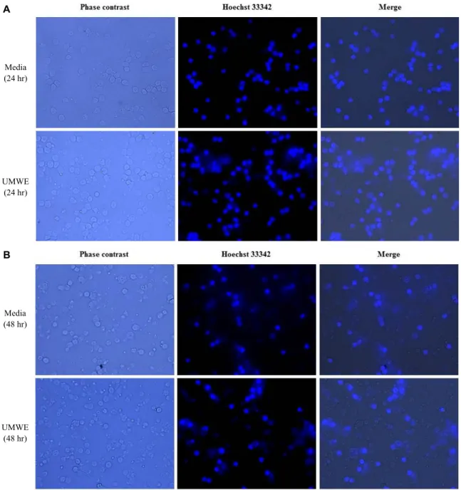
UMWE improved the survival rate of splenocytes in general cell culture condition
Tumor cells or self-renewal cells survive in vitro cell cul- ture condition. Those cells have proliferative ability within in vitro culture condition using general cell culture media.
A
B
Media (24 hr)
UMWE (24 hr)
Media (48 hr)
UMWE (48 hr)
Fig. 1. UMWE augment survival rate in live splenocyte staining by Hoechst 33342. Splenocytes were treated with media (negative control), 100 μg/ml UMWE for 24 hr, 48 hr, 72 hr and 96 hr. The cells were fixed, stained with Hoechst 33342 and observed under phase contrast and fluorescent microscope at 40X. Representative images of cells after treatment for 24 hr, 48 hr, 72 hr and 96 hr. Live cell morphology was confirmed by merge between phase contrast and Hoechst 33342 image.
However, natural cell apoptosis occur in cell culture media in case of general primary cell. In other words, splenocytes isolated from spleen in complete cell culture media also start to die out. In our previous research, we found that UMWE was able to prolong cell life span via Incucyte analysis using general cell culture media. Thus, in order to confirm delayed natural cell apoptosis of UMWE, live cells were stained with Hoechst dye at various time intervals. The viability of sple- nocytes with 100 μg /ml UMWE was observed under fluo-
roscence microscope for 24 hr (Fig. 1A), 48 hr (Fig. 1B), 72 hr (Fig. 1C), and 96 hr (Fig. 1D) by Hoechst 33342 stain.
Splenocyte numbers were gradually decreased by time de- pendent manner in natural cell culture condition and UMWE-treated culture condition. However, the number of splenocytes was enhanced by the UMWE at a dose of 100 μg/ml for 24 hr, 48 hr, 72 hr and 96 hr compared to that of control at each time (Fig. 1). Furthermore, UMWE-treated cells showed a healthy morphology via phase contrast image
C
D
Media (72 hr)
UMWE (72 hr)
Media (96 hr)
UMWE (96 hr)
Fig. 1. Continued.
and a longer-lasting survival rate than the group with no treatment (Fig. 1). This result suggested that active com- pounds related to cell viability were contained in the UMWE and led us to perform further study on prolonged splenocyte.
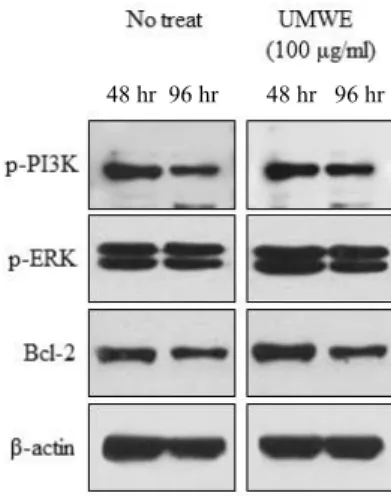
UMWE activated various signalling pathways such as PI3K, ERK, and Bcl-2 to improve the cell survival
Cell survival requires the activity inhibition of natural apoptosis. To confirm the UMWE and signal mechanism for inhibition of time-mediated apoptotic cell death in general cell culture condition, the expression levels of survival and
apoptotic marker proteins were estimated by Western blot analysis. The activation of PI3K is important to improve the cell survival and prevent the apoptosis [1]. As shown in Fig.
3, PI3K phosphorylation was slightly elevated at 48 hr and 96 hr in UMWE-treated cells. The MAPKs are an essential part of signal transduction machinery involved in the gene expression associated with the regulation of cell survival [9].
The ERK signalling pathway, also known as the p42/p44 MAPK pathway, is a major determinant of cell survival.
Thus, to estimate the level of ERK1/2 phosphorylation of UMWE, cells were stimulated with 100 μg/ml UMWE. 100
48 hr 96 hr 48 hr 96 hr
Fig. 2. Effect of UMWE on cell survival signaling pathway.
Splenocytes (2×10⁷ cell/ml) were cultured with 100 μg/
ml UMWE for 42 hr and 96 hr and whole cell lysates were prepared. The phospho- or total protein levels of PI3K, ERK and Bcl-2 were analyzed by immunoblotting analysis. β-actin was used as internal control to normal- ize gel loading.
48 hr 96 hr 48 hr 96 hr
Fig. 3. Effect of UMWE on induction of pro-apoptosis related proteins. Splenocytes (2×10⁷ cell/ml) treated with 100 μg/
ml for 42 hr and 96 hr were introduced to prepare whole cell lysates and total and active form levels of caspase 3and ICAD. β-actin were then detected by immunoblot- ting analysis.
ug /ml UMWE was shown to cause increases in the level of ERK1/2 phosphorylation used at 48 hr and 96 hr (Fig.
2). ERK1/2 is usually associated with pro-survival signalling [21], the up-regulation of the anti-apoptotic protein Bcl-2, and non-transcriptional inhibition of Bcl-xL/Bcl-2-associated death promoter [23]. To determine the mechanism involved, we examined the effect of the UMWE on Bcl-2 anti-apoptotic signaling molecule. Bcl-2 protein level in splenocytes with UMWE compared to the pro-survival protein Bcl-2 under resting splenocytes were elevated at 48 hr and 96 hr (Fig.
2). Thus, UMWE via MAPK-related signalling may regulate the prevention of apoptosis and promotion of cell survival.
UMWE activated various signalling pathways such as caspase 3 and ICAD
Among the numerous proteins and genes involved, mem- bers of the Bcl-2 and caspase play important roles in inhibit- ing apoptosis [8]. In view of the roles of caspase and Bcl-2 in the apoptotic pathway, we tried to examine the expression of the key caspase in UMWE-treated cells. It is well known that in caspase family, caspase-3 plays the central role.
Western blot assay was used to detect protein level of the caspase-3. Splenocytes were cultured with UMWE (100 μg/
ml) for 42 hr and 96 hr, then the cell lysate was used. In natural cell culture condition at 48 hr and 96 hr, the cas- pase-3 level was maintained to the control level. However, UMWE decreased caspase-3 level at 48 hr and 96 hr (Fig.
3). In fact, caspase-3 is responsible for ICAD cleavage, with this nuclease giving rise to the typical apoptotic nuclei [6].
Then, the expression of ICAD protein was further examined by Western blot. ICAD protein increased at 48 hr culturing time (Fig. 3). Together, these observations indicated that UMWE promoted anti-apoptosis in splenocyte involving caspase-3 reduced activation and weak cleavage of its sub- strate ICAD.
UMWE increased the expression level of hema- topoietin cytokine family in cell culture supernatant
Throughout life, lymphocytes are maintained at fairly sta- ble numbers by various homeostatic mechanisms. These mechanisms are mainly governed by cytokines. These sig- nals do not induce proliferation but instead allow the cells to survive for prolonged periods in a quiescent state. Of sev- eral cytokine families, hematopoietin cytokine family plays a role to maintain lymphocyte homeostsis. To evaluate the effects of UMWE on soluble hematopoietin cytokine pro- duction (IL-2 and IL-4) in cell culture media, the cytokine levels were measured by ELISA. UMWE reduced the cyto- kines IL-2 at 96 hr (p<0.0001) but increased IL-4 at 96 hr compared with control in UMWE-treated-cells (Fig. 4). T cell proliferation and survival are regulated by cytokines [5].
IL-2, IL-7, and IL-15 are particularly critical for T cell homeostasis. IL-7 is important for T lymphopoiesis and pro- motes survival of naive, activated, and memory T cells [12], while IL-15 selectively promotes proliferative renewal of ac- tivated/memory CD8+ T cells [20]. The role of IL-2 is more complex. IL-2- develop T lympho-proliferation because IL-2 is required for survival of T regulatory cells that limit con-
Times (hr) Times (hr)
Fig. 4. Effect of UMWE on the production of hematopoietin cytokine. After 42 hr and 96 hr, IL-2 and IL-4 cytokine levels in cell culture media were measured using ELISA assay. 100 μg/ml UMWE increased the expression level of IL-4 cytokines related to B and T cell immunity. The levels of IL-2 were slightly decreased by UMWE. P values are indicated by ***p<0.0001 compared to the control.
Times (hr) Times (hr)
Fig. 5. Effect of UMWE on the production of IL-12 and IFN-γ family. After 42 hr and 96 hr, IL-12 and IFN-γ cytokine levels in cell culture media were measured using ELISA assay. 100 μg/ml UMWE increased the expression level of IL-12 and IFN-γ cytokines. P values are indicated by ***p<0.0001 compared to the control.
ventional T cell proliferation [13]. However, increased levels of IL-2 can also stimulate conventional T cells [4]. All of these cytokines bind to receptors that contain cytokine receptor common γ-chain (γc) 3 [15]. IL-4, another γ-chain-associated cytokine, has a direct activating effect that potently promotes proliferation and survival of T lymphocyte. IL-2 is Th1-type cytokine and IL-4 is Th2-type cytokine. It suggests that UMWE can extend splenocyte life span and facilitate Th2 immune response for humoral immunity.
UMWE increased the expression level of other cy- tokine family IL-12 and IFN-γ in cell culture super- natant
We investigated whether UMWE could produce other cy- tokine family IL-12 and IFN-γ during cell cultivation.
Increased IFN-γ levels were verified in the supernatant of UMWE-treated cells in all periods (48 hr and 96 hr).
However, higher levels of IFN-γ were observed in late peri- od (p<0.0001) (Fig. 5). Increased patterns in the production
of IL-12 cytokine occurred as compared with control after 48 and 96 hr in UMWE-treated-cell cultures (Fig. 5). In the innate immune response, macrophages are activated by IFN- γ made by NK cells and in turn produce the cytokine IL-12 [3]. This binds to IL-12 receptors on the NK cells, including further secretion of IFN-γ and maintenance of macrophage activation. In an adaptive immune response, IL-12 secreted by activated macrophages acts on activated Th1 cells, includ- ing their differentiation into IFN-γ secreting Th1 cells, which interact with the macrophage to strengthen its activation.
CD8 cytotoxic T cells are also responsive to the IL-12 pro- duced by the macrophage and produce more IFN-γ. Thus, our results suggest that UMWE has a sufficient role to cause mutual activation of macrophages and effector lymphocytes in the innate and adaptive immune responses to intracellular stimulation.
In conclusion, our results suggest that the effect of UMWE on splenocytes possess its ability to increase cell longevity against loss of immune cells such as an immune-senescence
condition and UMWE contains potent components that could be used to modulate immune cell homeostasis in the manner required by these therapies.
Acknowledgements
We are grateful to Sang Ho Lee and Seung-Ju Kim for technical assistance. This work was supported by the R&D program of MOTIE/KIAT (N0000697, Establishment of Infra Structure for Anti-aging Industry Support).
References
1. Abu Eid, R., Friedman, K. M., Mkrtichyan, M., Walens, A., King, W., Janik, J. and Khleif, S. N. 2015. Akt1 and -2 in- hibition diminishes terminal differentiation and enhances central memory CD8+ T-cell proliferation and survival.
Oncoimmunology 4, e1005448.
2. Beug, S. T., Cheung, H. H., LaCasse, E. C. and Korneluk, R. G. 2012. Modulation of immune signalling by inhibitors of apoptosis. Trends Immunol. 33, 535-545.
3. Biron, C. A. and Tarrio, M. L. 2015. Immunoregulatory cyto- kine networks: 60 years of learning from murine cytomega- lovirus. Med. Microbiol. Immunol. 204, 345-354.
4. Boyman, O., Kovar, M., Rubinstein, M. P., Surh, C. D. and Sprent, J. 2006. Selective stimulation of T cell subsets with antibody-cytokine immune complexes. Science 311, 1924- 1927.
5. Bradley, L. M., Haynes, L. and Swain, S. L. 2005. IL-7: main- taining T-cell memory and achieving homeostasis. Trends Immunol. 26, 172-176.
6. Errami, Y., Naura, A. S., Kim, H., Ju, J., Suzuki, Y., El- Bahrawy, A. H., Ghonim, M. A., Hemeida, R. A., Mansy, M. S., Zhang, J., Xu, M., Smulson, M. E., Brim, H. and Boulares, A. H. 2013. Apoptotic DNA fragmentation may be a cooperative activity between caspase-activated deoxy- ribonuclease and the poly (ADP-ribose) polymerase-regu- lated DNAS1L3, an endoplasmic reticulum-localized endo- nuclease that translocates to the nucleus during apoptosis.
J. Biol. Chem. 288, 3460-3468.
7. Guarrera, P. M. and Savo, V. 2013. Perceived health proper- ties of wild and cultivated food plants in local and popular traditions of Italy: A review. J. Ethnopharmacol. 146, 659-680.
8. Han, B. J., Li, W., Jiang, G. B., Lai, S. H., Zhang, C., Zeng, C. C. and Liu, Y. J. 2015. Effects of daidzein in regards to cytotoxicity in vitro, apoptosis, reactive oxygen species lev- el, cell cycle arrest and the expression of caspase and Bcl-2 family proteins. Oncol. Rep. 34, 1115-1120.
9. Hommes, D. W., Peppelenbosch, M. P. and Von Deventer, S. J. H. 2003. Mitogen activated protein (MAP) kinase signal transduction pathways and novel anti-inflammatory targets.
Gut 52, 144-151.
10. Kim, K. W., Park, J. S., Kim, K. S., Jin, U. H., Kim, J. K., Suh, S. J. and Kim, C. H. 2008. Inhibition of Ulmus da- vidiana Planch (Ulmaceae) on bone resorption mediated by processing of cathepsin K in cultured mouse osteoclasts.
Phytother. Res. 22, 511-517.
11. Kwon, J. H., Kim, S. B., Park, K. H. and Lee, M. W. 2011.
Antioxidative and anti-inflammatory effects of phenolic compounds from the roots of Ulmus macrocarpa. Arch.
Pharm. Res. 34, 1459-1466.
12. Lee, S. K. and Surh, C. D. 2005. Role of interleukin-7 in bone and T-cell homeostasis. Immunol. Rev. 208, 169-180.
13. Malek, T. R. 2003. The main function of IL-2 is to promote the development of T regulatory cells. J. Leukocyte Biol. 74, 961-965
14. Medeiros, M. C., Frasnelli, S. C., Bastos Ade, S., Orrico, S.
R. and Rossa, C. 2014. Modulation of cell proliferation, sur- vival and gene expression by RAGE and TLR signaling in cells of the innate and adaptive immune response: role of p38 MAPK and NF-KB. J. Appl. Oral Sci. 22, 185-193.
15. Nakajima, H., Shores, E. W., Noguchi, M. and Leonard, W.
J. 1997. The common cytokine receptor gamma chain plays an essential role in regulating lymphoid homeostasis. J. Exp.
Med. 185, 189-195.
16. Ndhlala, A. R., Finnie, J. F. and Van Staden, J. 2011. Plant composition, pharmacological properties and mutagenic evaluation of a commercial Zulu herbal mixture: Imbiza ephuzwato. J. Ethnopharmacol. 133, 663-674.
17. Oh, K. S., Ryu, S. Y., Oh, B. K., Seo, H. W., Kim, Y. S. and Lee, B. H. 2008. Antihypertensive, vasorelaxant, and anti- oxidant effect of root bark of Ulmus macrocarpa. Biol.
Pharm. Bull. 31, 2090-2096.
18. Rajput, S. and Mandal, M. 2012. Antitumor promoting po- tential of selected phytochemicals derived from spices: a review. Eur. J. Cancer Prev. 21, 205-215.
19. Strickland, L. R., Pal, H. C., Elmets, C. A. and Afaq, F. 2015.
Targeting drivers of melanoma with synthetic small mole- cules and phytochemicals. Cancer Lett. 359, 20-35.
20. Surh, C. D. and Sprent, J. 2005. Regulation of mature T cell homeostasis. Semin. Immunol. 17, 183-191.
21. Vauzour, D., Vafeiadou, K., Rice-Evans, C., Williams, R. J.
and Spencer, J. P. 2007. Activation of pro-survival Akt and ERK1/2 signalling pathways underlie the anti-apoptotic ef- fects of flavanones in cortical neurons. J. Neurochem. 103, 1355-1367
22. Yang, W. K., Lee, J. J., Sung, Y. Y., Kim, D. S., Myung, C.
S. and Kim, H. K. 2013. Extract of Ulmus macrocarpa Hance prevents thrombus formation through antiplatelet activity.
Mol. Med. Rep. 8, 726-730.
23. Zhanga, Y. and Liu, D. 2011. Flavonol kaempferol improves chronic hyperglycemia-impaired pancreatic beta-cell via- bility and insulin secretory function. Eur. J. Pharmacol. 670, 325-332.
초록:
Ulmus Macrocarpa
열수 추출물에 의한 비장세포 수명 연장강경화2,5․현숙경3,5․황혜진3,5․김병우5․김철민6․정경태4,5․이종환1,5*
(1동의대학교 생명공학과, 2동의대학교 한의학과, 3동의대학교 식품영양학과, 4동의대학교 임상병리학과, 5동의대학
교 항노화 기술개발 사업단/블루바이오 RIC, 6부산대학교 항노화 기술개발 사업단)
Ulmus macrocarpa 자양강장 및 생리활성 물질로 이용되어 왔다. U, macrocarpa 열수 추출물(UMWE)이 일반적인 세포배양 조건에서 비장세포 수명연장에 미치는 효과에 대한 연구를 진행하였다. 100 μg/ml UMWE를 비장세포 에 처리하여 실험을 진행하였다. 살아있는 세포확인은 Hoechst 33342 염색법과 세포생존관련 인자의 변화는 Western blot으로 확인하였다. 사이토카인 변화는 ELISA로 검증하였다. UMWE는 비장세포에 대하여 향상된 세 포 생존력을 보였다. UMWE를 48시간과 96시간째 처리된 비장세포의 PI3K 및 ERK1/2의 인산화를 증가시켰다.
더욱이, 48시간과 96시간때에 Bcl-2의 발현량도 증가하였다. 반면, UMWE는 48시간과 96시간에 caspase-3의 활성 이 줄어들었다. ICAD 단백질은 48시간에 증가하였다. UMWE는 조혈 및 세포생존력에 영향을 미치는 IL-2 cyto- kine량은 줄었지만 반면, IL-4 hematopoietin cytokine의 양은 증가하였다. UMWE는 48시간과 96시간에 증가된 IFN-γ level을 나타내었고 IL-12의 경우는 증가패턴을 보이는 효과를 발휘하였다. 이러한 결과는 UMWE가 다양한 신호전달 및 사이토카인 조절을 통해 비장세포 수명연장을 할 수 있다는 것으로 사료된다.