- 39 -
R eceived R e v i s e d A ccepted
: December 19, 2017
: October 17, 2018(1차) / October 23, 2018(2차) : October 30, 2018
+Corresponding author: Jeong Kyu Kim, MD,PhD
Department of Otolaryngology-Head and Neck Surgery, Catholic University of Daegu school of Medicine, 33 Duryugongwon-ro 17-gil, Nam-gu, Daegu 42472, Republic of Korea.
Tel: +82-53-650-4071, Fax: +82-53-650-4533 E-mail: [email protected]
대한두경부종양학회지, 제34권 제2호, 2018. pp.39-41 Korean Journal of Head & Neck Oncology, Vol.34, No.2
https://doi.org/10.21593/kjhno/2018.34.2.39 ISSN 1229-5183(Print) / ISSN 2586-2553(Online)
정상 면역 기능을 가진 환자에서 성대에 국한되어 발생한 후두 칸디다증 1예
김보문⋅김정규+⋅손호진⋅길부관
대구가톨릭대학교 의과대학 이비인후과학교실
A case of laryngeal candidiasis confined to vocal cord in an immunocompetent patient
Bo Mun Kim, MD, Jeong Kyu Kim, MD, PhD+, Ho Jin Son, MD, Bu Kwan, Kil, MD
Department of Otolaryngology-Head and Neck Surgery, Catholic University of Daegu school of Medicine, Daegu, Korea
= Abstract =
Primary laryngeal candidiasis is rare in immunocompetent patients and is prone to confusion with early glottic carcinoma or leukemia. We experienced a case of 74-year-old man who has 3- month history of hoarseness. The pathologic diagnosis was laryngeal candidiasis. He was treated with antifungal agents for 4 weeks after vocal cord stripping under general anesthesia. After treatment, the patient had no candidiasis or discomfort with his voice. We report this case with a review of literature.
Key W ords : Larynx, Vocal cords, Candidiasis, Leukoplakia
서 론
진균 감염은 흔히 면역 저하자, 장기간의 흡입형 스테 로이드 및 광범위 항생제 사용자, 만성 질환자에서 호흡 기 및 인두 식도 부위에서 발생하는 반면 정상 면역 기능 을 가진 성인에서 후두 부위에 국한된 진균 감염은 흔하 지 않다.1) 칸디다는 정상적으로 숙주의 피부, 구강 및 질 등에 상재하며, 숙주의 면역이 떨어지는 경우에 통제 를 벗어나 증식하여 통증과 염증 등을 유발한다.2) 후두 에 국한된 칸디다증은 매우 드물며 폐, 인두 혹은 식도의 칸디다 균이 면역 저하 상태에서 객담을 통해 확산되거 나 흡입형 스테로이드 치료 후에 발생한다고 알려져 있
다.3,4)게다가 성문부에 국한되어 발생한 칸디다증은 백 반증, 조기 성문암, 인후두 역류 및 육아종성 질환과 단순 후두경상으로는 감별이 어려워 불필요한 진단 및 수술적 치료가 시행될 수 있으며 항진균제 치료가 지연되는 경 우가 많다. 이에 본 저자들은 정상 면역 환자에서 백반증 및 조기 성문암과 유사한 성대에 국한된 후두 칸디다증 을 경험하였기에 문헌고찰과 함께 보고하고자 한다.
증 례
74세 남자 환자가 내원 3개월 전부터 발생한 쉰 목소리 를 주소로 본원 이비인후과에 내원하였다. 연하통, 호흡 곤란 등의 증상은 없었다. 환자는 30갑년의 흡연력이 있 었으나, 20년 전부터 흡연하지 않았으며 음주력은 없었 다. 과거력상 다른 기저 질환이나 면역 이상 소견은 보이 지 않았다. 또한 환자의 혈액 및 흉부 엑스선 검사에서도 특이 소견을 보이지 않았다. 경성 후두내시경 검사에서 성대의 움직임은 정상이었으나 양측 진성대 및 전교련에 국한된 비교적 경계가 명확한 백반증 (leukoplakia) 소견 을 보였으며 경부종괴는 만져지지 않았다(Fig. 1). 환자는
- 40 -
Fig. 4. Postoperative 2 month endoscopic finding of larynx;
Both true vocal cord and anterior commissure are clear with- out evidence of disease.
Fig. 1. Endoscopic finding of larynx; whitish and irregular sur- face lesion with anterior commissure extension in both true vocal cord.
Fig. 2. Postoperative 1 week endoscopic finding of larynx;
Both true vocal cord lesion removed except anterior commissure. Whitish lesion presents in anterior commissure.
Granulation tissue presents in right true vocal cord.
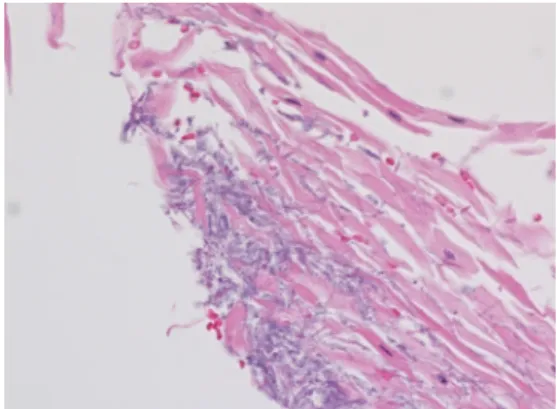
Fig. 3. Histopathological examination of laryngeal mucosal surface; Fungal septate hyphae visible in a hyperplastic en- dolaryngeal epithelium (H&E stain, X400)
전신 마취하에서 후두내시경 조직검사를 시행하였고 후 두 종물은 양측 성대 및 전교련에 있었고 성문하 및 성문 주위 침범은 보이지 않았다. 조기 성문암 및 백반증 의심 하에 성대 박리술 (vocal cord stripping)을 시행 하였으며 성대 막 (vocal cord web)을 예방하기 위하여 전교련의 일부 병변은 남겨두었다(Fig. 2). 병리조직검사에서 후두 점막의 과각화 (hyperkeratosis) 및 이상각화 (parakeratosis)
소견을 보였으며 악성 세포는 보이지 않았으며 많은 유격 이 있는 (septated hyphae) 효모 양의 균사들이 관찰되어 후두 칸디다증으로 진단 받았다(Fig. 3). 환자는 4 주간의 nystatin suspension 2,000,000 IU (20 ml)/일 을 투여 받았고 2 개월 후 시행한 경성 후두내시경 검사에서 재발의 소견 보이지 않고 추적관찰 중이다(Fig. 4).
고 찰
후두 진균 감염은 면역 기능 저하자 또는 광범위 항생 제 사용, 흡입용 스테로이드, 방사선 치료, 흡연, 외상 및 위식도역류로 인하여 점막 방어 작용이 약화된 사람에게 주로 발생하여 진균이 과도 증식하게 되어 후두가 영향을 받게 되나 면역 기능이 정상인 환자에게도 드물지만 발생 이 가능하다.1)본 증례를 고려하였을 때도 위와 같은 병 력이 없는 환자이나 후두 칸디다증의 원인이 기회감염이 주원인이라는 점과 환자의 나이가 고령임을 고려하였을 때 면역 기능이 정상이라 하더라도 후두 진균 감염이 가 능할 것으로 여겨진다.
진균 감염의 원인균은 칸디다 및 아스페르길루스가 있 다.5)후두 칸디다증의 증상은 병의 경중에 따라 연하곤 란, 연하통, 애성, 흡기성 천명 및 호흡곤란으로 다양하게 나타날 수 있다.6)후두 칸디다증은 흔하게 관찰되는 기 회 감염 균주인 Candida albicans에 의한 감염으로 후두에 만 생길 수도 있지만 주로 호흡기, 인두, 식도의 파종성 질환으로 나타난다.
후두 칸디다증은 내시경에서 일반적으로 성대의 부종, 홍반, 궤양, 백색판 및 위막 형성 등으로 나타날 수 있으 며 일부에서는 돌출성 병변으로 나타나 후두 유두종이나 악성 종양과 감별해야 될 필요가 있다.7)본 증례의 경우 에는 양측 성대 및 전교련에 백색판의 소견을 보였고
- 41 - 백반증이나 조기 성문암과의 감별이 필요하였다. 병리 조직검사에서 후두 칸디다증은 과다상피증식 및 비특이 적 염증 소견을 보이며 이는 구강 칸디다증에서 나타나 는 소견과 같다. 확진을 위해서는 헤마톡실린-에오신 염 색에서 칸디다 속에 속하는 분할된 균사 및 포자체를 발견하여야 하며 PAS (periodic acid schiff) 염색, 고모리 메테나민 은 염색에서 더 쉽게 발견할 수 있다.8)병변을 확진하기 위해 조직 검사를 시행하는 것은 논란의 여지 가 있다. 그러나 악성 종양이 의심되거나 적절한 치료에 도 반응이 없을 경우에 조직 검사를 시행하여야 하며 덜 침습적인 방법으로는 기관지 브러시 세포생검이나 배양 을 시행할 수 있다. 그러나 적절한 항진균제 치료에 반응 이 없거나 악성 종양이 강하게 의심될 경우에는 조직 검 사가 필수적이다.9)구강 칸디다증 연구에 따르면 칸디다 감염은 상피 세포 증식, 이형성 및 악성 변화를 유발하였 는데 이는 진균의 니트로사민이 발암유전자를 활성화시 켜 구강 상피세포를 악성화 시킨다고 하였다.10)후두 칸 디다증은 주로 보조적으로 항진균제 치료를 시행하게 되는데 항진균제인 Nystatin 구강정제 (Mycosatin pastilles, 200,000~400,000units, 1일 4회), fluconazole 경구투여 (200mg 1일 1회, 심한 경우 400mg 1일 1회) 등을 병의 범위 및 경중에 따라 10-30 일간 시행할 수 있으며 전신적인 투약 이 필요한 경우 amphotericin 정맥 투여 또한 가능하다.4) 병의 범위 및 경중에 따라 이와 같이 후두 칸디다증은 항진균제 및 보조적 치료가 주된 치료이므로 후두 조직검 사를 시행할 경우 후두 칸디다증이 의심되면 본 증례와 같이 성대 막 등 술 후 목소리와 관련된 합병증을 예방하
기 위하여 성문 전교련부를 수술 범위에서 제외하는 등 최대한 음성을 고려한 수술을 시행하면 술 후 역효과를 예방할 수 있으리라 생각한다.
References
1) Nair AB, Chaturvedi J, Venkatasubbareddy MB. A case of iso- lated laryngeal candidiasis mimicking laryngeal carcinoma in an immunocompetent individual. Malays J Med Sci. 2011;18:75-78.
2) Sulica L. Laryngeal thrush. Ann Otol Rhinol Laryngol. 2005;
114:369.
3) Kameswaran M, Anand Kumar RS, Natarajan K. Laryngeal thrush: merf experience. Indian J Otolaryngol Head Neck Surg.
2006;58:329-331.
4) Nunes FP, Bishop T, Prasad ML, Madison JM, Kim DY.
Laryngeal candidiasis mimicking malignancy. Laryngoscope.
2008;118:1957-1959.
5) Vrabec DP. Fungal infections of the larynx. Otolaryngol Clin North Am. 1993;26(6):1091-1114.
6) Ganesan S, Harar RP, Dawkins RS. Invasive laryngeal candi- diasis: a cause of stridor in the previously irradiated patient. J Laryngol Otol. 1998;112:575–578.
7) DelGaudio JM. Steroid inhaler laryngitis: dysphonia caused by inhaled fluticasone therapy. Arch Otolaryngol Head Neck Surg.
2002;128:677-681.
8) Pabuççuog U, Tuncer C, Sengiz S. Histo pathology of candidal hyperplastic lesions of the larynx. Pathol Res Pract. 2002;198:
675-678.
9) Neuenschwander MC, Cooney A, Spiegel JR, Lyons KM, Sataloff RT. Laryngeal candidiasis. Ear Nose Throat J. 2001;80(3):138-139.
10) Field EA, Field JK, Martin MV. Does Candida have a role in oral epithelial neoplasia?. J Med Vet Mycol. 1989;27:277-294.