ISSN 1225-6552, eISSN 2287-7630 https://doi.org/10.7853/kjvs.2020.43.4.251
< Case Report >
Veterinary Service
Available online at http://kjves.org
*Corresponding author: Jae-Hoon Kim, E-mail. [email protected] ORCID https://orcid.org/0000-0002-4410-9126
Co-infection of Toxoplasma gondii and porcine reproductive and respiratory syndrome virus in suckling piglets in Jeju, Korea
Young-Min Choi
1, Hyoung-Seok Yang
2, Jae-Hoon Kim
3*
1
Jeju Animal Pharm Hospital, Jeju 63069, Korea
2
Jeju Self-Governing Provincial Veterinary Research Institute, Jeju 63344, Korea
3
College of Veterinary Medicine and Veterinary Medical Research Institute, Jeju National University, Jeju 63243, Korea
(Received 16 September 2020; revised 15 December 2020; accepted 15 December 2020)
Abstract
Two suckling piglets, 4 days and 10 days of age, showed lethargy and dyspnea after birth and mortality had been increased after incoming gilts from breeding farm. At necropsy, the lungs showed diffuse fail to collapse with rubbery consistency, edematous dilatation of interlobular septa, and lobular consolida- tion with purple red color. Heart was diffuse pale in color and had several irregular linear-shaped mac- ules or patches. Histopathologically, diffuse interstitial pneumonia with the proliferation of type II pneu- mocytes was present in the lungs of 2 piglets. Alveolar lumens contained necrotic cellular debris derived from neutrophils and macrophages. Multifocal hemorrhage and necrotizing pneumonia with protozoan tachyzoites were observed in the lungs. Severe multifocal to confluent necrotic myocarditis, necrotic en- cephalitis, and necrotic adrenalitis with intralesional protozoan tachyzoites were observed in piglets.
According to immunohistochemical analysis (IHC), Toxoplasma (T.) gondii tachyzoites antigens were confirmed in lung, heart, brain, and adrenal gland. And porcine reproductive and respiratory syndrome virus (PRRSV) antigens were also detected in the cytoplasm of macrophages in lungs using IHC. Based on the gross, histopathologic and immunohistochemical features, two suckling piglets were diagnosed as co-infection of T. gondii and PRRSV.
Key words : Congenital infection, Interstitial pneumonia, Necrotic encephalitis, PRRSV, Toxoplasma gondii
INTRODUCTION
Toxoplasmosis is a parasitic disease caused by in- fection with Toxoplasma (T.) gondii (Dubey, 1986, 1998).
This disease can occur in all mammals, rodents, birds, and humans. Cats are the only definitive host which can spread oocysts through their feces. Humans can be in- fected by ingesting water, vegetables and meats conta- minated with T. gondii oocysts. In many countries, the pork is considered a source of T. gondii infection for humans (Lindsay et al, 2012). Therefore, it is important to rapidly diagnosis and control toxoplasmosis in pigs.
In most case, infections of T. gondii do not show clin-
ical sign and may develop the latent infection for years (Dubey, 1986). Infection occurs in sow during preg- nancy by ingesting oocysts or tachyzoites, which can be transmitted to fetus through placenta. Although abortions due to T. gondii are uncommon, postnatal pigs may be born premature or die soon after birth (Lindsay et al, 2012).
Porcine reproductive respiratory syndrome (PRRS) is commonly considered of major wasting disease in pigs and so it is affecting economically severe damage to the global pig industry. According to recent study, annual losses in the United States of America were reported at
$560.32 million and ₩100 billion was estimated in Korea
(Neumann et al, 2005; Holtkamp et al, 2013). The sow
infected with PRRS virus (PRRSV) show clinical sign
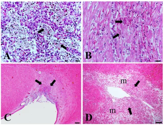
Fig. 1. Histopathologic findings in piglets. (A) The alveolar wall is lined by type II pneumocytes and the lumen contained with necrotic macrophages and cell debris (ar- rows). Bar=20 µm. (B) Note my- ocardial muscular degeneration with intra-lesional tachyzoites (arrows) of T. gondii. Bar=20 µm. (C) Note focal necrotic encephalitis (arrows) around para-ventricular area of ce- rebrum. Bar=200 µm. (D) Note ne- crotic adrenalitis (arrows) in adre- nal medulla (m). Bar=100 µm. H&E.
such as abortion, stillborn, mummification and weak born piglets and also respiratory sign can distress to pigs of all ages. Porcine abortion associated with T.
gondii and toxoplasmosis in piglets were previously re- ported in Korea (Roh et al, 1997; Kim et al, 2009).
This study reports congenital co-infection of T. gondii and PRRSV in suckling piglets in Jeju, Korea.
CASE
At a pig farm with a scale of 600 pigs in Jeju, the sudden death of suckling piglets had been increased af- ter incoming 40 ∼50 gilts from breeding farms. The dead piglets were delivered from these newly introduced gilts, and showed clinical signs such as dyspnea, anorexia and lethargy before death. Two suckling piglets with 4 days and 10 days of age were referred to the Pathology Laboratory at the College of Veterinary Medicine in Jeju National University. After the necropsy, collected tissue was fixed in 10% buffered formalin, embedded in paraffin, sectioned at 3 µm and stained with hematox- ylin & eosin (H&E).
At necropsy, piglets were mild emaciated. The lungs
showed diffuse fail to collapse with rubbery consistency, edematous dilatation of interlobular septa, and lobular consolidation with purple red color. Heart was diffuse pale in color and had several irregular linear-shaped macules or patches. Histopathologically, diffuse inter- stitial pneumonia was present in the lungs of 2 piglets.
Most alveolar walls were thickened due to the pro- liferation of type II pneumocytes, and alveolar lumens contained necrotic cellular debris derived from neutro- phils and macrophages (Fig. 1A). As mentioned in pre- vious literature (Dubey, 1986), multifocal necrotizing in- flammations suspected protozoal infection were observed in various internal organs. Multifocal hemorrhage and necrotizing pneumonia with protozoan tachyzoites char- acterized by round, ovoid or crescent shape were ob- served in the lungs. Severe multifocal to confluent ne- crotic myocarditis with intralesional protozoan tachy- zoites were observed throughout the heart (Fig. 1B). In the brain, necrotic encephalitis with intralesional proto- zoan tachyzoites was present at the adjacent area of lat- eral ventricle (Fig. 1C). Focal necrotic lesion with many tachyzoites also noted in the medulla of adrenal gland (Fig. 1D).
To confirm the protozoal tachyzoites, replicate sec-
Fig. 2. Immunohistochemistry for T. gondii and PRRSV in the lungs of piglets. (A) Positive re- actions for tachyzoites of T. gondii (arrow) in the lungs. (B) Antigens of PRRSV (arrows) were detected in the lungs. IHC, Bar=20 µm.
Fig. 3. Immunohistochemistry for T. gondii in piglets. (A) The heart. Note brown tachyzoites of T. gondii (arrows) in cardiac muscle. (B) Note dif- fuse positive reactions for tachyzoites of T. gondii (arrows) in necrotic foci of brain. (C) The adrenal gland. Note brown tachyzoites of T. gondii (arrows) in the medulla of adrenal gland. Bar=20 µm. IHC.