ISSN 1225-6552, eISSN 2287-7630 http://dx.doi.org/10.7853/kjvs.2014.37.1.1
< Original Article >
Veterinary Service
Available online at http://kjves.org
*Corresponding author: Won-Il Kim, Tel. +82-63-270-3981, Fax. +82-63-270-3780, E-mail. [email protected]
Different immunological features of two genetically distinct type 2 porcine reproductive and respiratory syndrome (PRRS) viruses
Nadeem Shabir, Amina Khatun, Won-Il Kim*
College of Veterinary Medicine, Chonbuk National University, Jeonju 561-756, Korea (Received 24 January 2014; revised 3 March 2014; accepted 10 March 2014)
Abstract
Although it has been generally accepted that porcine reproductive and respiratory syndrome virus (PRRSV) induces weak and delayed protective immunity after infection, it is unclear that the same im- munological features can be applicable to all PRRS viruses because huge genetic variation exists even among the same genotypes of PRRSV (Type 1 and 2). In the current study, two genetically distinct type 2 PRRSV strains (VR-2332 and JA142) which showed approximately 90% nucleotide homology based on ORF5 sequences were characterized by both in vitro and in vivo assessments to determine the immunological features of the viruses. For in vitro assessment, porcine alveolar macrophages (PAM) were infected with the viruses at 10−3 multiplicity of infection (MOI) and then supernatants and cells were collected separately at 36 hrs post infection to determine the relative expression levels of IL-1α, IL-12, TNF-α and INF-α/β by quantitative RT-PCR. In addition, five PRRSV-free pigs were inoculated with either of JA142 or VR2332 for in vivo assessment. Serum samples were collected every week until 6 weeks post challenge. The serum samples were analyzed for the levels of viremia, PRRSV nucleocap- sid-specific antibody and virus neutralizing antibody. Based on those assessments, the two viruses showed different patterns of cytokine expression in PAM and immune responses in pigs after infection.
These results indicate that genetically distinct PRRSV strains have different immunological features, which might be criteria for virus classification and selection of candidate virus strains for vaccine devel- opment in the future.
Key words : PRRSV, Porcine alveolar macrophages, Cytokine expression, Serum virus neutralizing antibody, ELISA
INTRODUCTION
Porcine reproductive and respiratory syndrome (PRRS) is characterized by reproductive disorder in breeding an- imals and respiratory distress in all ages of pigs (Bilodeau et al, 1991; Goyal, 1993). Its emergence was first reported in the United States in 1987 (Keffaber, 1989) and subsequently reported in Europe in 1990 (Paton et al, 1991). Isolation of PRRS virus (PRRSV), the causal agent of the disease, was first reported in Europe in 1991 (Wensvoort et al, 1991) and later in
North America in 1992 (Collins et al, 1992). Since then, the virus has been identified in most of pig-producing regions worldwide and has caused significant economic losses in swine industries (Dea et al, 1992; Paton et al, 1992; Murakami et al, 1994). PRRSV is a member of the family Arteriviridae in the order Nidovirales (Cavanagh, 1997) and an enveloped virion containing a single-stranded positive-sense RNA genome. PRRSV has been classified into two genetically and antigenically distinct groups, European (Type 1) and North American (Type 2) genotype (Meng et al, 1995; Murtaugh et al, 1995). Remarkable genetic and antigenic variations have been observed between the genotypes and within the
same genotype (Yang et al, 1999; Cheon and Chae, 2000; Key et al, 2001; Forsberg et al, 2002; Pesch et al, 2005; Fang et al, 2007; Zhou et al, 2009). Such genetic and antigenic variation has hampered effective pre- vention and control of PRRS.
In general, PRRSV induces anti-inflammatory cyto- kines (Suradhat et al, 2003; Charerntantanakul et al, 2006; Gimeno et al, 2011; Dwivedi et al, 2012) and weak proinflammatory cytokines (López -Fuertes et al, 2000; Thanawongnuwech et al, 2004). Thus, it stim- ulates delayed and weak protective immunity [i.e., se- rum virus neutralizing (SVN) antibody and interferon (IFN)-γ secreting T cells] which seldom appears until 3 to 4 weeks after infection (Meier et al, 2003; Faaberg et al, 2006; Vanhee et al, 2009). Passively acquired SVN antibody alone was proven to prevent viremia and re- productive failure in pigs challenged with virulent PRRSV strains (Osorio et al, 2002), suggesting the crit- ical role of SVN antibody in the control of virus infection. Nonetheless, cell-mediated immunity (CMI) appears to be essential for the clearance of PRRSV since PRRSV has been detected in lungs and lymph no- des despite the presence of SVN antibodies in serum or bronchoalveloar lavage fluid (Wills et al, 1997;
Labarque et al, 2000; Bierk et al, 2001). The mecha- nisms for the weak induction of protective immunity by PRRSV have been explored in previous studies. The structural and non-structural proteins (nsp) are believed to modulate host immune responses (Wang et al, 2009).
It has been recently shown that nsp1 of PRRSV modu- lates the expression of type I interferon (Beura et al, 2010; Beura et al, 2012; Chen et al, 2010; Wang et al, 2013; Kim et al, 2010) and TNF-α (Subramaniam et al, 2012), which leads to persistent infection (Lamontagne et al, 2003). In addition, PRRSV infects and destroys porcine alveolar macrophage (PAM), which is an active producer of cytokines and leukotrienes and has im- portant roles in inducing pro- and anti-inflammatory re- sponses in the alveolus (Gordon and Read, 2002) and innate immune response against respiratory infections (Pribul et al, 2008).
As described above, the weak and delayed protective immunity after PRRSV infection has been reported by many previous studies, it is still unclear that the same
immunological features can be applicable to all PRRS viruses because huge genetic variation exists among PRRSV strains and only a few PRRSV strains have been well characterized for their immune responses after infection. In the current study, two genetically distinct type 2 PRRS viruses (VR-2332 and JA142) which showed approximately 90% nucleotide homology based on ORF5 sequences were characterized by both in vitro and in vivo assessments to determine the immunological features of the viruses.
MATERIALS AND METHODS
Cells and viruses
PAM cells were harvested from ten, 6-week old, PRRS negative pigs as described. Pigs were euthanized and the lungs along with trachea and bronchus were aseptically collected from pigs. The lungs were lavaged thrice with 0.01 M phosphate buffer saline (PBS, pH 7.4) and the harvested wash fluid was centrifuged for 10 minutes at 1,000×g. The resulting pellet was washed three times with PBS and resuspended in 5 mL of PBS.
To evaluate cell viability, the cells were diluted 100 times in PBS and mixed with 0.4% trypan blue in 1:1 ratio and counted by the CountessTM Automated Cell Counter (Invitrogen, Carlsbad, CA, USA). After count- ing, cells adjusted to 5×10⁷/mL were dispensed in cryo- vials and saved in −80oC until used. For culturing PAM, the cells were briefly thawed in a waterbath at 37oC with gentle stirring and diluted 10 times in RPMI-1640 (Sigma Aldrich, St. Louis, USA) containing 10% FBS, 1% L-glutamine and 1% antibiotics (10,000 units/mL of penicillin, 10,000 μg/mL of streptomycin, and 25 μg/mL of Amphotericin B) (Gibco, Carlsbad, CA, USA) so as to maintain a final cell number of 5×10⁶/mL. The cells were seeded in each well of a 6-well plate (BD Falcon, Franklin Lakes, NJ, USA) and cultured overnight. Two North American (Type 2) PRRSV strains, JA142 and VR2332, were selected for used in this study and propagated in MARC-145 cells.
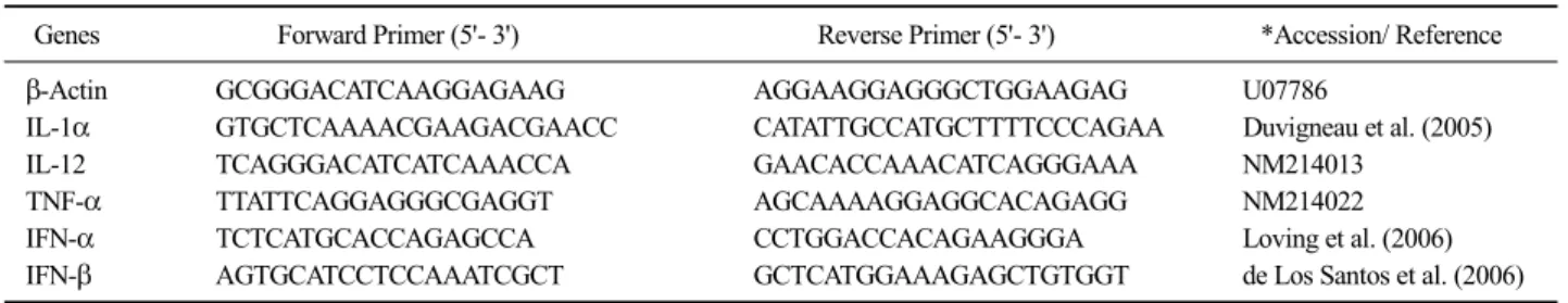
Table 1. The primer information for real-time RT-PCR used to measure the mRNA expression levels of various cytokines
Genes Forward Primer (5'- 3') Reverse Primer (5'- 3') *Accession/ Reference
β-Actin GCGGGACATCAAGGAGAAG AGGAAGGAGGGCTGGAAGAG U07786
IL-1α GTGCTCAAAACGAAGACGAACC CATATTGCCATGCTTTTCCCAGAA Duvigneau et al. (2005)
IL-12 TCAGGGACATCATCAAACCA GAACACCAAACATCAGGGAAA NM214013
TNF-α TTATTCAGGAGGGCGAGGT AGCAAAAGGAGGCACAGAGG NM214022
IFN-α TCTCATGCACCAGAGCCA CCTGGACCACAGAAGGGA Loving et al. (2006)
IFN-β AGTGCATCCTCCAAATCGCT GCTCATGGAAAGAGCTGTGGT de Los Santos et al. (2006)
Evaluation of transcriptional activation of cytokines in PAM cells
PAM cells were infected with 10−3 MOI of JA142 or VR2332. Cells were harvested at 36 hrs after infection and cellular RNA was extracted using GeneAllⓇ Hybrid-RTM kit (GeneAll Biotechnology, Seoul, Korea) following the manufacturer’s instructions. RNA was re- verse transcribed into cDNA using high-capacity cDNA reverse transcription kits (Applied Biosystems, Foster city, CA) and real-time polymerase chain reaction (PCR) was then performed on a 7500 Fast Real-time PCR sys- tem (Applied Biosystems) using various cytokine specif- ic primers by following the manufacturer’s instructions.
Primer sequences used in this study are shown in Table 1. Ten μL of 2× Power SYBGR (Applied Biosystems), 2 μL of cDNA and 1 μL of each forward primer (10 pm/μL) and reverse primer (10 pm/μL) were used for PCR amplification. All the samples were tested in dupli- cate and the cycling conditions were as follows: (a) Holding for 10 min at 95oC; (b) 40 cycles of 15 s at 95oC and 1 min at 60oC and (c) Melt curve stage for 15 s at 95oC, 1 min at 60oC, 15 s at 95oC and 15 s at 60oC. Relative quantities of cytokine mRNA in infected and non-infected cells were normalized to β-actin mRNA and the amounts were determined by the 2-ΔΔCt method.
Quantitative PCR for virus titration
The virus titers in the supernatant and PAM cell ly- sates were measured by a real-time reverse transcription- polymerase chain reaction (RT-PCR) using TaqManⓇ chemistry. The primer and probe sequences designed are:
Forward primer: 5’-TGTCAGATTCAGGGAGRATAA- GTTAC-3’;
Probe: 5’-FAM-TTTTGCACCACMGCCAGCCC-BHQ-3’;
Reverse primer: 5’-ATCARGCGCACAGTRTGATGC-3’
Viral RNA was extracted from supernatant or cells using GeneAllⓇ viral RNA extraction kit (GeneAll Biotechnology) or GeneAllⓇ Hybrid-RTM kit (GeneAll Biotechnology), respectively, following the manufac- turer’s instructions. Viral quantification was carried out by real-time RT-PCR using AgPath-IDTM One-Step RT-PCR Kit (Ambion, Austin, TX, USA) in a 25 μL reaction volume using 5 μL of extracted template. The final concentration of each primer or probe was 0.8 or 1 pmol, respectively. The PCR amplification was per- formed as follows: (a) reverse transcription for 10 min at 45oC; (b) a 10 min activation step at 95oC; and (c) 40 cycles of 15 s at 95oC and 45 s at 60oC. Samples with a threshold cycle (Ct) of 35 cycles or less were considered positive. A set of serially diluted virus sam- ples with known titers was used to calculate the amount of PRRSV in each sample by converting Ct value to vi- rus titer (TCID50/mL).
Animal study
A total of 15, three-week-old PRRSV-free pigs were randomly divided into 3 groups and housed separately.
After acclimated for 3 days, 5 pigs in each group were challenged with VR2332, JA142 (2 mL of 103 TCID50/ mL per pig) or sham inoculum (RPMI-1640) and housed for 6 weeks. All pigs were bled at 0, 7, 14, 21, 28, 35 and 42 days post challenge (dpc). Sera were sep- arated immediately after bleeding and stored in a −80oC freezer until used.
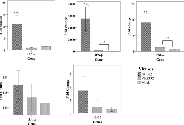
Fig. 1. Transcriptional expression of IL-1α, IL-12, TNF-α and INF-α/β in naïve PAM infected with either JA142 or VR2332. The relative ex- pression was determined by quantitative RT-PCR. The values represent the relative expression of each cytokine normalized with β-actin. Asterisks indicate the significant differences in the cytokine expression induced by each virus as compared with mock infection (* indicates P≤0.05, ** in- dicates P≤0.01).
ELISA
All collected sera were tested by a commercial ELISA kit (Anigen PRRS Ab ELISA 4.0, Bionote, Hwasung, Korea) as per the manufacturer’s instructions to determine IgG antibody specific for nucleocapsid pro- tein of PRRSV (both type 1 and type 2).
Virus neutralization assay
Fluorescent focus neutralization (FFN) assay was per- formed to assess the titer of SVN antibodies in the sera as previously described (Kim and Yoon, 2008). The se- rum samples were first heat inactivated at 56oC for 45 min and then 2-fold serially diluted with RPMI-1640 medium growth medium. One hundred μL of each di- luted serum were mixed with an equal volume of
PRRSV (VR2332 or JA142) at 103 fluorescent focus forming unit per mL (FFU/mL). The mixtures were in- cubated at 37oC for 1 hr in a humidified atmosphere with 5% CO2 and then transferred into MARC-145 cell monolayers prepared in 96-well plates (Corning Inc., Corning, NY, USA) and incubated for another 1 hr at 37oC in a humidified atmosphere with 5% CO2 supply.
After the removal of the inoculum, cells were re- plenished with 200 μL of fresh RPMI-1640 growth me- dium per well and further incubated at 37oC for 20 hr.
Afterwards, cells were fixed with ice cold 80% acetone aqueous solution for 5 min. The acetone was removed and cells were air dried for 30 min. Then, cells were re- acted with 1:10000 diluted PRRSV nucleocapsid specif- ic monoclonal antibody SDOW-17 (Rural Technologies, Brookings, SD, USA) and stained with 1:250 diluted fluorescein isothiocyanate (FITC) labeled goat an-
Fig. 2. Virus titers of PAM cell lysates (A) and supernatants (B) at 36 hrs after infection with either JA142 or VR2332. The virus titers were quantified by real-time RT- PCR. Asterisks indicate the sig- nificant difference in virus titers as compared with VR2332 (* indi- cates P≤0.05, ** indicates P≤0.01).
Fig. 3. The levels of viremia in sera collected from pigs challenged with JA142, VR2332 or sham inoculum. Three-week-old pigs were challenged with VR2332, JA142, or sham inoculums at 0 dpc and bled to measure viremia levels at indicated time points.
ti-mouse IgG (H+L) (KPL, Gaithersburg, MD, USA).
Before observation under an invert fluorescent micro- scope, plates were washed three times with PBS and then number of virus-specific fluorescent foci in each well was counted. SVN antibody titer was expressed as reciprocal of the highest dilution in which 90% or high- er reduction in the number of FFU was observed.
Data Analysis
Statistical analysis was done by SPSS Advanced Statistics 17.0 (SPSS Inc., Chicago, USA). The cytokine induction by each virus in PAM was compared with that by VR2332 using Man Whitney U test. The repeated measurements of viremia and PRRSV nucleocapsid-spe- cific antibody levels in the challenged pigs were analyzed using repeated measures ANOVA to determine the overall difference and pairwise comparisons between groups.
RESULTS
Different PRRSV strains displayed various levels of transcriptional activation of cytokines PAM cells were infected with JA142 or VR2332 and the transcriptional activation of cytokines, viz. TNF-α, IFN-α, IFN-β was measured by real-time PCR. JA142 induced a significantly higher mRNA expression of IFN-α, IFN-β and TNF-α (Fig. 1) compared with mock or VR2332 though the mRNA accumulation of TNF-α and IFN-β induced by VR2332 was also significantly higher compared with mock. Other cytokines, viz. IL-1α and IL-12 did not display any significant differences in
their levels induced by either of viruses.
Viral titers in the supernatants and PAM cell lysates
Viral titers detected from PAM cell lysates (Fig. 2) did not show any significant differences between JA142 and VR2332 infected cells, however, the viral titers in the supernatants (Fig. 2) were significantly higher in JA142-infected cells when compared with VR2332-in- fected cells.
Viremia levels after challenge with JA142 or VR2332
Viremia was detected in all of the sera collected from the challenged pigs at 7 dpc (Fig. 3) when most viruses reached their highest titer. The mean levels of viremia in the pigs challenged with VR2332 or JA142 were 4.6 or 5.2 log10 TCID50/mL, respectively. At 42 dpc, the last
Fig. 4. The levels of PRRSV nucleocapsid-specific antibody in sera collected from pigs challenged with JA142, VR2332 or sham inoculum. The cut-off for positive is 0.4 S/P ratios.
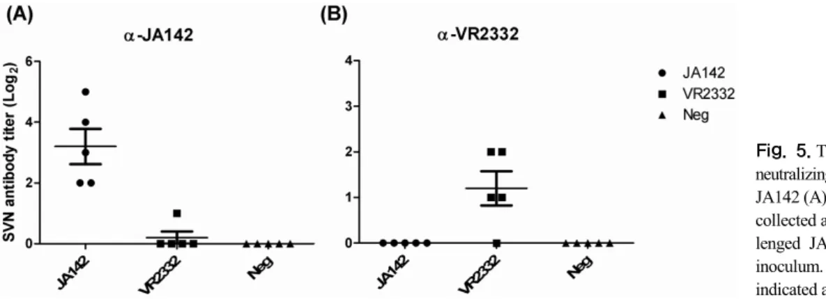
Fig. 5. The levels of serum virus neutralizing (SVN) antibody against JA142 (A) and VR2332 (B) in sera collected at 42 dpc from pigs chal- lenged JA142, VR2332 or sham inoculum. Mean of each group was indicated as a horizontal bar.
bleeding time point, viremia was not detected in most of the challenged pigs. There were no significant differ- ences in viremia levels between the groups challenged with JA142 or VR2332.
PRRSV nucleocapsid-specific antibody re- sponses
Pigs seroconverted to PRRSV at 2 weeks after the exposure to PRRSV and remained seropositive until the end of the study (Fig. 4). The negative control group re- mained seronegative until the end of the study. No sig- nificant difference was observed between the groups challenged with JA142 or VR2332.
The induction of SVN antibodies after challenge
Levels of SVN antibodies in collected serum samples
were determined by FFN. At 42 dpc (the last bleeding time point), only homologous SVN antibody was in- duced and no significant cross-neutralizing antibody was induced either by VR2332 or JA142 (Fig. 5). However, JA142 induces significantly higher levels of homologous SVN antibodies (1:4 to 1:32) as compared with VR2332 (<1:4).
DISCUSSION
The aberrant immune response by PRRSV infection has been one of the major hurdles in understanding the immunobiology of PRRS. Although it is known that PRRSV primarily replicates in PAM (Benfield et al, 1992; Wensvoort et al, 1992), local and systemic im- mune responses mediated by PAM infected with various PRRSV strains remains largely unexplored. In the pres- ent study, the immunological features of two genetically distinct type 2 PRRSV strains were characterized by in vitro and in vivo assessments. Even though the two strains showed approximately 90% nucleotide homology based on ORF5 sequences, the transcriptional activation of various cytokines induced by these two PRRSV strains varied significantly; VR2332 induced sig- nificantly lower mRNA expression of INF-α/β and TNF-α than JA142 in PAM though it induced still high- er levels of IFN-β and TNF-α expression compared with mock controls. In special, VR2332 failed to induce a significant expression of INF-α, an important type I interferon and it might indicate that weak protective im- mune responses induced by VR2332 could be resulted from poor type I interferon expression after VR2332
infection. The differential cytokine expression levels in- duced by JA142 and VR2332 is in agreement with a re- cently published report demonstrating that various PRRSV isolates induce different cytokine-expression profiles on antigen presenting cells (Gimeno et al, 2011).
In animal study, there were no differences in viremia and nucleocapsid-specific antibody levels detected from the pigs infected with either JA142 or VR2332 until 42 dpc, suggesting that JA142 and VR2332 replicate equal- ly well and produce similar levels of non-virus neutral- izing antibody (i.e., nucleocapsid-specific antibody) upon infection. However, the pigs infected with JA142 pro- duced significantly higher levels of SVN antibody at 42 dpc as compared with those infected with VR2332.
Because JA142 induced also higher levels of pro-in- flammatory cytokine than VR2332 in PAM after in- fection, it was speculated that different expression levels of pro-inflammatory cytokines in PAM could play an important role in the production of SVN antibodies and protective immune responses.
The present study gives an insight into the differential immunological features of two PRRSV strains though genetic determinants and mechanisms for the modulation of host immune response by PRRSV should be explored further in the future. In addition, the current study could be useful in classifying PRRSV based on immunological characteristics and selecting candidate strains for vaccine development against PRRSV.
ACKNOWLEDGMENTS
The study was supported by Technology Development Program for Bio-industry (Project No. 311023-3), Ministry for Food, Agriculture, Forestry and Fisheries, Republic of Korea.
REFERENCES
Benfield DA, Nelson E, Collins JE, Harris L, Goyal SM, Robison D, Christianson, WT, Morrison RB, Gorcyca D, Chladek D. 1992. Characterization of swine infertility and respi- ratory syndrome (SIRS) virus (isolate ATCC VR2332).
J Vet Diagn Invest 4: 127-133.
Beura LK, Sarkar SN, Kwon B, Subramaniam S, Jones C, Pattnaik AK, Osorio FA. 2010. Porcine reproductive and respiratory syndrome virus nonstructural protein 1beta modulates host innate immune response by antagonizing IRF3 activation. J Virol 84: 1574-1584.
Beura LK, Subramaniam S, Vu HL, Kwon B, Pattnaik AK, Osorio FA. 2012. Identification of amino acid residues important for anti-IFN activity of porcine reproductive and respiratory syndrome virus non-structural protein 1.
Virology 433: 431-439.
Bierk MD, Dee SA, Rossow KD, Otake S, Collins JE, Molitor TW. 2001. Transmission of porcine reproductive and respiratory syndrome virus from persistently infected sows to contact controls. Can J Vet Res 65: 261-266.
Bilodeau R, Dea S, Sauvageau RA, Martineau GP. 1991. 'Porcine reproductive and respiratory syndrome' in Quebec. Vet Rec 129: 102-103.
Cavanagh D. 1997. Nidovirales: a new order comprising Corona- viridae and Arteriviridae. Arch Virol 142: 629-633.
Charerntantanakul W, Platt R, Roth JA. 2006. Effects of porcine reproductive and respiratory syndrome virus-infected an- tigen-presenting cells on T cell activation and antiviral cytokine production. Viral Immunol 19: 646-661.
Chen Z, Lawson S, Sun Z, Zhou X, Guan X, Christo- pher-Hennings J, Nelson E A, Fang Y. 2010.
Identification of two auto-cleavage products of non- structural protein 1 (nsp1) in porcine reproductive and respiratory syndrome virus infected cells: nsp1 function as interferon antagonist. Virology 398: 87-97.
Cheon DS, Chae C. 2000. Antigenic variation and genotype of isolates of porcine reproductive and respiratory syn- drome virus in Korea. Veterinary Rec 147: 215-218.
Collins JE, Benfield DA, Christianson WT, Harris L, Hennings JC, Shaw DP, Goyal SM, McCullough S, Morrison RB, Joo HS, Gorcyca D, Chladek D. 1992. Isolation of swine infertility and respiratory syndrome virus (isolate ATCC VR-2332) in North America and experimental re- production of the disease in gnotobiotic pigs. J Vet Diagn Invest 4: 117-126.
de Los Santos T, de Avila Botton S, Weiblen R, Grubman MJ.
2006. The leader proteinase of foot-and-mouth disease virus inhibits the induction of beta interferon mRNA and blocks the host innate immune response. J Virol 80:
1906-1914.
Dea S, Bilodeau R, Athanaseous R, Sauvageau RA, Martineau GP. 1992. PRRS syndrome in Quebec: isolation of a vi- rus serologically related to Lelystad virus. Vet Rec 130:
167.
Duvigneau JC, Hartl RT, Groiss S, Gemeiner M. 2005. Quantita- tive simultaneous multiplex real-time PCR for the de- tection of porcine cytokines. J Immunol Methods 306:
16-27.
Dwivedi V, Manickam C, Binjawadagi B, Linhares D, Murtaugh MP, Renukaradhya GJ. 2012. Evaluation of immune re- sponses to porcine reproductive and respiratory syn-
drome virus in pigs during early stage of infection under farm conditions. Virol J 9: 45.
Faaberg KS, Hocker JD, Erdman MM, Harris DL, Nelson EA, Torremorell M, Plagemann PG. 2006. Neutralizing anti- body responses of pigs infected with natural GP5 N-gly- can mutants of porcine reproductive and respiratory syn- drome virus. Viral Immunol 19: 294-304.
Fang Y, Schneider P, Zhang WP, Faaberg KS, Nelson EA, Rowland RR. 2007. Diversity and evolution of a newly emerged North American type 1 porcine arterivirus:
analysis of isolates collected between 1999 and 2004.
Arch Virol 152: 1009-1017.
Forsberg R, Storgaard T, Nielsen HS, Oleksiewicz MB, Cordioli P, Sala G, Hein J, Botner A. 2002. The genetic diversity of European type PRRSV is similar to that of the North American type but is geographically skewed within Europe. Virology 299: 38-47.
Gimeno M, Darwich L, Diaz I, de la Torre E, Pujols J, Martin M, Inumaru S, Cano E, Domingo M, Montoya M, Mateu E. 2011. Cytokine profiles and phenotype regulation of antigen presenting cells by genotype-I porcine re- productive and respiratory syndrome virus isolates. Vet Res 42: 9.
Gordon SB, Read RC. 2002. Macrophage defences against respi- ratory tract infections. Br Med Bull 61: 45-61.
Goyal SM. 1993. Porcine reproductive and respiratory syndrome.
J Vet Diagn Invest 5: 656-664.
Keffaber K. 1989. Reproductive failure of unknown etiology. Am Assoc Swine Prac News 1: 1-9.
Key KF, Haqshenas G, Guenette DK, Swenson SL, Toth TE, Meng XJ. 2001. Genetic variation and phylogenetic anal- yses of the ORF5 gene of acute porcine reproductive and respiratory syndrome virus isolates. Vet Microbiol 83: 249-263.
Kim O, Sun Y, Lai FW, Song C, Yoo D. 2010. Modulation of type I interferon induction by porcine reproductive and respiratory syndrome virus and degradation of CREB- binding protein by non-structural protein 1 in MARC- 145 and HeLa cells. Virology 402: 315-326.
Kim WI, Yoon K J. 2008. Molecular assessment of the role of envelope-associated structural proteins in cross neutrali- zation among different PRRS viruses. Virus Genes 37:
380-391.
Labarque GG, Nauwynck HJ, Van Reeth K, Pensaert MB. 2000.
Effect of cellular changes and onset of humoral im- munity on the replication of porcine reproductive and respiratory syndrome virus in the lungs of pigs. J Gen Virol 81: 1327-1334.
Lamontagne L, Pagé C, Larochelle R, Magar R. 2003. Porcine re- productive and respiratory syndrome virus persistence in blood, spleen, lymph nodes, and tonsils of ex- perimentally infected pigs depends on the level of CD8high T cells. Viral Immunol 16: 395-406.
López-Fuertes L, Campos E, Doménech N, Ezquerra A, Castro JM, Domínguez J, Alonso F. 2000. Porcine reproductive
and respiratory syndrome (PRRS) virus down-modulates TNF-alpha production in infected macrophages. Virus Res 69: 41-46.
Loving CL, Brockmeier SL, Ma W, Richt JA, Sacco RE. 2006.
Innate cytokine responses in porcine macrophage pop- ulations: evidence for differential recognition of dou- ble-stranded RNA. J Immunol 177: 8432-8439 Meier WA, Galeota J, Osorio FA, Husmann RJ, Schnitzlein WM,
Zuckermann FA. 2003. Gradual development of the in- terferon-gamma response of swine to porcine reproduc- tive and respiratory syndrome virus infection or vacci- nation. Virology 309: 18-31.
Meng XJ, Paul PS, Halbur PG, Lum MA. 1995. Phylogenetic analyses of the putative M (ORF 6) and N (ORF 7) genes of porcine reproductive and respiratory syndrome virus (PRRSV): implication for the existence of two gen- otypes of PRRSV in the U.S.A. and Europe. Arch Virol 140: 745-755.
Murakami Y, Kato A, Tsuda T, Morozumi T, Miura Y, Sugimura T. 1994. Isolation and serological characterization of porcine reproductive and respiratory syndrome (PRRS) viruses from pigs with reproductive and respiratory dis- orders in Japan. J Vet Med Sci 56: 891-894.
Murtaugh MP, Elam MR, Kakach LT. 1995. Comparison of the structural protein coding sequences of the VR-2332 and Lelystad virus strains of the PRRS virus. Arch Virol 140: 1451-1460.
Osorio FA, Galeota JA, Nelson E, Brodersen B, Doster A, Wills R, Zuckermann F, Laegreid WW. 2002. Passive transfer of virus-specific antibodies confers protection against re- productive failure induced by a virulent strain of porcine reproductive and respiratory syndrome virus and estab- lishes sterilizing immunity. Virology 302: 9-20.
Paton DJ, Brown IH, Edwards S, Wensvoort G. 1991. 'Blue ear' disease of pigs. Vet Rec 128: 617.
Paton DJ, Brown IH, Scott AC, Done SH, Edwards S. 1992.
Isolation of a Lelystad virus-like agent from British pigs and scanning electron microscopy of infected macrophages. Vet Microbiol 33: 195-201.
Pesch S, Meyer C, Ohlinger VF. 2005. New insights into the ge- netic diversity of European porcine reproductive and res- piratory syndrome virus (PRRSV). Vet Microbiol 107:
31-48.
Pribul PK, Harker J, Wang B, Wang H, Tregoning JS, Schwarze J, Openshaw PJM. 2008. Alveolar macrophages are a major determinant of early responses to viral lung in- fection but do not influence subsequent disease develop- ment. J Virol 82: 4441-4448.
Subramaniam S, Beura LK, Kwon B, Pattnaik AK, Osorio FA.
2012. Amino acid residues in the non-structural protein 1 of porcine reproductive and respiratory syndrome virus involved in down-regulation of TNF-α expression in vi- tro and attenuation in vivo. Virol 432: 241-249.
Suradhat S, Thanawongnuwech R, Poovorawan Y. 2003.
Upregulation of IL-10 gene expression in porcine periph-
eral blood mononuclear cells by porcine reproductive and respiratory syndrome virus. J Gen Virol 84: 453- 459.
Thanawongnuwech R, Thacker B, Halbur P, Thacker EL. 2004.
Increased production of proinflammatory cytokines fol- lowing infection with porcine reproductive and respira- tory syndrome virus and Mycoplasma hyopneumoniae.
Clin Diagn Lab Immunol 11: 901-908.
Vanhee M, Delputte PL, Delrue I, Geldhof MF, Nauwynck HJ.
2009. Development of an experimental inactivated PRRSV vaccine that induces virus-neutralizing antibo- dies. Vet Res 40: 63.
Wang R, Nan Y, Yu Y, Zhang YJ. 2013. Porcine reproductive and respirator syndrome virus NSP1β inhibits interfer- on-activated JAK/STAT signal transduction by inducing karyopherin-α1 degradation. J Virol 87: 5219-5228.
Wang X, Li J, Jiang P, Li Y, Zeshan B, Cao J, Wang X. 2009.
GM-CSF fused with GP3 and GP5 of porcine re- productive and respiratory syndrome virus increased the immune responses and protective efficacy against viru- lent PRRSV challenge. Virus Res 143: 24-32.
Wensvoort G, de Kluyver EP, Pol JM, Wagenaar F, Moormann RJ, Hulst MM, Bloemraad R, den Besten A, Zetstra T, Terpstra C. 1992. Lelystad virus, the cause of porcine
epidemic abortion and respiratory syndrome: a review of mystery swine disease research at Lelystad. Vet Microbiol 33: 185-193.
Wensvoort G, Terpstra C, Pol JM, ter Laak EA, Bloemraad M, de Kluyver EP, Kragten C, van Buiten L, den Besten A, Wagenaar F, Broekhuijsen JM, Moonen PLJM, Zetstra T, de Boer EA, Tibben HJ, de Jong MF, van ‘t Veld P, Greenland GJR, van Gennep JA, Voets MT, Verheijden JHM, Braamskamp J. 1991. Mystery swine disease in the Netherlands: the isolation of Lelystad virus. Vet Q 13: 121-130.
Wills RW, Zimmerman JJ, Yoon KJ, Swenson SL, McGinley MJ, Hill HT, Platt KB, Christopher-Hennings J, Nelson EA.
1997. Porcine reproductive and respiratory syndrome vi- rus: a persistent infection. Vet Microbiol 55: 231-240.
Yang L, Yoon KJ, Li Y, Lee JH, Zimmerman JJ, Frey ML, Harmon KM, Platt KB. 1999. Antigenic and genetic var- iations of the 15 kD nucleocapsid protein of porcine re- productive and respiratory syndrome virus isolates. Arch Virol 144: 525-546.
Zhou L, Chen S, Zhang J, Zeng J, Guo X, Ge X, Zhang D, Yang H. 2009. Molecular variation analysis of porcine re- productive and respiratory syndrome virus in China.
Virus Res 145: 97-105.