• 검색 결과가 없습니다.

Preliminary assessment of correlation between T-lymphocyte responses and control of porcine reproductive and respiratory syndrome virus (PRRSV) in piglets born after in-utero infection of a type 2 PRRSV

N/A
N/A
Protected

Academic year: 2021

Share "Preliminary assessment of correlation between T-lymphocyte responses and control of porcine reproductive and respiratory syndrome virus (PRRSV) in piglets born after in-utero infection of a type 2 PRRSV"

Copied!
8
0
0

로드 중.... (전체 텍스트 보기)

전체 글

(1)

Korean J Vet Res(2018) 58(1) : 9~16 https://doi.org/10.14405/kjvr.2018.58.1.9

9

<Original Article>

Preliminary assessment of correlation between T-lymphocyte responses and control of porcine reproductive and respiratory syndrome virus (PRRSV)

in piglets born after in-utero infection of a type 2 PRRSV

Sang-Ho Cha

1

, Carey Bandaranayaka-Mudiyanselage

2

, Chandima B. Bandaranayaka-Mudiyanselage

2

, Dharani Ajiththos

3

, Kyoung-Jin Yoon

4

, Kathleen A. Gibson

4

, Ji-Eun Yu

1

, In-Soo Cho

1

,

Stephen S. Lee

5

, Chungwon J. Chung

2,3,

*

,†

1

Animal and Plant Quarantine Agency, Gimcheon 39660, Korea

2

VMRD Inc., Pullman, WA 99163, USA

3

Department of Veterinary Microbiology and Pathology, Washington State University, Pullman, WA 99163, USA

4

Department of Veterinary Diagnostic and Production Animal Medicine, College of Veterinary Medicine, Iowa State University, Ames, IA 50011, USA

5

University of Idaho, Moscow, ID 83843, USA

(Received: November 11, 2017; Revised: February 10, 2018; Accepted: February 14, 2018)

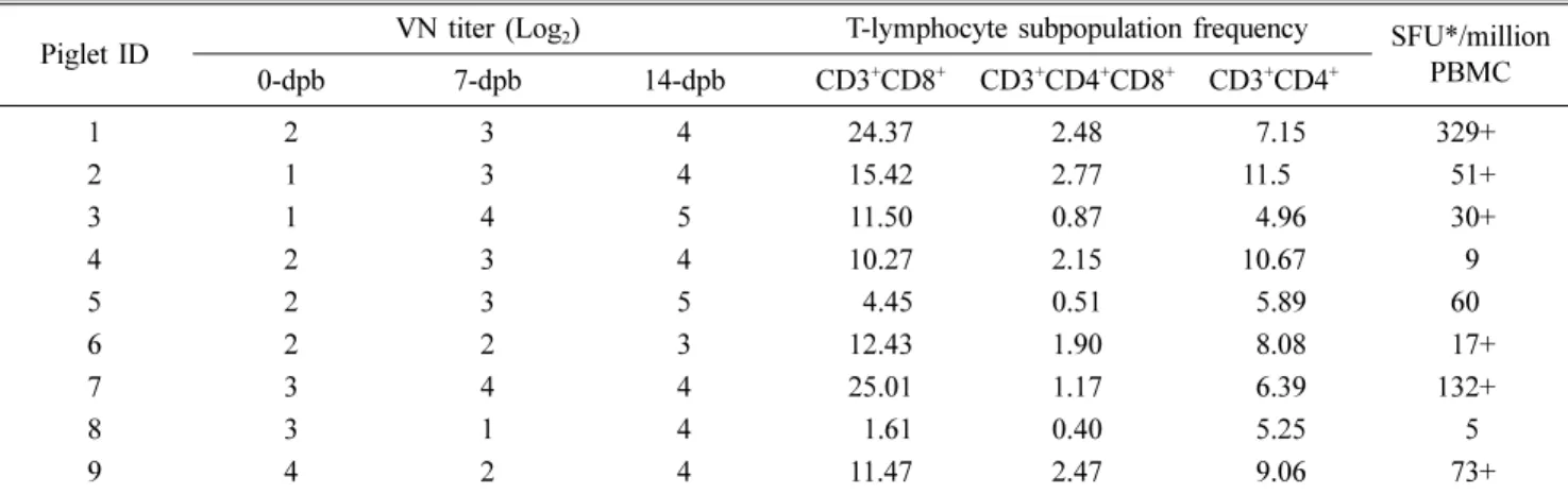
Abstract: A preliminary study into the protective mechanisms of adaptive immunity against porcine reproductive and respiratory syndrome virus (PRRSV) in piglets (n = 9) born to a gilt challenged intranasally with a type-2 PRRSV.

Immune parameters (neutralizing antibodies, CD3

+

CD4

+

, CD3

+

CD8

+

, CD3

+

CD4

+

CD8

+

T-lymphocytes, and PRRSV- specific interferon (IFN)- γ secreting T-lymphocytes) were compared with infection parameters (macro- and microscopic lung lesion, and PRRSV-infected porcine alveolar macrophages (CD172α

+

PRRSV-N

+

PAM) as well as with plasma and lymphoid tissue viral loads. Percentages of three T-lymphocyte phenotypes in 14-days post-birth (dpb) peripheral blood mononuclear cell (PBMC) had significant negative correlations with percentages of CD172 α

+

PRRSV-N

+

PAM ( p < 0.05) as well as with macroscopic lung lesion (p < 0.01). Plasma and tissue viral loads had significant (p < 0.05) negative correlations with CD3

+

CD4

+

CD8

+

T-lymphocyte percentage in PBMC. Frequencies of CD3

+

CD8

+

and CD3

+

CD4

+

T-lymphocytes in 14-dpb PBMC had significant negative correlations with of lymph node (p = 0.04) and lung ( p = 0.002) viral loads. IFN-γ-secreting T-lymphocytes frequency had a significant negative correlation with gross lung lesion severity ( p = 0.002). However, neutralizing antibody titers had no significant negative correlation (p > 0.1) with infection parameters. The results indicate that T-lymphocytes contribute to controlling PRRSV replication in young piglets born after in-utero infection.

Keywords: T-lymphocyte, neutralizing antibodies, porcine reproductive and respiratory syndrome virus

Introduction

Porcine reproductive and respiratory syndrome (PRRS) was first recognized in the United States and Europe as

‘Mystery Swine Disease’ in mid-1980s. The disease charac- terized by manifestation of reproductive failure and respira- tory distress in swine herds had been subsequently reported from Asia and other pig-producing areas between the late 1980s and early 1990s [7, 31]. The causative agent of this globally epidemic, economically important porcine disease was then identified as a member of the Arteriviridae family, which contains a single-stranded positive-sense RNA genome

of approximately 15 Kb in the order of Nidovirales. Porcine reproductive and respiratory syndrome virus (PRRSV) is divided into two major genotypes: type-1 PRRSV (European type) and type-2 PRRSV (North American type).

Vaccination with attenuated live viruses has been used to control clinical PRRS in many countries [2, 26]. However, this conventional vaccine formulation using a few selected PRRSV isolates has not been satisfactory to protect swine herds affected with various PRRSV isolates, possibly due to induction of ineffective immune response, high antigenic variation among isolates and a possible reversion to viru- lence of attenuated vaccine strains [1, 24]. Up to date, neu-

*Corresponding author

Tel: +1-631-323-3429, Fax: +1-631-323-3097 E-mail: [email protected]

Present address: Plum Island Animal Disease Center, Greenport, NY 11944, USA

(2)

tralizing antibodies have been considered as an indicator of protective immunity in vaccinated pigs. However, neutraliz- ing antibodies are often induced in a delayed mode in vacci- nated pigs [21, 34] and not efficacious enough to prevent pigs from being affected by PRRS [3, 13]. Furthermore, neu- tralizing antibodies are protective only against antigenically homologous viruses, while partial or no protective against heterologous strains of PRRSV [17].

Cell-mediated immunity (CMI) against PRRSV appears in two to four weeks after infection reaching to a peak response in four to seven weeks when measured by interferon (IFN)- γ secretion and lymphocyte proliferation [22, 33]. The infor- mation on the efficacy and mechanism of PRRSV-specific T- lymphocytes is lacking with conflicting reports, particularly on protective effect of cytotoxic T-lymphocytes (CTL) in PRRSV-infected pigs [20, 33].

In-utero infection of PRRSVs resulted in still-birth and death of fetuses, and varying degree of clinical syndromes accompanying with viremia and development of PRRSV-spe- cific-antibodies in live piglets [12, 29]. The present study was conducted to characterize adaptive immune responses associated with control of lung lesion, virus replication in tis- sues and viremia pattern in piglets born to a gilt exposed to a type-2 PRRSV strain at the late gestation.

Materials and Methods

Preparation of PRRSV

A Type 2 PRRSV

SD23983

[11] was used in this study. Propa- gation and titration of this virus were carried out as previ- ously described [5, 15, 27].

Animals and viral challenge

A 12-month-old sexually mature gilt negative for PRRSV, porcine circovirus and porcine epidemic diarrhea virus was purchased and transferred into an isolation facility at Wash- ington State University, and bred by artificial insemination using the commercial semen from a PRRSV- and PCV-free boar. The pig was maintained in the facility for the entire study period. At 98 days of gestation, the pregnant gilt was intranasally inoculated with 1.58 × 10

4

TCID

50

of PRRSV

SD23983

. Nine piglets live born to the gilt were bled at 0 (before feed- ing colostrum), 7- and 14-days post-birth (dpb) for serum and peripheral blood mononuclear cells (PBMCs). At 14-dpb (33-day post-inoculation), the piglets were euthanized and pulmonary alveolar macrophage (PAM) was obtained from lungs of the all nine piglets. Macroscopic and microscopic lung lesions were examined per previously described meth- ods [12]. At necropsy, several regional lymph nodes (medial retropharyngeal, tracheobronchial, mesenteric, inguinal, sub- mandibular, and mediastina lymph nodes), tonsil, lung and spleen were collected, and homogenate was made from each tissue for viral load quantitation. Animal handling and care was performed as per the animal use protocol (IACUC No.

04372) approved by Institutional Animal Care and Use Com-

mittee at Washington State University.

Real-time reverse transcriptase-polymerase chain reaction (RT-PCR)

Real-time RT-PCR on various samples was performed at Iowa State University Veterinary Diagnostic Laboratory as per the established protocol using VetMax NA and EU PRRSV reagents (Applied Biosystems, USA). PCR reac- tions were done per the manufacturer’s instructions using a 7500 Fast Real-Time PCR System (Applied Biosystems).

The thresholds for NA and EU PRRSVs were set at 0.10 and 0.05, respectively. A cycle threshold (C

T

) of < 37 cycles was used as cut-off for positive on the assay. A negative control without RNA and a positive control provided in the kit were added to each PCR plate along with positive and negative extraction controls.

Preparation of PBMCs and storage

Swine PBMCs were isolated from fresh venous blood mixed with 5 mM EDTA (final concentration) by centrifuga- tion on a discontinuous gradient using Lymphoprep (1.077 g/

mL, STEMCELL Technologies, Canada) as previously described [19, 5].

Analysis of T-lymphocyte subpopulations and PRRSV- infected alveolar macrophages by flow cytometry

To analyze T-lymphocyte subpopulations in PBMC col- lected at 14-dpb, staining of T-lymphocytes was performed using phenotype-specific monoclonal antibodies and second- ary antibody conjugates per the method previously described [11]. Monoclonal antibodies (MAbs) used in surface staining were anti-CD3 (8E6 clone, IgG1 isotype; VMRD, USA), anti-CD8 (PT81B clone, IgG2b clone; VMRD) and anti- CD4-PerCP-Cy5.5 (BD Pharmingen, USA). The secondary antibody conjugates for anti-CD3 and anti-CD8 MAbs were goat anti-mouse IgG1 labeled with fluorescein isothiocyan- ate (FITC) (Life Technologies, USA) and goat anti-mouse IgG2b conjugated with R-phycoerythrin (R-PE) (Life Tech- nologies), respectively. To determine the proportion of PAM cells harboring PRRSV collected from lung of piglets, sur- face staining of CD172 α molecule as a PAM cell marker and intracellular staining of PRRSV nucleocapsid (N) protein were performed using CD172 α- and PRRSV-N-specific mono- clonal antibodies (clones 74-22-15 and 2D6, respectively) and secondary anti-mouse antibodies labelled with FITC or R-PE per the method previously described [6]. The stained cells were analyzed with a FACSCalibur analytical flow cytometer (BD, USA).

IFN- γ enzyme-linked immunospot (ELISpot) assay

ELISpot assay was carried out to assess PRRSV-specific T-

lymphocyte responses using a commercial assay (Mabtech,

Sweden) as per manufacturer’s instruction. The detailed meth-

ods for porcine PBMC stimulation and calculating signifi-

cant spot-forming unit (SFU) were described previously [5].

(3)

Fluorescent focusing neutralization assay

The PRRSV neutralizing antibody response in serum was measured using fluorescent focus neutralizing assay as previ- ously described [5, 32].

Statistical analysis

Statistical correlation between immune parameters and infection parameters measured on the 9 piglets was analyzed by Spearman’s rank correlation coefficient. Hypothesis tests based on these correlation coefficients were carried out by determining if different parameters had a significant (p <

0.05) nonzero correlation each other. All statistical analyses were performed using R software from the R foundation for Statistical Computing (Austria).

Results

Piglets developed PRRSV-specific immune responses after in-utero infection of PRRSV

SD23983

Nine piglets born to a gilt intranasally inoculated with

type-2 PRRSV

SD23983

at the late gestation were viremic at the day of birth before suckling colostrum, clearly demonstrat- ing in-utero infection. The proportion of CD172α

+

PRRSV- N

+

PAM harboring PRRSV ranged from 8.0% to 42.2%

among the piglets as determined by surface and intracellular staining and flow cytometry analysis. The macroscopic lung lesion score varied among the piglets, ranging between 5%

and 50% of total lung surface. Similarly, the degree of micro- scopic lesion in lung tissue was varying from mild to severe lesion caused by the replication of PRRSV (Fig. 1). Besides lung tissues, PRRSV RNA was also detected in spleen and mixed lymph nodes of all nine piglets (Table 1).

Six of nine piglets had significantly ( ≥ 5 SE of negative control SFU) high PRRSV-specific T-lymphocyte responses after in-utero infection as determined by ELISpot assay (Table 2). PRRSV-specific IFN- γ secreting T-lymphocyte response was particularly highly significant (> 100 SFU and

> 5 SE) in piglets 1 and 7, moderately significant (50–100 SFU and 5 SE) in piglets 2 and 9, and weakly significant (< 50 SFU) in piglets 3 and 6. Frequencies of three different phe-

Fig. 1. Varying severity in microscopic lung lesion of piglets born to a gilt inoculated with PRRSV

SD23983.

Piglets were euthanized at 14-dpb and subjected to necropsy and histological analysis. Representative pictures from piglets with mild to normal (piglet 1), moderate (piglet 2) and severe (piglet 8) lung lesions were presented as a supplementary data to the summary in Table 1. Scale bars = 50 μm.

Table 1. Analysis on infection parameters in piglets born after in-utero porcine reproductive and respiratory syndrome virus (PRRSV) infection

Piglet ID

CD172α

+

PRRSV-N

+

PAM

Lung lesion size (%)

Lung histology

Viral load (Ct) in plasma Viral load (Ct) in tissue

0-dpb 7-dpb 14-dpb Lung Spleen Lymph node

1 18.44 55 mild 19.3 22.8 24.1 23.4 30.7 23.9

2 22.92 25 moderate 20.9 24.7 23.8 31.5 29.8 24.6

3 36.75 30 moderate 37 18 24.8 21.3 29.5 23.8

4 25.94 50 severe 23.2 21.1 23.0 22.2 28.9 22.3

5 42.25 40 severe 20.7 19.0 17.1 21.1 24 21.1

6 18.86 30 severe 22.4 19.7 21.9 21.5 28.5 22.8

7 17.72 10 severe 22.1 18.9 19.9 21.5 25.9 22.0

8 34.23 50 severe 37 20.1 18.5 15.6 19.6 18.7

9 58.04 55 mild 17.8 19.5 24.8 23.6 30.1 21.9

Ct value was obtained by real time RT-PCR. CD172 α

+

PRRSV-N

+

PAM, macroscopic lung lesion, lung histology and tissue viral loads were

analyzed using PAMs and tissues collected at 14 days post-birth (dpb). RT-PCR, reverse transcriptase polymerase chain reaction; PAM, pul-

monary alveolar macrophage.

(4)

notypes of T-lymphocyte varied among the piglets (Table 2).

Neutralizing antibody titer was 1:2 to 1:16 at 0-dpb before colostrum consumption, and was 1:2 to 1:16 and 1:8 to 1:32 at 7- and 14-dpb, respectively.

Correlation between different infection parameters Correlation between different infection parameters was analyzed in Figure 2 and summarized in Table 3. The viral load in lung tissue had a significantly high positive correla- Table 2. Analysis on immune parameters in piglets born after in-utero PRRSV infection

Piglet ID VN titer (Log

2

) T-lymphocyte subpopulation frequency SFU*/million 0-dpb 7-dpb 14-dpb CD3

+

CD8

+

CD3

+

CD4

+

CD8

+

CD3

+

CD4

+

PBMC

1 2 3 4 24.37 2.48 57.15 329+

2 1 3 4 15.42 2.77 11.55 551+

3 1 4 5 11.50 0.87 54.96 530+

4 2 3 4 10.27 2.15 10.67 559

5 2 3 5 54.45 0.51 55.89 560

6 2 2 3 12.43 1.90 58.08 517+

7 3 4 4 25.01 1.17 56.39 132+

8 3 1 4 51.61 0.40 55.25 555

9 4 2 4 11.47 2.47 59.06 573+

*Spot-forming unit (SFU) numbers are mean SFU/10

6

peripheral blood mononuclear cell (PBMC) minus the mean of the appropriate unstimulated control; those adjusted SFU that were greater than the unstimulated control SFU mean plus 5 times the SE were scored as pos- itive and marked with a + in the table. T-lymphocyte subpopulation frequency and SFU of PRRSV specific interferon (IFN)- γ secreting cells were analyzed using PBMC collected at 14 days post-birth.

Fig. 2. Statistical correlation analysis between tissue viral loads (A, lung; B, spleen; and C, lymph nodes), PRRSV-infected PAM fre-

quency, macroscopic and microscopic lung lesions. Statistical analysis was performed with data in Table 1.

(5)

tion with PRRSV-infected PAM frequency (p = 0.01), and low positive correlations with gross lung lesion score (p = 0.06) and severity of microscopic lesion in lung (p = 0.06).

The viral load in spleen tissue was also significantly highly correlated with PRRSV-infected PAM frequency (p = 0.006), gross lung lesion score (p = 0.02) and severity of micro- scopic lesion in lung (p = 0.0008). The viral load in lymph node had a significantly high positive correlation (p = 0.03) with PRRSV-infected PAM frequency, but not with gross lesion score (p = 0.2) and histological severity in lung tissue (p = 0.2).

Relationship between infection parameters and immune parameters

Frequencies of T-lymphocyte subpopulations including CD3

+

CD4

+

, CD3

+

CD8

+

and CD3

+

CD4

+

CD8

+

in total PBMC were analyzed for correlation with infection parameters including PRRSV-infected PAM frequency, macroscopic lung lesion score, severity of microscopic lesion in lung, and viral loads in plasma and tissues (Table 3). CD3

+

CD4

+

CD8

+

T-lymphocytes had significantly high inverse correlations with CD172 α

+

PRRSV-N

+

PAM (p = 0.003), and viral load in all tissues (p < 0.03), but low inverse correlations with mac- roscopic (p = 0.07) and microscopic (p = 0.06) lung lesions.

CD3

+

CD4

+

T-lymphocytes had significantly high inverse correlations with various infection parameters including CD172 α

+

PRRSV-N

+

PAM (p = 0.017), macroscopic lung lesion score (p = 0.01), and viral loads of lung (p = 0.002). CD3

+

CD8

+

T-lymphocytes had significant inverse correlations with CD172 α

+

PRRSV-N

+

PAM frequency (p = 0.024), macroscopic lung lesion score (p = 0.01), and viral load in lymph nodes (p = 0.04). The frequency of PRRSV-specific IFN-γ secret- ing T-lymphocytes had a significant inverse correlation with macroscopic lung lesion score (p = 0.002) and a significant (p = 0.05) positive correlation with CD3

+

CD8

+

T-lymphocytes (Table 2).

Interestingly, the titer of virus neutralizing (VN) antibod- ies had no significant inverse correlations with viral loads in plasma (p = 0.89), lung (p = 0.37), spleen (p = 0.80) and lymph nodes (p = 0.61) at 14-dpb, and other infection parameters including CD172 α

+

PRRSV-N

+

PAM frequency (p = 0.78), macroscopic lung lesion score (p = 0.65) and severity of

microscopic lesion in lung (p = 0.79) at 14-dpb.

Discussion

Defining immune protective mechanisms in PRRSV-infected pigs have been focused on neutralizing antibody responses. It has been reported that neutralizing antibodies play a role in protecting pigs from PRRS to a certain extent [25, 34].

Therefore, neutralizing antibody titer has been used in most studies as a criterion to evaluate protective immunity in vac- cinated or naturally infected pigs. However, PRRSV-specific neutralizing antibodies were slowly induced, strain-specific and, therefore, not efficacious for controlling antigenically diverse PRRSV isolates including heterogeneous viruses of the same genotype or viruses of different genotypes [4, 23].

Up to date, neutralizing antibody-based evaluation of PRRS vaccines have not resulted in a commercial vaccine effica- cious against diverse virus isolates yet. Despite unsuccessful development of efficacious vaccines inducing protective neu- tralizing antibody responses against heterologous isolates in pigs, systematic approaches to define other protective mech- anisms including innate immunity and CMI have been lack- ing. Therefore, this study was conducted to define immune parameters associated with disease protection in piglets born to a gilt infected with a pathogenic type-2 PRRSV, SD23983 isolate.

Fetal pathogenicity of PRRSV

SD23983

after in-utero infec- tion varied in different studies. In this study, the gilt infected with PRRSV

SD23983

had two still-births (18.2% of piglets) and 9 live PRRSV-infected births. A previous study using the identical strain reported that 43–58% of piglets were born live, 24–43% stillborn and 6–18% mummified when sows were infected at 98 days of gestation [11]. In contrast, another study reported that the PRRSV infection of sows at 90 days of gestation resulted in 4.5% of dead piglets [28]. A possible explanation for higher fetal pathogenicity in two of three studies may be related to infection timing in gestation period. Eight-day delayed infection in gestation period may be related to higher rates of reproductive failure [10, 14, 16].

Another possible explanation is that different passage history of PRRSV stocks in green monkey cell line (Marc 145) may Table 3. Summary on statistical correlation analysis ( p value) between immune parameters and infection parameters in piglets born

after in utero infection

Piglet ID

CD172 α

+

PRRSV-N

+

PAM

Gross lung lesion size

(%)

Lung his- tology

Viral load (Ct) in plasma Viral load (Ct) in tissue

0-dpb 7-dpb 14-dpb Lung Spleen Lymph

node

T cell

CD3

+

CD4

+

0.017* 0.01 0.3 0.09 0.41 0.14 0.002 0.07 0.47

CD3

+

CD8

+

0.024 0.01 0.3 0.21 0.45 0.27 0.11 0.1 0.04

CD3

+

CD4

+

CD8

+

0.003 0.07 0.06 0.05 0.03 0.02 0.000006 0.006 0.03

IFN- γ

2

T cell SFU 0.13 0.002 0.09 0.01 0.39 0.46 0.25 0.11 0.35

VN titer 0.78 0.65 0.79 0.43 0.26 0.89 0.37 0.8 0.61

*Statistical analysis was performed with data of Tables 1 and 2. VN, virus neutralizing.

(6)

cause varying virulence [9]. Despite severe pathogenicity of PRRSV

SD23983

in in-utero infected piglets in this study and other previous studies [11, 28], manifestation in the gilt after intranasal infection of PRRSV

SD23983

was limited to short period of viremia without clinical symptom such as lung lesion and nasal discharge.

All of piglets born to the gilt infected with PRRSV

SD23983

by intranasal route developed PRRS-specific diseases includ- ing clinical symptoms (anorexia, depression, and respiratory syndrome), viremia, viral loads in various lymph nodes, and macroscopical and microscopical lung lesions. PRRSV-spe- cific neutralizing antibody measured by VN titer and PRRSV-specific T-lymphocytes measured by IFN- γ T-cell SFU were detectable at varying levels. VN titers in infected live birth piglets were ranging from 1:2 to 1:16 (1–4 log

2

) at 0-dpb before colostrum consumption, and reached to 3–5 log

2

at 14-dpb. Despite weak VN titers of in-utero infected piglets, higher VN positive rate (100%) at birth was notice- able than a previous report [28] with positive VN titers in only 22% of piglets in-utero infected at 90 days of gestation.

Increasing VN titers in 9 piglets at later dpbs suggest that neutralizing antibody responses in the infected piglets are not solely due to passive transfer through colostrum of the gilt.

No significant difference in VN titer among piglets with var- ious levels in disease parameters may support that neutraliz- ing antibodies may not be a protection correlate in in-utero PRRSV-infected piglets.

Significantly high positive correlations between various PRRSV infection parameters in piglets born after in-utero infection were demonstrated in this study. The viral loads in lung, spleen and lymph nodes had significant positive corre- lations with PRRSV-infected PAM frequency. Viral loads in lung were also significantly correlated with macroscopic lung lesion score of PRRS-infected piglets. Since PAM in PRRSV- infected pigs is the main target cell in which virulent PRRSVs actively replicate [9, 35], significant correlation between PRRSV-infected PAM frequency and viral loads in lymph nodes in in-utero infected piglets demonstrates that PAM infectivity is a good indicator for respiratory virulence of PRRSV SD23983 strain than microscopic lesion using focal tissue samples in PRRS affected lung.

Significantly high inverse correlations between PRRS dis- ease parameters and protective CMI parameters induced in piglets born after in-utero infection were demonstrated as a major finding in this study. Percentages of three T-lympho- cyte subpopulations in the in-utero infected piglets had a sig- nificant inverse correlation with macroscopic lung lesion score. Particularly, CD3

+

CD4

+

CD8

+

T-lymphocytes were sig- nificantly associated with lower viral load in plasma and tis- sues. Previous reports on IFN- γ secreting cells in lung of PRRSV-infected pigs [29, 30], and the protective correlation between IFN- γ secreting cells in PBMC and control of pseu- dorabies [36] led to expectation on contribution of CMI in the control of PRRSV in infected pigs. PRRSV-specific T- lymphocyte response determined by IFN- γ ELISPOT assay

was significantly associated with reduction of reproductive failures in swine farms [20]. IFN- γ response was first reported in PRRSV-infected fetuses, suggesting the capabil- ity of fetuses to develop virus-specific T-lymphocyte responses [28]. However, the protective role of the IFN- γ secreting cells has not been systematically studied yet. In this study, the number of IFN- γ secreting T-lymphocytes in PBMC of the piglets in-utero infected with type-2 PRRSV

SD23983

had a highly significant (p = 0.002) inverse correlation with macro- scopic lung lesion of those infected piglets, suggesting that IFN- γ secreting cells developed in the in-utero PRRSV- infected fetuses may be associated with protection against neonatal PRRS development after birth. In addition, number of IFN- γ secreting cells had a significant (p = 0.05) positive correlation with percentage of CD3

+

CD8

+

T lymphocytes in PBMC, indicating that IFN- γ secreting cells may be mainly composed of CD3

+

CD8

+

T lymphocytes. Therefore, even though previous reports [8, 18] could not define the clear functional roles of T-lymphocytes in controlling PRRS and PRRSV replication in tissues, the positive correlation between CD3

+

CD8

+

T-lymphocytes and PRRSV-specific IFN- γ secre- tion in in-utero infected piglets in this study suggests the potential protective role of the T-lymphocyte subpopula- tions. Simple correlation results based on frequencies of T- lymphocyte subpopulations and PRRSV-specific total T-lym- phocytes induced in piglets after intra-uterine infection of PRRSV in this study need to be further characterized using other functional T-lymphocyte assays directly related to elim- ination of RRSV-infected target cells in infected pigs as reported in lentivirus infection [6]. One of our ongoing studies to further characterize protective T-lymphocyte responses demonstrated cytotoxic responses by both CD8

+

and CD4

+

T- lymphocytes in three PRRSV

SD23983

-infected gilts (data not shown).

This study reports that the disease severity of in-utero PRRSV

SD23983

-infected piglets is inversely associated with the numbers of major T-lymphocyte subpopulations and PRRSV- specific IFN- γ secreting T-cells, but not with the level of PRRSV-specific neutralizing antibodies. Further study will be needed to define protective mechanisms of PRRSV-spe- cific T-lymphocyte responses and their efficacies in various age groups of pig.

Acknowledgments

The authors are grateful to Ms. Grace Chung for critical review of manuscript. This study was funded by Animal and Plant Quarantine Agency, Republic of Korea (QIA I- 1543083-2015-16-01).

References

1. Bøtner A, Strandbygaard B, Sørensen KJ, Have P,

Madsen KG, Madsen ES, Alexandersen S. Appearance of

acute PRRS-like symptoms in sow herds after vaccination

with a modified live PRRS vaccine. Vet Rec 1997, 141, 497-

499.

(7)

2. Cano JP, Dee SA, Murtaugh MP, Trincado CA, Pijoan C.

Effect of vaccination with a modified-live porcine reproductive and respiratory syndrome virus vaccine on dynamics of homologous viral infection in pigs. Am J Vet Res 2007, 68, 565-571.

3. Choi K, Lee J, Park C, Jeong J, Chae C. Comparison of the pathogenesis of single or dual infections with type 1 and type 2 porcine reproductive and respiratory syndrome virus. J Comp Pathol 2015, 152, 317-324.

4. Choi K, Park C, Jeong J, Chae C. Comparison of protection provided by type 1 and type 2 porcine reproductive and respiratory syndrome field viruses against homologous and heterologous challenge. Vet Microbiol 2016, 191, 72-81.

5. Chung CJ, Cha SH, Grimm AL, Chung G, Gibson KA, Yoon KJ, Parish SM, Ho CS, Lee SS. Recognition of highly diverse type-1 and -2 porcine reproductive and respiratory syndrome viruses (PRRSVs) by T-lymphocytes induced in pigs after experimental infection with a type-2 PRRSV strain. PLoS One 2016, 11, e0165450.

6. Chung C, Lee W, Loffredo, JT, Burwitz B, Friedrich TC, Giraldo Vela JP, Napoe G, Rakasz EG, Wilson NA, Allison DB, Watkins DI. Not all cytokine-producing CD8

+

T cells suppress simian immunodeficiency virus replication. J Virol 2007, 81, 1517-1523.

7. Collins JE, Benfield DA, Christianson WT, Harris L, Hennings JC, Shaw DP, Goyal SM, McCullough S, Morrison RB, Joo HS, Gorcyca D, Chladek D. Isolation of swine infertility and respiraoty syndrome virus (isolate ATCC VR-2332) in North America and experimental reproduction of the disease in gnotobiotic pigs. J Vet Diagn Invest 1992, 4, 117-126.

8. Costers S, Lefebvre DJ, Goddeeris B, Delputte PL, Nauwynck HJ. Functional impairment of PRRSV-specific peripheral CD3

+

CD

8high

cells. Vet Res 2009, 40, 46.

9. Delputte PL, Nauwynck HJ. Porcine arterivirus infection of alveolar macrophages is mediated by sialic acid on the virus.

J Virol 2004, 78, 8094-8101.

10. Done SH, Paton DJ. Porcine reproductive and respiratory syndrome: clinical disease, pathology and immunosuppression.

Vet Rec 1995, 136, 32-35

11. Feng W, Laster SM, Tompkins M, Brown T, Xu JS, Altier C, Gomez W, Benfield D, McCaw MB. In utero infection by porcine reproductive and respiratory syndrome virus is sufficient to increase susceptibility of piglets to challenge by Streptococcus suis type II. J Virol 2001, 75, 4889-4895.

12. Halbur PG, Paul PS, Frey ML, Landgraf J, Eernisse K, Meng XJ, Lum MA, Andrews JJ, Rathje JA. Comparison of the pathogenicity of two US porcine reproductive and respiratory syndrome virus isolates with that of the Lelystad virus. Vet Pathol 1995, 32, 648-660.

13. Han K, Seo HW, Shin JH, Oh Y, Kang I, Park C, Chae C. Effect of the modified live porcine reproductive and respiratory syndrome virus (PRRSV) vaccine on European and North American PRRSV shedding in semen from infected boars. Clin Vaccine Immunol 2011, 18, 1600-1607.

14. Karniychuk UU, De Spiegelaere W, Nauwynck HJ.

Porcine reproductive and respiratory syndrome virus infection is associated with an increased number of Sn-positive and CD8-positive cells in the maternal-fetal interface. Virus Res

2013, 176, 285-291.

15. Kim HS, Kwang J, Yoon IJ, Joo HS, Frey ML. Enhanced replication of porcine reproductive and respiratory syndrome (PRRS) virus in a homogeneous subpopulation of MA-104 cell line. Arch Virol 1993, 133, 477-483.

16. Kranker S, Nielsen J, Bille-Hansen V, Bøtner A. Experi- mental inoculation of swine at various stages of gestation with a Danish isolate of porcine reproductive and respiratory syndrome virus (PRRSV). Vet Microbiol 1998, 61, 21-31.

17. Li X, Galliher-Beckley A, Pappan L, Trible B, Kerrigan M, Beck A, Hesse R, Blecha F, Nietfeld JC, Rowland RR, Shi J. Comparison of host immune responses to homologous and heterologous type II porcine reproductive and respiratory syndrome virus (PRRSV) challenge in vaccinated and unvaccinated pigs. Biomed Res Int 2014, 2014, 416727.

18. Lohse L, Nielsen J, Eriksen L. Temporary CD8

+

T-cell depletion in pigs does not exacerbate infection with porcine reproductive and respiratory syndrome virus (PRRSV). Viral Immunol 2004, 17, 594-603.

19. López Fuertes L, Doménech N, Alvarez B, Ezquerra A, Domínguez J, Castro JM, Alonso F. Analysis of cellular immune response in pigs recovered from porcine respiratory and reproductive syndrome infection. Virus Res 1999, 64, 33-42.

20. Lowe JE, Husmann R, Firkins LD, Zuckermann FA, Goldberg TL. Correlation of cell-mediated immunity against porcine reproductive and respiratory syndrome virus with protection against reproductive failure in sows during outbreaks of porcine reproductive and respiratory syndrome in commercial herds. J Am Vet Med Assoc 2005, 226, 1707- 1711.

21. Meier WA, Husmann RJ, Schnitzlein WM, Osorio FA, Lunney JK, Zukermann FA. Cytokines and synthetic double-stranded RNA augment the T helper 1 immune response of swine to porcine reproductive and respiratory syndrome virus. Vet Immunol Immunopathol 2004, 102, 299- 314.

22. Molina RM, Cha SH, Chittick W, Lawson S, Murtaugh MP, Nelson EA, Christopher-Hennings J, Yoon KJ, Evans R, Rowland RRR, Zimmerman JJ. Immune response against porcine reproductive and respiratory syndrome virus during acute and chronic infection. Vet Immunol Immunopathol 2008, 126, 283-292.

23. Murtaugh MP, Xiao Z, Zuckermann F. Immunological responses of swine to porcine reproductive and respiratory syndrome virus infection. Viral Immunol 2002, 15, 533-547.

24. Nielsen HS, Oleksiewicz MB, Forsberg R, Stadejek T, Botner A, Storgaard T. Reversion of a live porcine reproductive and respiratory syndrome virus vaccine investigated by parallel mutations. J Gen Virol 2001, 82, 1263-1272.

25. Osorio FA, Galeota JA, Nelson E, Brodersen B, Doster A, Wills R, Zuckermann F, Laegreid WW. Passive transfer of virus-specific antibodies confers protection against reproductive failure induced by a virulent strain of porcine reproductive and respiratory syndrome virus and establishes sterilizing immunity. Virology 2002, 302, 9-20.

26. Park C, Seo HW, Kang I, Jeong J, Choi K, Chae C. A

new modified live porcine reproductive and respiratory

syndrome vaccine improves growth performance in pigs

(8)

under field conditions. Clin. Vaccine Immunol 2014, 21, 1350-1356.

27. Reed LJ, Muench H. A simple method of estimating fifty percent endpoints. Am J Hygiene 1938, 27, 493-497.

28. Rowland RRR. The interaction between PRRSV and the late gestation pig fetus. Virus Res 2010, 154, 114-122.

29. Thanawongnuwech R, Rungsipipat A, Disatian S, Saiyasombat R, Napakanaporn S, Halbur PG. Immunohistochemical staining of IFN-γ positive cells in porcine reproductive and respiratory syndrome virus-infected lungs. Vet Immunol Immunopathol 2003, 91, 73-77.

30. Thanawongnuwech R, Thacker EL. Interleukin-10, interleukin-12, and interferon-γ levels in the respiratory tract following Mycoplasma hyopneumoniae and PRRSV infection in pigs. Viral Immunol 2003, 16, 357-367.

31. Wensvoort G, Terpstra C, Pol JMA, ter Laak EA, Bloemraad M, de Kluyver EP, Kragten C, van Buiten L, den Besten A, Wagenaar F, Broekhuijsen JM, Moonen PLJM, Zetstra T, de Boer EA, Tibben HJ, de Jong MF, van ‘t Veld P, Greenland GJR, van Gennep JA, Voets MTh, Verheijden JHM, Braamskamp J. Mystery swine disease in The Netherlands: the isolation of Lelystad virus.

Vet Q 1991, 13, 121-130.

32. Wu WH, Fang Y, Farwell R, Steffen-Bien M, Rowland RRR, Christopher-Hennings J, Nelson, EA. A 10-kDa structural protein of porcine reproductive and respiratory syndrome virus encoded by ORF2b. Virology 2001, 287, 183-191.

33. Xiao Z, Batista L, Dee S, Halbur P, Murtaugh MP. The level of virus-specific T-cell and macrophage recruitment in porcine reproductive and respiratory syndrome virus infection in pigs is independent of virus load. J Virol 2004, 78, 5923- 5933.

34. Yoon IJ, Joo HS, Goyal SM, Molitor TW. A modified serum neutralization test for the detection of antibody to porcine reproductive and respiratory syndrome virus in swine sera. J Vet Diagn Invest 1994, 6, 289-292.

35. Zhang Q, Yoo D. PRRS virus receptors and their role for pathogenesis. Vet Microbiol 2015, 177, 229-241.

36. Zuckermann FA, Martin S, Husmann RJ, Brandt J. Use

of interleukin 12 to enhance the cellular immune response of

swine to an inactivated herpesvirus vaccine. Adv Vet Med

1999, 41, 447-461.

수치

Table 1. Analysis on infection parameters in piglets born after  in-utero porcine reproductive and respiratory syndrome virus (PRRSV) infection Piglet  ID  CD172α + PRRSV-N + PAM Lung lesion size (%) Lung  histology
Fig. 2. Statistical correlation analysis between tissue viral loads (A, lung; B, spleen; and C, lymph nodes), PRRSV-infected PAM fre- fre-quency, macroscopic and microscopic lung lesions

참조

관련 문서

First, the general characteristics of the two groups were analyzed by frequency analysis and correlation analysis, which is technical statistics, and the

For establishing appropriate treatment plans and prognosis, the estimated value of the correlation between the change in hard and soft tissues is required,

Sixth, there was no significant correlation between lower extremity muscle thickness, static stability, and dynamic stability of excellent athletes in college

2) In between-group comparison before and after elastic band exercise, a significant difference was found in the percentage of body fat, fat-free

Correlation between Results ofthe Multiple Allergosorbent Test-Chemiluminescent Assay(MAST-CLA) and Clinical Severity of Chronic Urticaria. A study of food allergy

These results demonstrated that a positive role of Slug and Twist in tumor invasion in CRA, and inverse correlation between Twist and Slug expression

Results: The results demonstrated a significant positive correlation between the muscle atrophy and the tendon tear.(p&lt;0.05) And there was statistically

• Theory can extent to molten polymers and concentrated solutions.. The single-molecule bead spring models.. a)