41(1) : 21 25 (2010)
21
3T3-L1 세포에 대한 옻나무 추출물의 지방축적 억제효과
김세건·류동영1·김도국·고다형·김윤경·이영미·정현주
*
원광대학교 약학대학한약학과및원광한약연구소
,
1목포대학교한약자원학과Inhibitory Effect of Heartwood of Rhus verniciflua Stokes on Lipid Accumulation in 3T3-L1 Cells
Se-Gun Kim, Dong-Young Rhyu
1, Do-Kuk Kim, Da-Hyung Ko, Yun-Kyung Kim, Young-Mi Lee and Hyun-Ju Jung
*Department of Oriental Pharmacy and Wonkwang-Oriental Medicines Research Institute, Wonkwang University, Sinyong-Dong, Iksan, 570-749, Korea
1
Department of Medicinal Plant Resources, Mokpo National University, 61 Dorim-ri, Chonggye-myon, Muan-gun, Jeonnam 534-729, Korea
Abstract −
The MeOH extract of Rhus verniciflua heartwood inhibited lipid accumulation in 3T3-L1 adipocytes. Chro- matographic methods including of silica gel, RP-18 and high-pressure liquid chromatography isolated sulfuretin and fisetin from the extract as active constituents. The isolated compounds, especially sulfuretin, strongly inhibited lipid accumulation in adipocytes. The treatment of sulfuretin and fisetin led to decreased expression of peroxisome proliferator-activated receptor- gamma (PPAR
γ), as an important transcription factor in fat cell differentiation, which was equal to the decrease in the quercetin positive control. The presence of a hydroxyl group (C-5) in quercetin compared to fisetin, and the presence of C-2 double bonds in fisetin compared with fustin increased the inhibitory effect of lipid accumulation.
Key words −
Rhus verniciflua , flavonoid, sulfuetin, adipocyte, PPAR
γ비만은현재인류에게가장위협적인질병중하나로1)급 격한속도로증가하고있으며
,
공중보건에서도중요한질환으로여겨지고있다
.
2)비만은과잉의지방이체내축적되는것을뜻하며
,
체질량지수(BMI)
가25-29 kg/m
2인사람은과체중
, 30 kg/m
2이상인사람은비만으로정의된다.
3)또한비만은전구지방세포가지방세포
(adipocyte)
로분화하는지방세포의이상발달이라고도하며이러한지방세포에대한지 방의축적은지방세포수의증가나지방세포크기가증가 됨에따라증상이심해지는것으로알려져있다
.
인체에지방을축적하는세포는
hepatocyte (
단기축적)
와adipocyte
(
장기축적)
로알려져있으나비만과관련이있는것은주로
adipocyte
로알려져있다. adipocyte
는preadipocyte
상태로존재하다가적절한환경하에서분화되어
adipocyte
가되어세포내지방을축적하는데
adipocyte
의크기또는수적증가가비만의정도를결정짓는하나의요인으로간주되고 있다
.
따라서preadipocyte
의분열및adipocyte
의생성을억제하는물질은비만을예방하거나치료할수있을것으로 생각되어지고있다
.
비만과관련된질환은당뇨병,
심혈관질환및이상지질혈증등이보고되어있다
.
4-5)현재폴리페놀성화합물을포함한식이성항산화제들은 암
,
심장질환,
죽상동맥경화증등과같이산화적스트레스와관련된질병에 예방효과를가지고있으며
,
특히폴리페놀성화합물인플라보노이드는항바이러스
,
항염,
항암효과를포함한많은질병과
,
6-7)지질과산화,
혈소판응집,
모세관투과성과리포옥시게네이즈
(lipoxygenase)
를포함하는 효소계의활동을크게억제한다고보고되어있다
.
8)참옻나무
(
Rhus vernicifluaStokes, Anacardiaceae)
는낙엽교목으로서높이가
20 m
에달하며수피는혈액순환약및통경의목적으로사용되었다
.
9-10)수피의활성성분인urushiol
은강력한항암효과및항산화효과가알려져있지만
,
단백질과비특이적인결합및피부앨러지를발생시키는문제
*교신저자(E-mail):[email protected] (Tel): +82-63-850-6814
점을가지고있어서의약품으로개발하기에는문제점을가 지고있다
.
하지만수피를제거한심재부분에는Urushiol
이존재하지않고
,
주성분으로는플라보노이드계열의물질들이항산화및항류마티스활성이나타나는것으로보고되 어있다
.
11)본연구에서는옻나무심재의메탄올추출물이
3T3-L1
preadipocyte
의분화및지방축적을억제하므로그활성물질을분리동정하였으며그작용기전을추정할목적으로 지방세포의분화를촉진하는
PPAR
γ의발현정도를측정하였다
.
재료 및 방법
식물재료 − 실험에사용한옻나무심재는충청북도제천 에소재한황금들녘나라에서구입하여원광대학교약학대 학한약학과권동렬교수가동정한후사용하였다
.
기기 및 시약 −1
H-NMR (500 MHz)
및 13C-NMR (125 MHz) spectra
는JEOL Eclipse 500 FT-NMR spectrometer
으로측정하였고
, silica gel
과TLC plate
는Merck
사의것을사용하였으며
, RP-18 gel
은YMC
사의제품을사용하였다.
세포실험에 사용한 시약인
dexamethasone, 3-Isobutyl-1- methylxanthine (IBMX), Oil red-O
및insulin
은Sigma
사Dulbecco's modified Eagle's medium (DMEM), trypsin- EDTA, fetal bovine serum (FBS), bovine calf serum (BCS)
는Hyclone
사의제품을사용하였다.
추출 및 분획 − 수피를벗기고음건한참옻나무목질부
10 kg
을MeOH
로3
회환류추출하고,
이를감압농축하여MeOH ex. 380 g
을얻었다.
이중MeOH ex. 322 g
을3
차증류수에현탁시킨다음계통분획법에의하여
chloroform (31 g), ethyl acetate (251 g), butanol (16 g), H
2O (24 g)
으로분획하였다
.
화합물의 분리 및 정제 −
Ethyl acetate fraction(100 g)
을silica gel 744 g
을충전시킨내경10 cm
의컬럼에서CHCl
3: MeOH : H
2O = 8 : 2 : 1 (
하층)
의이동상으로전개하였다
.
용출된 시료는250 ml
씩 소분획하고, thin layer
chromatography
를실시하여분획들을유사한그룹으로 합쳐용출된 순서대로
RVSE-1, RVSE-2, RVSE-3, RVSE-4,
RVSE-5
의 소분획으로 나누고,
소분획RVSE-1
을octadecylsilane (ODS) column chromatography (MeOH : H
2O = 55 : 45)
를실시하여순수한백색의화합물 1, 149
㎎ 을분리하였다
.
위에서얻어진소분획RVSE-2
를ODS column chromatography (MeOH : H
2O = 60 : 40)
를 실시하고
HPLC
로정제하여순수한황색의화합물2, 66 mg
을분리하였다
.
소분획
RVSE-3
을ODS column chromatography (MeOH : H
2O = 60 : 40)
를실시한것으로부터는순수한오렌지색의화합물 3
, 158 mg
을분리하였다.
화합물 1 −
White amorphous powder,
1H-NMR (CD
3OD, 500 MHz)
δ: 4.47 (1H, d,
J= 11.9 Hz, H-3), 4.93 (1H, d,
J= 11.9 Hz, H-2), 6.32 (1H, d,
J= 2.3 Hz, H-8), 6.52 (1H, dd,
J= 2.3, 8.7 Hz, H-6), 6.80 (1H, d,
J= 8.3 Hz, H-5'), 6.85 (1H, dd,
J= 1.8, 8.3 Hz, H-6'), 6.98 (1H, d,
J= 1.8 Hz, H-2'), 7.71 (1H, d,
J= 8.7 Hz, H-5),
13C-NMR (CD
3OD, 125 MHz)
δ: 84.3 (C-2), 73.2 (C-3), 193.1 (C- 4), 128.8 (C-5), 110.8 (C-6), 163.8 (C-7), 112.1 (C-8), 165.6 (C-9), 102.4 (C-10), 128.8 (C-1'), 114.6 (C-2'), 145.0 (C-3'), 145.8 (C-4'), 114.7 (C-5'), 120.0 (C-6').
화합물 2 −
Yellow amorphous powder,
1H-NMR (CD
3OD, 500 MHz)
δ: 6.89 (1H, d,
J= 8.7 Hz, H-5'), 6.90 (1H, d,
J
= 2.3 Hz, H-8), 6.90 (1H, dd,
J= 2.3, 9.6 Hz, H-6), 7.66 (1H, dd,
J= 1.8, 8.7 Hz, H-6'), 7.76 (1H, d,
J= 1.8 Hz, H-2'), 7.97 (1H, d,
J= 9.6 Hz, H-5).
13C-NMR (CD
3OD,125 MHz)
δ: 146.2 (C-2), 137.2 (C-3), 173.1 (C- 4), 126.2 (C-5), 114.7 (C-6), 162.9 (C-7), 101.6 (C-8), 157.2 (C-9), 114.1 (C-10), 123.0 (C-1'), 114.9 (C-2'), 144.9 (C-3'), 147.3 (C-4'), 114.7 (C-5'), 120.3 (C-6').
화합물 3 −
Orange amorphous powder,
1H-NMR (DMSO-
d6
, 500 MHz)
δ: 6.64 (1H, s, H-2), 6.70 (1H, dd,
J= 1.9, 8.7 Hz, H-6), 6.74 (1H, d,
J= 1.9 Hz, H-8), 6.83 (1H, d,
J
= 8.2 Hz, H-5'), 7.24 (1H, dd,
J= 1.9, 8.2 Hz, H-6'), 7.44 (1H, d,
J= 1.9 Hz, H-2'), 7.60 (1H, d,
J= 8.7 Hz, H-5).
13C-NMR (DMSO-
d6, 125 MHz)
δ: 112.3 (C-2), 146.2 (C-3), 148.6 (C-4), 126.3 (C-5), 125.1 (C-6), 168.0 (C-7), 98.9 (C-8), 166.7 (C-9), 113.8 (C-10), 123.9 (C-1'), 118.5 (C-2'), 146.1 (C-3'), 148.6 (C-4'), 116.6 (C-5'), 125.1 (C-6').
세포배양 − 활성실험에 사용한
3T3-L1
세포는ATCC (American Type Culture Collection, Rockville, MD)
에서구입하여
,
인큐베이터(37
oC, 5% CO
2)
에서10% BCS
가포함된
DMEM
배지를사용하여배양하였다. 3T3-L1
세포를지방세포로분화시키기위하여
4
일동안10% BCS
를사용하였고
,
분화시작점에는10% FBS
배지내에1
µM dexame- thasone, 0.5 nM IBMX, 10
µg/ml insulin
을넣어주고,
분화시작점으로부터
2
일후에10
µg/ml insulin
인포함된10%
FBS
로갈아주고, 4
일후와6
일후에는10% FBS
배지로갈아주었다
.
샘플처리는분화시작일과2
일째, 4
일째되던날3
회처리해주었고
, 8
일째되던날Oil red O
로염색하여세포내지방을확인하였다
.
Oil red O 염색 − 분화시작일로부터
8
일째되던날배지를제거하고
PBS
로세척한후,
상온에서30
분동안10%
포르말린으로세포를고정하고
, Oil red O
로염색시킨후60% isopropanol
로 세척하고 세포내의 지방은Olympus (Tokyo, Japan)
현미경을이용하여사진촬영하였다.
염색된지방들은
isopropanol
에 녹여96 well plate
에 옮긴 후490 nm
에서흡광도를측정하였다.
Western blot 분석 − 실험이종료된세포를
PBS
용액으로
1
회세척한후lysis buffer (20 mM Tris, pH 7.0, 5 mM EDTA, 1 mM EGTA, 1% Triton X-100, 0.2 mM PMSF, 1
µg/
µl aprotinin, 20
µg/
µl leupeptin, 1 mM Na3VO4, 10mM NaF, 1mM pyrophosphate, 1mM
β-glycerophosphate)
를넣고얼음위에서
10
분간용해시킨다.
세포를수거하여13,000 rpm, 4
oC
에서15
분간원심분리하여상층액만취하여단백질을정량하였다
. Lysate
는loading buffer (1 M Tris, 50% glycerol, 10% SDS, 2-mercaptoethanol, 1% bromophenol blue)
와혼합하여95
oC
에서10
분간가열시킨후, 10% SDS polyacrylamide gel
에서 전기영동 한 후nitrocellulose membrane
에전이시켰다.
그후membrane
은5%
무지분유가첨가된
TTBS (1 M Tris, 5 M NaCl, tween 20)
용액에넣고상온에서
1
시간동안blocking
시킨다. TTBS
용액으로
3
회(
각각10
분씩)
세척한후일차항체(PPAR
γ1 : 250)
로
2
시간동안반응시킨다음에다시TTBS
용액으로3
회(
각각7
분씩)
세척한다.
그다음에peroxidase
가포함된이차항체
(goat anti mouse 1 : 2000)
로1
시간동안반응시켰고항체의검출은
enhanced chemiluminescence
시약을이용하여가시화하였고
UVP (Image acquisition and analysis software, Visionwork TMLS)
로정량하였다.
통계처리 − 실험결과는
SPSS ver. 11.5
의one-way
ANOVA
분석을통하여수행하였고,
각처리군간의유의성검정은
Duncan's multiple test
에의하여p<0.05
에서실시하였다
.
결과 및 고찰
옻나무심재는항산화활성이우수한
flavonoid
성분이다량함유되어있으며피부앨러지반응으로의약품개발을
저해시키는
urushiol
성분이거의존재하지않는다고보고되어왔다
.
11)특히,
옻나무심재의메탄올추출물은3T3-L1 cell
을지방세포로유도하여도cell
내지방축적이억제되는항비만활성을나타내므로활성성분을규명하기위하여물 질분리를시도하였다
.
옻나무MeOH
추출물을클로로포름,
에틸아세테이트
,
부탄올그리고잔류물로분획할때,
주성분및활성이에틸아세테이트층에집중되었기에에틸아세 테이트분획물에대한물질분리를반복적으로실시하였다
.
물질분리는
silica gel
및ODS column chromatography
와HPLC (High Performance Liquid Chromatography)
를이용하였으며
,
그결과3
종의flavonoids
를분리할수있었다.
분리한물질은
MP., IR, UV,
1H-NMR
및 13C-NMR
을측정하여 문헌과 비교한 결과 화합물 1은
dihydroflavonol
인fustin,
12) 화합물 2는flavonol
계열의fisetin,
13) 화합물 3은aurone
계열의sulfuretin
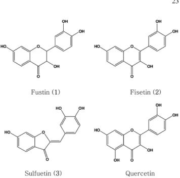
인것으로확인되었다(Fig. 1).
14)Fustin
과fisetin
은C-2
에이중결합의유무만이차이가있었고
, fisetin
은3T3-L1 cell
에지방분화능이있다고보고되어진
quercetin
과비교할때15)C-5
위치에-OH
기만이없는화합물이었다
.
3T3-L1 cell
을이용하여분리된화합물의지방생성억제활성을실험한결과는
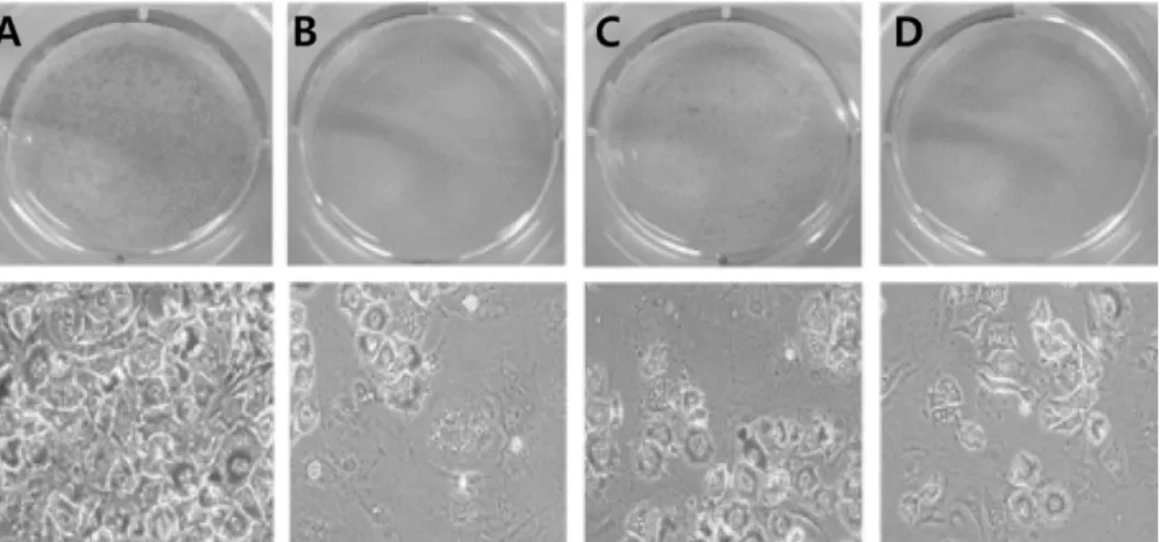
Fig. 2
와같이관찰되었다.
화합물의농도는 박희준등이16)
HL-60, HepG2, Primary hepatocyte
등의
cell
에IC
50가300
µM
이상이라보고한 것을참고로3T3-L1 cell
에세포독성이관찰되지않는20
µM
의안전한농도를사용하였으며
quercetin
을positive control
로하여실험하였다
.
그결과fustin
은활성이관찰되지않았으며fisetin
은
negative control
에비하여약40%
정도지방생성이감소하였음을알수있었다
.
이것은fustin
과fisetin
의구조적차이점이이중결합의유무에만있으므로
C-2
의이중결합은adipocyte
의지방축적능에중요한구조부분임을확인할수있었다
. Fisetin
은positive control
로사용한quercetin
의구조중
C-5
의-OH
기가없는구조로이러한구조적차이는adipocyte
의생성에도영향을미쳐지방축적억제효과를약10%
정도감소시키는것으로관찰되었다.
Aurone
계열의sulfuretin
은분리한화합물중3T3-L1 cell
에지방축적을가장강하게억제시켰으며
,
활성의크기는positive control
인quercetin
과동일정도인약50%
정도인것으로확인되었다
.
Fig. 1.
The structures of the isolated compounds from the
heartwood of R. verniciflua .
지방조직에서주로나타나는
PPAR
γ는발현이증가할수록세포의인슐린에대한감수성및전구지방세포에서지 방세포로분화를크게증가시키는것으로알려져있다
.
17-18)지방세포생성의
key regulator
로알려진PPAR
γ(peroxisome proliferator-activated receptor)
의발현에대한분리한화합물의작용을조사한결과는
Fig. 3
과같다.
전구지방세포에서지방세포로분화억제활성을나타낸
fisetin
과sulfuretin
에대한실험결과
, fisetin
은대조군과비교하여PPAR
γ의발현을약
50%
감소시켰으며sulfuretin
은약75%
정도매우강하게억제시키는것으로관찰되었다
.
이것은앞에서관찰된
3T3-L1 cell
의지방생성능억제효과가PPAR
γ의발현과크게연관되어있음을나타내고있다고생각되어졌다
.
이상의결과로부터옻나무심재로부터
3T3-L1 cell
이adipocyte
로분화되는것을억제시키는물질로
fisetin
과sulfuretin
이분리되었으며이는
PPAR
γ의발현이억제되어나타나는결과로추정되었다
.
사 사
본연구는사단법인진안군친환경홍삼한방산업클러스터 사업단의지원에의해수행되었기에이에감사드립니다
.
인용문헌
1. Bray, G. A. (2006) Obesity: The disease. J. Med. Chem.
49: 4001-4007.
2. Friedman, J. M. (2000) Obesity in the new millennium.
Nature
404: 632-634.
3. Scott, M. and Grundy (2004) Obesity, metabolic syndrome, and cardiovascular disease. J. Clin. Endocr. Metab.
89: 2595- 2600.
4. Kopelman, P. G. (2000) Obesity as a medical problem. Nature
404
: 635-643.
5. Spiegelman, B. M. and Flier, J. S. (1996) Adipogenesis and Obesity: Rounding Out the Big Picture. Cell
87: 377-389.
6. Bravo, L. (1998) Polyphenols: chemistry, dietary sources,
Fig. 2.
The effects of the isolated compounds on lipid accumulation in 3T3-L1 adipocytes. Media was removed and washed with PBS. Cells were fixed with 10% formalin and incubated 30 min at room temperature. After incubation, lipid droplets stained with Oil red O for 1 h and washed wells with 60% isopropanol. Stained lipid droplets took pictures with Olympus (Tokyo, Japan) microscope. (A) negative control, (B) positive control (quercetin 20
µM), (C) fisetin 20
µM, (D) sulfuretin 20
µM.
Fig. 3.
Effects of fisetin and sulfuretin on expression of PPAR
γin adipocytes. Total protein after lysis were isolated by 10%
SDS-PAGE and transferred to PVDF membrane, blocked for 1 h at room temperature in 5% skim milk. Anti-PPAR
γantibody were added to 1% BSA in PBS-T and incubated with
the membrane for 1 h at ambient temperature. The membranes
were washed three times for 7 min each in TBST and then
incubated with HRP-conjugated secondary antibody for 1 h at
room temperature. The membranes were developed with an
enhanced chemiluminescence detection system. Prepared cell
lysates were subjected to western blot analysis to detect the
expression of key transcription factor in fat cell. Statistical
significance: p <0.05.
metabolism, and nutritional significance. Nutr. Rev.
56: 317- 7. Hertog, M. G. and Hollman, P. C. (1996) Potential health 333.
effects of the dietary flavonol quercetin. Eur. J. Clin. Nutr.
50: 63-71.
8. Aherne, S. A. and O’Brien, N. M. (1999) Protection by the flavonoids myricetin, quercetin, and rutin against hydrogen peroxide-induced DNA damage in Caco-2 and Hep G2 cells.
Nutr. Cancer
34: 160-166.
9.
이창복(2003)
대한식물도감. p. 687.
향문사,
서울. 10.
김만조,
김갑태,
최태봉,
현정오(1998)
기상요인과채취시기가옻나무칠액채취량및칠액의질에미치는영향
.
한 국자원식물학회지 11: 70-79.
11. Choi, J. W., Yoon, B. J., Huh, K., Park, K. U., Lee, K. T. and Park, H. J. (2002) Anti-rheumatoidal effect of sulfuretin iso- lated from the heartwood of Rhus veniciflua in rats and Mice.
Nutraceuticals & Food
7: 347-352.
12. Anees, A. A. (2008)
1H NMR, spectroscopic and molecular modeling studies on paramagnetic lanthanide(III)-quercetin complexes. Main Group Chemistry
7: 15-30.
13. Yoshioka, T., Inokuchi, T., Fujioka, S. and Kimura, Y. (2004) Phenolic compounds and flavonoids as plant growth regu-
lators from fruit and leaf of Vitex rotundifolia . Z. Naturforsch.
59
: 509-514.
14. V. Ju'nior, G. M., De M. Sousa C. M., J. Cavalheiro, A., G.
Lago, J. H. and H. Chaves, M. (2008) Phenolic derivatives from fruits of Dipteryx lacunifera Ducke and evaluation of their antiradical activities. Helvetica Chimica Acta
91: 2159- 2167.
15. Ahn, J. Y., Lee, H. J., Kim, S. N., Park, J. H. and Ha, T. Y.
(2008) The anti-obesity effect of quercetin is mediated by the AMPK and MAPK signaling pathways. Biochem. Biophys.
Res. Commun.
373: 545-549.
16. Park, H. J., Kwon, S. H., Kim, G. T., Lee, K. T., Choi, J. H., Choi, J. and Park, K. Y. (2000) Phsicochemical and biological characteristics of flavonoids isolated from the heartwoods of Rhus verniciflua . Kor. J. Pharmacogn.
31: 345-350.
17. Rosen, E. D. (2005) The transcriptional basis of adipocyte development. Prostaglandins Leukot Essent Fatty Acids
73: 31-34.
18. Evans, R. M., Barish, G. D. and Wang, Y. X. (2004) PPARs and the complex journey to obesity. Nat. Med.
10: 355-361.
(2010년 1월 23일 접수)