당지질로 유도한 염증반응에서 Piceatannol의 항염증 기전 연구
조한진1․심재훈1․소홍섭2․윤정한1†
1
한림대학교 자연과학대학 식품영양학과
2
원광대학교 의과대학 전정와우기관연구센터 & 미생물학교실
Mechanism Underlying the Anti-Inflammatory Action of Piceatannol Induced by Lipopolysaccharide
Han Jin Cho1, Jae-Hoon Shim1, Hong-Seob So2, and Jung Han Yoon Park1†
1Dept. of Food Science and Nutrition, Hallym University, Gangwon-do 200-702, Korea
2Vestibulocochlear Research Center & Dept. of Microbiology, Wonkwang University School of Medicine, Jeonbuk 570-749, Korea
Abstract
3,4,3',5'-Tetrahydroxy-trans-stilbene (piceatannol) is a derivative of resveratrol with a variety of biological activities, including anti-inflammatory, anti-proliferative, and anti-cancer activities. We assessed the mecha- nisms by which piceatannol inhibits inflammatory responses using lipopolysaccharide (LPS)-treated Raw264.7 murine macrophages. Piceatannol (0~10 μmol/L) decreased LPS-induced release of nitric oxide, tumor necrosis factor (TNF)-α, interleukin (IL)-6, IL-1β, and inhibited LPS-induced protein expression of inducible nitric oxide synthase (iNOS). Activation of nuclear factor-kappaB (NF-κB), activator protein (AP)-1, and signal transducer and activator of transcription 3 (STAT3) are crucial steps during an inflammatory response. Piceatannol pre- vented LPS-induced degradation of inhibitor of κB (IκB), translocation of p65 to the nucleus, and phosphorylation of stress-activated protein kinase/c-Jun NH2-terminal kinase (SAPK/JNK). Additionally, piceatannol inhibited LPS-induced phosphorylation of STAT3 and IL-6-induced translocation of STAT3 to the nucleus. Furthermore, piceatannol increased the protein and mRNA levels of hemeoxygenase (HO)-1, the rate-limiting enzyme of heme catabolism that plays a critical role in mediating antioxidant and anti-inflammatory effects. Piceatannol further induced antioxidant response elements (ARE)-driven luciferase activity in Raw264.7 cells transfected with an ARE-luciferase reporter construct containing the enhancer 2 and minimal promoter region of HO-1. These re- sults suggest that piceatannol exerts anti-inflammatory effects via the down-regulation of iNOS expression and up-regulation of HO-1 expression.
Key words: piceatannol, iNOS, NF-κB, STAT3, HO-1
†
Corresponding author. E-mail: [email protected]
†
Phone: 82-33-248-2134, Fax: 82-33-256-0199
서 론
자극에 대한 생체조직의 방어반응의 하나인 염증반응은 감염에 의한 외인적인 요인 및 조직의 stress와 기능부전에 의한 내인적인 요인에 의해 유도(1)되어 다양한 질병의 생리 학적인 과정과 병리학적인 과정에 폭넓게 관여한다. 특히 만성질환이 만연한 현대에서 염증반응은 치매, 심혈관질환, 암, 비만, 대사성 증후군 등의 만성질환의 원인이 된다고 보 고되고 있다(2). 이는 염증반응을 촉진하는 인자들[예로 tu- mor necrosis factor-α(TNF-α), interleukin(IL)-6, IL-1β, inducible nitric oxide synthase(iNOS), cyclooxygenase-2 (COX-2)]과 신호전달경로[예로 nuclear factor kappa-B (NF-κB)]가 염증반응뿐 아니라 이들 만성질환에서도 깊숙 이 관여하고 있기 때문이며, 따라서 염증반응을 억제하는
물질은 만성질환을 예방하거나 억제하기 위한 기능성 물질 로 사용될 가능성이 높다고 볼 수 있다.
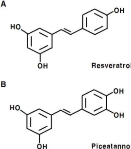
Piceatannol(3,4,3',5'-tetrahydroxy-trans-stilbene)은 포
도와 와인, 대황 등에서 발견되어지는 polyphenol로서 화학
적 암 예방 물질로 주목받는 resveratrol(3,5,4'-trihydroxy-
trans-stilbene)의 유도체이다(Fig. 1)(3,4). 많은 연구들을
통하여 piceatannol의 항암활성이 보고되고 있다. 유방암과
전립선암세포에서 piceatannol은 세포주기를 억제하고 세포
사멸을 증가시키며, 암세포의 이동과 전이를 억제하였다
(5-9). 항암활성 외에도 piceatannol의 항염증 활성이 보고
되었으며, lipopolysaccharide(LPS)로 염증반응을 유도시킨
대식세포에서 piceatannol은 염증반응의 주요 매개체인 ni-
tric oxide(NO)의 생성을 억제하였다(4,10). 또한 선행연구
를 통해 piceatannol이 dextran sulfate sodium으로 유도된
Fig. 1. Chemical structure of resveratrol (A) and piceatannol (B).
대장염을 억제함을 관찰하였다(11). 하지만 다양한 시각에 서 작용기전이 밝혀진 항암활성과 달리 piceatannol의 항염 증 기전은 여전히 제한적으로 보고되고 있다. 본 연구에서는 piceatannol의 항염증 기전을 염증반응에서 중요한 NF-κB 를 포함한 다양한 신호전달경로를 통해 밝히고자 한다.
재료 및 방법
실험재료
세포배양에 사용한 Dulbecco’s modified Eagle’s me- dium(DMEM)은 Welgene(Daegu, Korea)에서 구입하였고, fetal bovine serum(FBS)과 penicillin-streptomycin(P/S) 은 Lonza(Walkersville, MD, USA)에서 구입하였다. Pice- atannol, LPS, β-actin 항체, 3-(4,5-dimethylthiazol-2-yl)- 2,5-diphenyltetrazolium bromide(MTT), propidium iodide (PI)는 Sigma Chemical Co.(St. Louis, MO, USA)에서 구입 하였다. p65, Lamin-B, signal transducer and activator of transcription 3(STAT3), hemeoxygenase(HO)-1, NF-E2 related factor 2(Nrf2) 항체는 Santa Cruz Biotechnology (Santa Cruz, CA, USA)에서, iNOS와 COX-2, P-STAT3 (Ser
727), P-STAT3(Tyr
705) 항체는 BD Transduction La- boratories(Palo Alto, CA, USA)에서, 그리고 inhibitory κ B(IκB)-α, P-stress-activated protein kinase/c-Jun NH
2- terminal kinase(SAPK/JNK) 항체는 Cell Signaling Tech- nology(Beverly, MA, USA)에서 구입하였다. Anti-rabbit IgG-Alexa488 항체는 Invitrogen(Carlsbad, CA, USA)에서 구입하였다.
세포배양
본 연구에서 사용한 쥐의 대식세포인 Raw264.7 세포는 American Type Culture Collection(Rockville, MD, USA)에 서 구입하였다. Raw264.7 세포는 DMEM을 사용하여 37
oC 습윤한 CO
2incubator(5% CO
2/95% air)에서 배양하였다.
실험에 사용할 세포를 유지하기 위해서 DMEM에 10% FBS 와 100 kU/L penicillin, 0.17 mmol/L streptomycin을 첨가하 여 사용하였다. 세포가 80% confluent해지면 phosphate- buffered saline(PBS, pH 7.4)으로 세포의 단층을 씻어낸 후, scraper를 사용하여 세포를 긁어서 계대 배양하였다.
MTT 분석
Piceatannol의 농도에 따른 Raw264.7 세포의 독성을 조사 하기 위하여 MTT assay를 통해 살아있는 세포의 수를 조사 하였다. Raw264.7 세포를 10% FBS가 포함된 배지에 희석 하여 50,000 cells/well의 밀도로 24-well plate에 분주하였 다. 24시간 후, 1% FBS가 함유되어 있는 DMEM(serum- deprivation medium, SDM)으로 24시간 serum deprivation 하였다. 그 후, LPS(1 mg/L)와 다양한 농도(0~10 μmol/L) 의 piceatannol이 포함된 SDM으로 세포를 24시간 배양한 후, MTT(2.4 mmol/L)가 포함된 배양액으로 교체하였다. 3 시간 후에 배양액을 제거한 후, 형성된 푸른색의 formazan 을 isopropanol로 용해하여 570 nm 파장에서 흡광도를 측정 하였다.
NO 및 염증성 사이토카인의 분비 측정
Piceatannol이 LPS에 의해 유도되는 NO 및 염증성 사이 토카인의 분비에 미치는 영향을 조사하기 위하여 위에서 언 급한 바와 같이 Raw264.7 세포를 LPS와 piceatannol를 첨가 한 배양액으로 배양하였다. 그 후 24시간 conditioned me- dium을 수집한 후, 세포배양액에 분비된 NO의 양은 Griess reagent system(Promega, Madison, WI, USA)으로, TNF- α , IL-6, IL-1β의 양은 각각의 enzyme-linked immuno- sorbent assay(ELISA) kit(eBioscience, San Diego, CA, USA)를 사용하여 측정하였다.
Total cell lysates와 nuclear extracts의 준비
Raw264.7 세포를 100-mm 배양 접시에 1.5×10
6세포를 분주한 후, 위에서 언급한 바와 같이 LPS와 piceatannol를 첨가한 배양액으로 배양하였다. 세포를 1 mmol/L iodo- acetic acid와 1 mmol/L phenylmethanesulfonyl fluoride (PMSF)가 포함된 ice-cold PBS(washing buffer)로 헹구었 다. 배양 접시에 washing buffer 2 mL를 넣고 얼음 위에서 scraper를 사용하여 세포를 모은 후 원심분리(4,000×
g, 3 min, 4
oC)하였다. Pellet에 lysis buffer(20 mmol/L HEPES, 1% Triton X-100, 150 mmol/L NaCl, 1 mmol/L EDTA, 1 mmol/L EGTA, 100 mmol/L NaF, 10 mmol/L sodium pyrophosphate, 1 mmol/L iodoacetic acid, 0.2 mmol/L PMSF, 20 mg/L aprotinin, 15 μmol/L antipain, 21 μmol/L leupeptin, and 0.5 mmol/L benzamidine HCl)를 넣고 40분 동안 4
oC에서 세포를 용해하였다. Total cell lysates는 원심 분리(13,400×
g, 10 min, 4
oC)하여 cell debris를 제외한 상층 액을 수집하여 준비하였다.
핵분획을 준비하기 위하여 scraper를 사용하여 모은 세포
에 hypotonic buffer[10 mmol/L HEPES, pH 7.9, 1.5 mmol/
L MgCl
2, 10 mmol/L KCl, 0.2 mmol/L PMSF, 0.5 mmol/L dithiothreitol(DTT), 0.5% NP-40, 10 mmol/L iodoacetic acid, 20 mg/L aprotinin, 15 μmol/L antipain, 21 μmol/L leu- peptin, 0.5 mmol/L benzamidine HCl]를 첨가하여 얼음에서 10분간 용해하였다. 원심분리(2,300×
g, 15 min, 4
oC)하여 상 층액(세포질분획)을 수집한 후, NP-40가 포함되지 않은 hypotonic buffer로 pellet를 한차례 헹구었다. 이 pellet를 150 μ L low salt buffer(20 mmol/L HEPES, pH 7.9, 1.5 mmol/L MgCl
2, 10 mmol/L KCl, 0.2 mmol/L PMSF, 0.2 mmol/L EDTA, 0.5 mmol/L DTT, 10% glycerol)로 resuspension한 후, 50 μL high salt buffer(1.6 mol/L KCl이 포함)를 한 방울 씩 첨가하여 섞어주고 얼음에서 한 시간 동안 두었다. 핵분 획은 원심분리(25,000×
g, 30 min, 4
oC)하여 상층액을 수집 한 후 -70
oC에 보관하였다. Total cell lysates, 세포질과 핵 분획의 단백질의 양은 BCA protein assay(Pierce, Rockford, IL, USA)를 사용하여 정량하였다.
Western blot analysis
Cell lysates(50 μg protein)를 4~20% gradient sodium- dodecylsulfate-polyacrylamide gel electrophoresis(SDS- PAGE)에서 크기에 따라 분리한 뒤, polyvinylidene fluoride membrane(Millipore, Bedford, MA, USA)으로 이동시켰다.
Membrane은 5% milk-TBST(20 mmol/L Tris-HCl, 150 mmol/L NaCl, 0.1% Tween-20, pH 7.5)로 1시간 incubation 한 후, TBST로 10분간 3회 헹구었다. 각각의 항체를 5%
milk/TBST(iNOS, COX-2, β-actin, p65, Lamin-B, HO-1) 혹은 5% BSA/TBST(IκB-α, P-SAPK/JNK, P-STAT3, STAT3)로 희석(1:1,000)한 후, membrane을 1시간 incuba- tion하고 TBST로 10분간 3회 헹구었다. Anti-rabbit 또는 anti-mouse HRP-conjugated antibody를 5% milk/TBST 에 희석(1:5,000)하여 membrane을 1시간 동안 incubation 하고 TBST로 10분간 3회 헹구었다. Antibody에 결합된 단 백질들의 signal은 SuperSignal West Dura Extended Dur- ation Substrate(Pierce)를 사용한 chemiluminescence 방법 을 통하여 가시화하였다.
Reverse transcription-polymerase chain reaction (RT-PCR)
위에서 언급한 바와 같이 Raw264.7 세포를 LPS와 picea- tannol를 첨가한 배양액에서 배양한 후, RNeasy Plus Mini Kit(Qiagen, Valencia, CA, USA)를 사용하여 total RNA를 분리하였다. Oligo dT와 superscript II reverse transcrip- tase를 이용하여 template RNA로부터 cDNA를 합성하고 HO-1(12)의 primer(sense: 5'-TTACCTTCCCGAACAT- CGAC-3', antisense: 5'-GCATAAATTCCCACTGCCAC- 3') 혹은 β-actin(13)의 primer(sense: 5'-GTTTGAGACC- TTCAACACCCC-3', antisense: 5'-GTGGCCATCTCC-
TGCTCGAAGTC-3')를 사용하여 PCR을 수행하였다(an- nealing temperature: 60
oC). PCR product는 1% agarose gel 에서 전기영동하고 ethidium bromide를 사용하여 가시화하 였다.
Luciferase reporter gene assay
Raw264.7 세포는 실험 당일에 배양 접시를 80%로 덮도록 분주하고, P/S이 첨가되지 않은 10% FBS를 함유한 DMEM 배지에서 24시간 동안 배양하였다. Antioxidant response elements(ARE)의 transcriptional activity는 mouse HO-1 의 enhancer 2와 minimal promoter region을 포함하는 ARE reporter plasmid(14)를 이용하여 측정하였다. 먼저 Nucleo- fector-II(Amaxa, Gaithersburg, MD, USA)를 사용하여 ARE reporter plasmid를 pCMV-β-galactosidase control vector와 co-transfection 하였다. Transfection된 세포를 24-well plate에 분주하고, 다음날 SDM으로 24시간 동안 serum deprivation한 후, 다양한 농도의 piceatannol이 첨가 된 배양액에서 6시간 배양하였다. Transcriptional activity 는 luciferase assay system(Promega)를 이용하여 측정하였 고 β-galactosidase activity로 normalize 하였다.
Immunocytochemistry
Raw264.7 세포를 4-well chamber slide에 분주하고 다음 날 SDM으로 serum deprivation 하였다. 24시간 후 10 μmol/
L piceatannol이 첨가되거나 첨가되지 않은 배양액에서 4시 간 배양하였다. 배양액을 제거하고 4% paraformaldehyde로 15분간 고정한 후, 0.1% Triton X-100으로 30분간 per- meabilization 하였다. Nrf2 항체(1:100)로 4
oC에서 incuba- tion(overnight)한 후, TBST로 헹구었다. Anti-rabbit IgG- Alexa488 항체(1:1,000)로 상온에서 1시간 incubation한 후, TBST로 헹구었다. 핵은 PI(1.5 μmol/L)로 염색한 후, 봉합하 여 형광현미경(Carl Zeiss, Jena, Germany)으로 관찰하였다.
통계처리
본 연구의 모든 실험 분석 결과는 각 실험군의 평균과 표 준오차로 계산하고 각 실험 군들의 평균치간의 유의성은 SAS statistical software, version 8.12(SAS Institute, Cary, NC, USA)를 이용하여 ANOVA 분석 후 Duncan's multiple range test로 p<0.05 수준에서 검증하였다.
결과 및 고찰
Piceatannol이 NO의 생성에 미치는 영향
선천 면역을 담당하는 대식세포는 염증반응을 일으키는
대표적인 면역세포로서 활성산소종, 활성질소종, 사이토카
인과 같은 염증성 인자들의 분비를 통해 염증반응을 매개한
다. 쥐의 대식세포인 Raw264.7은 염증반응을 위한 세포실험
에 폭넓게 사용되는 세포주로서, 염증반응을 유도하기 위한
자극제로 그람음성균 세포막의 당지질인 LPS가 주로 사용
0 0.2 0.4 0.6 0.8 1 1.2 1.4
0 2.5 5 10
Piceatannol (μmol/L)
A b s o rb a n c e ( a t 5 7 0 n m ) .
LPS
Fig. 2. Effects of piceatannol on viable Raw264.7 cell number.
Raw264.7 cells were plated in 24-well plates at 50,000 cells/well.
After serum deprivation, cells were treated with various concen- trations (0~10 μmol/L) of piceatannol in the presence of LPS for 24 hr. Cell numbers were estimated by the MTT assay. Each bar represents the mean±SEM (n=4).
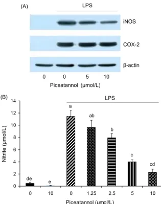
0 0 5 10 Piceatannol (μmol/L)
iNOS
COX-2
β-actin (A) LPS
LPS
0 2 4 6 8 10 12 14
0 10 0 1.25 2.5 5 10
Piceatannol (μmol/L)
N it ri te ( μ m o l/ L ) .
de
cd c b ab a
e
(B)
Fig. 3. Piceatannol decreases LPS-induced NO production via the inhibition of iNOS expression in Raw264.7 cells. (A) Raw264.7 cells were plated in 100 mm dishes at 1.5×10
6cells/dish.
After serum deprivation, cells were treated with various concen- trations (0~10 μmol/L) of piceatannol in the absence or presence of LPS. Cells were lysed, and the lysates were subjected to Western blotting with their relevant antibodies. Photographs of chemiluminescent detection of the blots, which were representa- tive of three independent experiments, are shown. (B) Raw264.7 cells were plated in 24-well plates at 50,000 cells/well. After se- rum deprivation, cells were treated with various concentrations (0~10 μmol/L) of piceatannol in the absence or presence of LPS.
24 hr conditioned media were collected and the NO concentrations were measured using the Griess reagent system. Each bar repre- sents the mean±SEM (n=4). Means without the same letter dif- fer, p<0.05.
된다. LPS 외에도 protein kinase C(PKC)의 활성화를 통해 암의 촉진인자로 작용하는 phorbol myristate acetate(PMA) 도 염증유도 물질로 사용되며, 이러한 유도물질에 따라 염증 성 인자들의 발현에 차이가 생기기도 한다. 예를 들어 대표 적인 염증성 사이토카인인 TNF-α, IL-6, IL-1β는 LPS에 의해 유도되지만 PMA는 IL-6와 IL-1β의 발현을 유도하지 못하였다(15). 이는 LPS가 세포막의 Toll-like receptor 4 (TLR4)에 결합하여 PKC를 포함한 다양한 신호전달경로를 활성화시키기 때문으로 사료된다. 본 연구에서는 LPS로 염 증반응을 유도한 Raw264.7 세포에서 piceatannol의 항염증 기전을 조사하였다.
먼저 MTT assay를 통해 Raw264.7 세포에 독성을 나타 내지 않는 piceatannol의 농도를 선정하고자 하였다. Fig. 2 에 나타난 바와 같이 piceatannol은 10 μmol/L의 농도에서 세포독성을 나타내지 않았다. Matsuda 등(4)의 연구에 따르 면 piceatannol은 peritoneal macrophages에서 100 μmol/L 의 높은 농도에서도 세포독성을 나타내지 않고 NO의 생성 을 억제하였다. 반면 Raw264.7 세포를 사용한 실험에서는 세포독성에 대한 IC50가 19 μmol/L였다고 보고되었다(10).
따라서 이후의 실험에서는 세포독성을 나타내지 않은 농도 인 10 μmol/L 이내로 piceatannol을 처리하였다.
염증반응에서 중요한 역할을 하는 염증 매개체인 NO와 prostaglandin(PG)E
2는 각각 NOS와 COX에 의해 생성된다.
NOS에는 3가지 isoform(neuronal NOS, inducible NOS, endothelial NOS)이 존재하며, 이들 중 iNOS가 염증 반응에 서 중요한 역할을 차지한다(16). 또한 COX는 2가지 isoform (COX-1, COX-2)이 존재하며, 대부분의 조직에서 발현되어 PGs의 항상성에 관여하는 COX-1과 달리 COX-2의 발현은 성장인자, 사이토카인 등의 자극에 의해 유도된다(17). Pic- eatannol이 iNOS와 COX-2의 발현에 미치는 영향을 조사하 기 위하여 Western blot를 수행하였다. iNOS와 COX-2의 발 현은 LPS 처리에 의해 유도되었으며, piceatannol은 iNOS
의 발현을 억제하였지만 COX-2의 발현에는 영향을 미치지 않았다(Fig. 3A). LPS와 piceatannol를 첨가한 배양액으로 세포를 배양하여 수집한 24시간 conditioned medium으로 세포가 분비한 NO의 양을 정량한 결과, piceatannol은 LPS 에 의해 증가된 NO의 생성을 농도 의존적으로 감소시켰다 (Fig. 3B). 이 결과를 통해 piceatannol은 iNOS의 발현 억제 를 통해 NO의 생성을 억제함을 알 수 있다. Peritoneal mac- rophages와 BV2 microglia를 이용한 실험에서 piceatannol 은 iNOS의 발현을 억제하여 NO의 생성을 억제함이 보고되 었다(4,18). 반면 Raw264.7 세포를 이용한 실험에서 picea- tannol은 iNOS의 발현에는 영향을 미치지 않고 NO의 생성 을 억제하였다(10). 이러한 결과의 차이는 실험방법(본 연구 에서는 24시간 동안 처리함)의 차이인 것으로 여겨진다.
Piceatannol이 염증관련 신호전달에 미치는 영향
iNOS gene promoter에는 NF-κB, activator protein-
1(AP-1), CCAAT/enhancer binding protein(C/EBP)과 같
LPS
0 200 400 600 800 1000 1200 1400
0 10 0 5 10
Piceatannol (μmol/L)
T N F -α ( p g /m L ) .
(A)
d
c b
a
d
LPS
0 1000 2000 3000 4000 5000
0 10 0 5 10
Piceatannol (μmol/L)
IL -6 ( p g /m L ) .
c
b b a
c
(B)
LPS
0 20 40 60 80 100
0 10 0 5 10
Piceatannol (μmol/L)
IL -1 β ( p g /m L ) .
d
b c a
d
(C)
Fig. 4. Piceatannol decreases LPS-induced production of TNF-α, IL-6, and IL-1β in Raw264.7 cells. Raw264.7 cells were treated with piceatannol and/or LPS as described in Fig.
3. 24 hr-conditioned media were collected, and the amount of TNF-α, IL-6, and IL-1β released within them was measured us- ing the relevant ELISA kits. Each bar represents the mean±SEM (n=4). Means without the same letter differ, p<0.05.
은 전사인자들의 결합을 위한 homologous consensus se- quence들이 포함되어 있다(19,20). Piceatannol은 Raw264.7 세포에서 LPS에 의해 유도된 C/EBPδ의 발현을 감소시킴이 보고(10)되었으나 NF-κB와 AP-1 신호전달계에 미치는 영 향에 대해서는 명확하게 보고되지 않았다. IκB는 NF-κB subunit(p65, p50)과 결합하여 세포질에 묶어두는 역할을 한 다. NF-κB 신호전달경로가 활성화되면 IκB의 인산화가 유 도되어 IκB의 분해가 이루어지고, 결과적으로 NF-κB sub- unit들은 IκB로부터 자유롭게 되어 핵으로 이동하여 전사인 자로 작용하게 된다(21). Raw264.7 세포를 18시간 동안 pi- ceatannol로 전처리한 후, LPS를 20분 동안 처리하여 염증 반응을 유도하였다. Western blot 결과, IκB의 단백질 수준은 LPS 처리에 의해 감소하였으며, piceatannol의 처리는 LPS 에 의해 유도된 IκB의 분해를 농도 의존적으로 억제하였다 (Fig. 4A). 또한 piceatannol의 처리는 LPS에 의해 유도된 p65의 핵으로 이동을 억제하였다(Fig. 4B). Piceatannol에 의한 NF-κB 신호전달의 억제는 다양한 연구를 통해 보고되 었다. Piceatannol은 Jurkat과 BV2 microglia와 같은 면역세 포 외에도 암세포(MCF-7과 Hela 세포) 및 정상상피세포 (MCF-10A)에서 염증유도 물질(LPS, TNF, PMA)에 의해 유도된 NF-κB의 활성화를 억제(18,22-24)하였으며, 이는 IKKβ를 통해 이루어짐이 관찰되었다(24).
또 다른 전사인자로서 AP-1은 c-Jun, c-Fos로 구성되며, 이들은 각각 SAPK/JNK와 extracellular signal-regulated kinase1/2의 영향을 받는다(25). c-Jun의 upstream 단백질로 작용하는 SAPK/JNK의 인산화는 LPS 처리에 의해 증가하 였으며, 증가된 SAPK/JNK의 인산화는 piceatannol 처리에 의해 유의적으로 감소하였다(Fig. 4A). 이 결과를 통해 pi- ceatannol은 C/EBP 뿐만 아니라 NF-κB와 AP-1의 신호전 달경로의 억제를 통해 iNOS의 발현을 억제함을 알 수 있다.
Piceatannol이 염증성 사이토카인의 생성에 미치는 영향 대식세포는 NO와 PGE
2외에도 염증성 사이토카인을 분 비하여 염증반응에 관여한다. Raw264.7 세포에서 LPS와 piceatannol이 염증성 사이토카인에 미치는 영향을 ELISA kit를 사용하여 조사하였다. Piceatannol의 처리는 LPS에 의 해 증가된 TNF-α, IL-6, IL-1β의 분비를 유의적으로 감소 시켰다(Fig. 5). NF-κB와 AP-1은 TNF-α, IL-6, IL-1β gene의 transactivation에 필요한 전사인자로서 작용한다 (26). 따라서 piceatannol에 의한 NF-κB와 AP-1 신호전달 경로의 억제는 iNOS의 발현 감소뿐만 아니라 염증성 사이 토카인의 생성을 감소시킴을 알 수 있다.
IL-6의 autocrine/paracrine action에 대한 piceatannol 의 영향
NF-κB는 LPS 외에도 TNF, IL-1, IL-6, H
2O
2등의 자극 에 의해서도 활성화된다(27). 이는 LPS에 의해 증가된 염증 성 사이토카인이 autocrine 및 paracrine 방식으로 NF-κB
신호전달 경로를 통해 염증반응에 관여할 수 있음을 보여준 다. Piceatannol은 TNF에 의해 유도된 NF-κB의 활성화를 억제함이 보고되었다(23). 최근 염증반응과 STAT3에 대한 관심이 증가하고 있다. STAT3는 다양한 사이토카인과 성 장인자의 신호전달을 매개하는 중요한 molecule로서 작용 하며, 이들 신호전달의 활성화로 인하여 각각 Tyr
705와 Ser
727잔기의 인산화가 이루어진다. STAT3의 인산화로 인하여 STAT3는 핵으로 이동하여 전사인자로서 작용한다(28).
STAT3는 염증과정 중 독성환경으로부터 세포를 보호하고
세포사멸을 막는 역할을 하며, 이는 STAT3의 downstream
(A) LPS
IκB-α
P-SAPK/JNK
β-actin
(B) LPS
p65 (Nuclear)
Lamin-B
0 0 5 10 Piceatannol (μmol/L)
Fig. 5. Piceatannol inhibits LPS-stimulated NF-κB and AP- 1 signaling in Raw264.7 cells. Raw264.7 cells were plated and serum-deprived. After serum deprivation, cells were incubated for 18 hr in 1% FBS medium containing various concentrations (0~10 μmol/L) of piceatannol. Cells were then treated with LPS for another 15 min. (A) Cells were lysed, and the cell lysates were subjected to Western blotting with their relevant antibodies. (B) Cells were subjected to subcellular fractionation. The resulting nuclear fraction were analysed by Western blotting with their rel- evant antibodies. Photographs of chemiluminescent detection of the blots, which were representative of three independent experi- ments, are shown.
P-STAT3 (Ser
727)
P-STAT3 (Tyr
705)
STAT3
β-actin (A) LPS
0 5 0 2.5 5 Piceatannol (μmol/L)
P-STAT3 (Tyr
705)
β-actin
(B) rm-IL-6
0 0 5 10 Piceatannol (μmol/L)
(C) rm-IL-6
STAT3 (Cytosol)
STAT3 (Nuclear)
0 0 5 10 Piceatannol (μmol/L)
Fig. 6. Piceatannol inhibits STAT3 signaling in Raw264.7 cells. Raw264.7 cells were plated and serum-deprived. (A) After serum deprivation, cells were treated with various concentrations (0~5 μmol/L) of piceatannol in the absence or presence of LPS for 24 hr. Cells were lysed, and the lysates were subjected to Western blotting with their relevant antibodies. (B, C) Cells were incubated for 1 hr in 1% FBS medium containing various concen- trations (0~10 μmol/L) of piceatannol. Cells were then treated with recombinant mouse IL-6 (rm-IL-6) for another 5 min. (B) Cells were lysed, and the lysates were subjected to Western blot- ting with their relevant antibodies. (C) Cells were subjected to subcellular fractionation. The resulting fraction were analysed by Western blotting with an anti-STAT3 antibody. Photographs of chemiluminescent detection of the blots, which were representa- tive of three independent experiments, are shown.
target gene과 관련이 있다(29).
Raw264.7 세포에서의 LPS의 처리는 STAT3의 Ser
727잔 기의 인산화를 증가시켰다(Fig. 6A). Mouse mesangial cells 에서 IL-1β와 LPS는 STAT3를 활성화시킨다고 보고되었 다(30). Piceatannol의 처리는 LPS에 의해 유도된 STAT3의 Tyr
705와 Ser
727잔기의 인산화를 억제하였다(Fig. 6A). LPS 의 작용은 수용체인 TLR4를 통해 이루어지는데, 최근 LPS 가 TLR4의 adapter protein인 myeloid differentiation pri- mary-response protein 88을 통해 STAT3를 활성화시킨다 고 보고되었다(31). 또한 LPS의 처리는 STAT3의 Tyr
705잔 기의 인산화도 증가시켰으며, 이로써 LPS에 의해 증가된 사이토카인에 의해 STAT3의 Tyr
705잔기의 인산화가 이루 어졌으리라 예상할 수 있다.
STAT3의 Tyr
705잔기의 인산화를 유도하는 대표적인 사 이토카인으로 IL-6를 들 수 있다. IL-6가 수용체에 결합하면 Janus family kinase와 SRC tyrosine kinase가 활성화되어 STAT3를 인산화 시킨다. Recombinant IL-6의 처리는 p- STAT3(Tyr
705)를 증가시키고 STAT3의 핵으로의 이동을 증가시켰다. 그리고 piceatanol의 처리는 IL-6에 의해 유도 된 STAT3의 Try
705잔기의 인산화를 억제하고 STAT3의 핵으로의 이동을 억제하였다(Fig. 6B, 6C). Yu 등(32)은 myocytes에서 IL-6에 의해 활성화된 STAT3가 iNOS의 발 현을 증가시킴을 보고하였다. Piceatannol은 STAT3 in- hibitor(33)로 잘 알려져 있는데, 위 결과는 piceatannol이
IL-6의 발현을 억제(Fig. 5)함으로써 STAT3 경로를 억제함 을 보여준다.
Piceatannol이 HO-1의 발현이 미치는 영향
염증반응에서 late-limiting enzyme으로 작용하는 heme oxygenase(HO)는 heme이 carbon monoxide, biliverdin, ferrous ion으로 산화되는 반응을 촉매 하여 세포내의 항상 성을 유지하고, 산화적 손상의 감소, 염증반응을 억제, 세포 사멸 억제, 세포증식을 조절함으로써 조직을 보호하는 중요 한 역할을 한다(34). 3가지 mammalian HO isoform이 존재 하며, 이중 HO-1은 stress-responsive protein으로서 Nrf2 에 매개되는 ARE의 transcriptional activation을 통하여 다 양한 산화제에 의해 유도된다.
Piceatannol이 HO-1의 발현에 미치는 영향을 Western
blot과 RT-PCR을 통하여 조사한 결과, HO-1의 단백질 발
현은 piceatannol 처리 4시간부터 증가하기 시작하여 처리
HO-1
β-actin HO-1
β-actin
(A) (B)
0 2 4 6 8 10 μmol/L Piceatannol (h)
0 2 4 6 8 10 μmol/L Piceatannol (h)
0 5000 10000 15000 20000 25000 30000 35000 40000 45000
0 10 0 5 10
Piceatannol (μmol/L) A R E -d ri v e n l u c if e ra s e a c ti v it y . (% o f p G L 3 -b a s ic c o n tr o l) .
c c
a a
b
pGL3-basic ARE-Luc
(C)
Nrf2 PI Merge (D)
Fig. 7. Piceatannol increases the expression of HO-1 in Raw264.7 cells. (A, B) Raw264.7 cells were plated and se- rum-deprived. After serum deprivation, cells were treated with 10 μmol/L piceatannol for indicated times. (A) Cells were lysed, and the lysates were subjected to Western blotting with their rel- evant antibodies. (B) Total RNA was isolated and RT-PCR was performed. Photographs of chemiluminescent detection of the blots or the ethidium bromide-stained gels, which were repre- sentative of three independent experiments, are shown. (C) Cells were co-transfected with ARE reporter gene construct containing the enhancer 2 and minimal promoter region of mouse HO-1 and pCMV-β-galactosidase control vector using Nucleofector-II, and the transfected cells were plated in 6-well plates at 2×10
5cells/
well. After serum deprivation, cells were treated with various concentrations (0~10 μmol/L) of piceatannol for 6 hr. Luciferase and β-galactosidase activities were measured with total cell lysates. Luciferase activity was normalized to β-galactosidase activity. Each bar represents the mean±SEM (n=4). Means with- out the same letter differ, p<0.05. (D) Cells were plated in 4-well chamber slides and treated with 10 μmol/L piceatannol and sub- jected to immunocytochemistry with Nrf2 antibody. Cell nuclear DNA was stained with PI (red color). Bright yellow coloration of the merged image represents Nrf2 translocated from the cyto- sol to the nucleus.
Piceatannol LPS
NF-?B AP-1 STAT3
iNOS TNF-a IL-1ß IL-6
Nrf2
HO-1 Anti-inflammatory effect NF-κB AP-1 STAT3
Fig. 8. Proposed schemata of the mechanisms by which pi- ceatannol inhibits LPS-stimulated inflammatory responses.
6시간에 최고치에 달하였으며, 이후 다소 감소하였다(Fig.
7A). 그리고 HO-1 transcripts는 단백질 발현보다 앞선 pi- ceatannol 처리 2시간부터 증가하였으며 처리 4시간에 최고 치에 달하였다(Fig. 7B). HO-1 transcription activity를 조 사하기 위하여 mouse HO-1의 enhancer 2와 minimal pro-
moter region을 포함한 ARE reporter plasmid를 사용하여 luciferase assay를 수행하였다. 5 μmol/L piceatannol의 처 리에서부터 ARE-driven luciferase의 transcriptional ac- tivity가 증가하였다(Fig. 7C). 또한 immunocytochemistry 결과, piceatannol은 Nrf2의 핵내 축적을 증가시켰다(Fig.
7D). HO-1과 carbon monoxide는 iNOS의 발현과 NO의 생 성을 억제한다고 보고되었다(35,36). 또한 HO-1은 IL-10의 항염증효과를 매개한다고 보고되었다(37). 따라서 이 결과 를 통해 piceatannol은 Nrf2-ARE를 통해 HO-1의 발현을 증가시킴으로 항염증 효과를 나타냄을 보여준다.
대표적인 stilbene으로서 화학적 암 예방 물질로 주목받는 resveratrol은 항암활성 외에도 항염증 효과가 있다고 보고 되었다(38). Resveratrol은 piceatannol과 마찬가지로 NF-κ B를 억제하고 iNOS와 COX-2의 발현을 감소시킴이 관찰되 었다(39). 반면 resveratrol과 trans-stilbene은 HO-1의 발현 을 유도하지 못하였다고 보고되었다(40). 이는 resveratrol 과 piceatannol의 구조적인 차이(Fig. 1)에 의한 것으로 생각 되며, 특히 OH기의 위치가 HO-1의 발현에 중요하게 작용하 는 것으로 여겨진다.
본 연구에서 piceatannol은 HO-1의 발현을 증가시켰으며, 이는 iNOS와 염증성 사이토카인의 발현 억제와 함께 picea- tannol의 항염증 활성의 주요 요인으로 사료된다(Fig. 8).
요 약
본 연구에서는 염증반응을 조절하는 다양한 신호전달체
계를 중심으로 분자생물학적 방법을 통해 piceatannol의 항
염증 기전을 규명하였다. LPS로 염증반응을 유도한 Raw
264.7 대식세포에서 piceatannol은 iNOS의 발현 억제를 통
해 NO의 생성을 감소시키고 염증성 사이토카인(TNF-α,
IL-6, IL-1β)의 생성을 감소시켰다. 염증반응을 조절하는 신
호전달체계 중 piceatannol은 LPS에 의해 유도된 IκB의 분
해와 p65의 핵으로의 이동을 억제하고, LPS에 의해 유도된
SAPK/JNK의 인산화를 억제하였다. 또한 piceatannol은
LPS와 IL-6(LPS에 의해 증가됨)에 의한 STAT3의 활성화
를 억제하였다. 뿐만 아니라 piceatannol은 Nrf2의 핵 내 축 적을 야기하고 ARE의 transcriptional activity를 증가시켜 HO-1의 발현을 증가시켰다. 본 연구의 결과, piceatannol은 NF-κB와 AP-1, STAT3 신호전달의 억제를 통해, 그리고 HO-1의 발현 증가를 통해 항염증 효과를 나타내었다(Fig.
8).
감사의 글
본 연구논문은 정부(교육과학기술부)의 재원으로 한국연 구재단의 중견연구자지원사업(2012-0004903)과 선도연구 센터육성사업(2012-0000642), 그리고 지식경제부 지역혁신 센터사업(한림대 식의약품의 효능평가 및 기능성소재개발 센터)에 의하여 수행되었으며, 이에 감사드립니다.
문 헌