• 검색 결과가 없습니다.

Anti-inflammatory Effect of Dactyloquinone B and Cyclospongiaquinone-1 Mixture in RAW264.7 Macrophage and ICR Mice

N/A
N/A
Protected

Academic year: 2022

Share "Anti-inflammatory Effect of Dactyloquinone B and Cyclospongiaquinone-1 Mixture in RAW264.7 Macrophage and ICR Mice"

Copied!
5
0
0

로드 중.... (전체 텍스트 보기)

전체 글

(1)

21(4) : 268-272 (2015)

http://dx.doi.org/10.20307/nps.2015.21.4.268

268

Anti-inflammatory Effect of Dactyloquinone B and Cyclospongiaquinone-1 Mixture in RAW264.7 Macrophage and ICR Mice

Dong-Sung Lee

1,#

, In Hyun Hwang

2,#

, Nam-Kyung Im

3

, Gil-Saeng Jeong

3,

*, and MinKyun Na

4,

*

1

Department of Biomedical Chemistry, Konkuk University, Chungju 380-701, Korea

2

Department of Chemistry, University of Iowa, Iowa City, IA 52242, United States

3

College of Pharmacy, Keimyung University, Daegu 704-701, Korea

4

College of Pharmacy, Chungnam National University; Daejeon 305-764, Korea

Abstract – Sesquiterpene-quinone is a class of secondary metabolites frequently encountered from marine sponge.

The present study was designed to examine the anti-inflammatory action of sponge-derived dactyloquinone B (DQB) and cyclospongiaquinone-1 (CSQ1) mixture using lipopolysaccharide (LPS)-induced inflammatory responses.

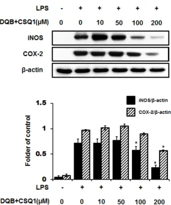
We measured the production of nitric oxide (NO), tumor necrosis factor-alpha (TNF-α), interleukin-1β (IL-1β), and interleukin-6 (IL-6) and expression of inducible nitric oxide synthase (iNOS) and cyclooxygenase-2 (COX-2) protein. TNF-α, IL-1β, and IL-6 production, which increased by treatment with LPS, were significantly inhibited by DQB and CSQ1 mixture. It also decreased the production of NO production, and iNOS and COX-2 expression. Furthermore, it reduced 12-O-tetradecanoylphorbol 13-acetate (TPA)-induced ear edema of ICR mice.

These results demonstrate that sesquiterpene-quinone, DQB and CSQ1 mixture, might serve as a chemical pipeline for the development of anti-inflammatory agent.

Keywords – Dactyloquinone B, Cyclospongiaquinone-1, Anti-inflammatory effect, Ear edema model

Introduction

Marine sponge has been served as a prolific source of diverse secondary metabolites.

1

In particular, sesquiterpene- quinone is a class of compounds frequently encountered as bioactive marine natural products.

2

A wide range of bioactivities have been described from this class, which included antimicrobial, anti-HIV, anti-inflammatory, and cytotoxic activities, and thereby a lot of synthetic attempts have been made for these compounds.

2

Ilimaquinone is one of the most well-known sesquiterpene-quinones comprised of sesquiterpene and benzoquinone subunits,

3,4

and its mechanism of action associated with Golgi membrane was reported.
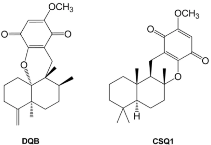
5

As a part of the chemical investigation of marine sponge, a series of cyclospongia- quinones were isolated from Stelospongia conulata.

6

Cyclospongiaquinones contain a characteristic ether linkage between the sesquiterpene and quinone units, forming an additional ring system relative to that of most other sesquiterpene-quinones such as ilimaquinone.

6

Recently, a short synthetic strategy for cyclospongiaquinone-1 was developed using commercial diterpene alcohol.

7

Another set of example showing the same type of ether connection is dactyloquinones obtained from Dactylospongia elegans, but the skeleton of their sesquiterpene unit is rearranged as was the case for ilimaquinone.

8-10

Despite the unique structural feature of these series of compounds, to our knowledge, previous reports of their bioactivity have been limited to moderate cytotoxicity of dactyloquinone B, neodactyloquinone, and cyclospongiaquinone-2.

10-12

Inflammatory response is a defense mechanism of the body to various stimuli, in which macrophages play an important role in regulating the reaction via production of pro-inflammatory mediators and cytokines, such as nitric oxide (NO), tumor necrosis factor- α (TNF-α), and interleukin-1 β (IL-1β).

13

Prolonged or deregulated inflam- matory responses can lead to various diseases, including arthritis, hepatitis, septic shock syndrome, neurodegenerative disorders, and sepsis.

14

Accordingly, many studies have focused on searching for the treatment of inflammatory-

*Author for correspondence

Professor Gil-Saeng Jeong, College of Pharmacy, Keimyung Uni- versity, 1095 Dalgubeol-daero, Daegu 704-701, Republic of Korea Tel: +82-53-580-6649; E-mail: [email protected]

Professor MinKyun Na, College of Pharmacy, Chungnam National University, 99 Daehak-ro,Yuseong, Daejeon 305-764, Republic of Korea

Tel: +82-42-821-5925; E-mail: [email protected]

#

These authors contributed equally to this work.

(2)

related diseases. In the course of our studies aiming to identification of anti-inflammatory metabolites from marine sponges, we investigated the anti-inflammatory activity of dactyloquinone B (DQB) and cyclospongiaquinone-1 (CSQ1) mixture obtained from the previous study.

15

The inhibitory mechanism of DQB and CSQ1 mixture was investigated through the inflammatory reactions in LPS- stimulated RAW264.7 macrophages and in 12-O-tetrade- canoylphorbol 13-acetate (TPA)-induced ear edema of ICR mice.

Experimental

Extraction and isolation – The collection and extraction of sponges and their extraction has been described in our earlier work.

15

The sponge extract (3.6 kg) was subjected to silica gel vacuum liquid chromatography (VLC) eluting with a stepwise gradient of hexane/acetone/methanol/

water to provide 13 fractions (Fr. 1 to 13). Fr. 10 (39.3 g) was further divided into 9 fractions (Fr. 10-1 to 10-9) using silica gel VLC with a gradient order of the same four solvents. C

18

MPLC (15.5 × 4 cm) was applied to Fr.

10-6 (2.7 g) with an isocratic solvent of methanol/water (85:15) to yield 7 subfractions (Fr. 10-6-1 to 10-6-7). A mixture of DQB and CSQ1 (4.3 mg) was obtained from repeated column chromatography of Fr. 10-6-2 (458 mg) using C

18

HPLC (250 × 20.0 mm, 5 μm) with isocratic methanol/water (78:22).

15

Materials – Dulbecco's modified Eagle's medium (DMEM), fetal bovine serum (FBS), and other tissue culture reagents were purchased from Gibco BRL Co. All other chemicals were obtained from Sigma Chemical Co. (St.

Louis, MO) unless indicated otherwise. RAW264.7 cells were obtained from the American Type Culture Collection (Manassas, VA). Primary antibodies, including those raised against COX-2 and iNOS appropriate secondary antibodies used for western blot analysis were purchased from Santa Cruz Biotechnology (Santa Cruz, CA).

Enzyme-linked immunosorbent assay (ELISA) kits for TNF- α, IL-6, and IL1-β were purchased from R&D Systems (Minneapolis, MN).

Cell culture and viability assay – RAW264.7 cells were maintained at 5 × 10

5

cells/mL in DMEM medium supplemented with 10% heat-inactivated FBS, penicillin G (100 units/mL), streptomycin (100 mg/mL), and L - glutamine (2 mM), and were incubated at 37

o

C in a humidified atmosphere containing 5% CO

2

and 95% air.

The effects of various experimental modulations on cell viability were evaluated by determining mitochondrial reductase function with an assay based on reduction of

tetrazolium salt, 3-[4,5-dimethylthiazol-2-yl]-2,5-dipheny- ltetrazolium bromide (MTT), into fomazan crystals. The formation of formazan is proportional to the number of functional mitochondria in living cells. For determination of cell viability, 50 mg/mL of MTT was added to cell suspension for 4 h. The formazan formed was dissolved in acidic 2-propanol and optical density was measured at 590 nm. The optical density of formazan formed in control (untreated) cells was taken as 100 % viability.

Nitrite (NO production) determination – Production of nitrite, a stable end product of NO oxidation, was used for measurement of iNOS activity. Nitrite present in conditioned media was determined by a method based on the Griess reaction. An aliquot of each supernatant (100 μL) was mixed with the same volume of Griess reagent (0.1% (w/v) N-(1-naphathyl)-ethylenediamine and 1% (w/

v) sulfanilamide in 5% (v/v) phosphoric acid) for 10 min at room temperature. The absorbance of the final product was measured spectrophotometrically at 525 nm using an ELISA plate reader and the nitrite concentration in samples was determined from a standard curve of sodium nitrite made up in phenol red-free DMEM.

TNF-α, IL-6, and IL-1β assay – Levels of TNF-α, IL- 6, and IL-1 β present in each sample were determined using a commercially available kit from R&D Systems (Minneapolis, MN, USA). The assay was performed according to the manufacturer’s instructions. Briefly, RAW264.7 cells were cultured in 24-well plates, pre- incubated for 12 h with different concentrations of DQB+

CSQ1 and then stimulated for 18 h with LPS. Cell culture supernatants were collected immediately after treatment and spun at 13,000 × g for 2 min to remove particulate matter. The medium was added to a 96-well plate pre- coated with affinity-purified polyclonal antibodies specific for mouse TNF- α, IL-6, or IL-1β. An enzyme-linked polyclonal antibody specific for mouse TNF- α, IL-6, or IL-1 β was added to the wells and left to react for 2 h, followed by a final wash to remove any unbound antibody- enzyme reagent. A substrate solution was added, and the intensity of color produced, measured at 450 nm (correction wavelength set at 540 nm or 570 nm), as proportional to the amount of TNF- α, IL-6, or IL-1β present.

Western blot analysis – Western blot analysis was performed by lysing cells in 20 mM Tris-HCl buffer (pH 7.4) containing a protease inhibitor mixture (0.1 mM PMSF, 5 mg/mL aprotinin, 5 mg/mL pepstatin A, and 1 mg/mL chymostatin). Protein concentration was determined using a Lowry protein assay kit (P5626; Sigma). An equal amount of protein for each sample was resolved by 12%

sodium dodecyl sulfate-polyacrylamide gel electrophoresis

(3)

(SDS-PAGE) and then electrophoretically transferred onto a Hybond enhanced chemiluminescence (ECL) nitro- cellulose membrane (Bio-Rad, Heracules, CA). The membrane was blocked with 5% skim milk and sequen- tially incubated with primary antibody (Santa Cruz Biote- chnology) and horseradish peroxidase-conjugated secondary antibody, followed by ECL detection (Amersham Pharmacia Biotech, Piscataway, NJ).

Animal experiment – The Institutional Animal Care and Use Committee at Johnson & Johnson approved all procedures used in these experiments. ICR mice (male, 25-30 g) were purchased from the Samtako Bio Korea Co. (Gyeonggido, Korea) and permitted free access to a standard chow diet and tap water. All mice were accli- matized for 1 week prior to the experiments and maintained at 22 ± 2°C with a relative humidity of 50 ± 5% and 12 h light–dark cycle.

TPA-induced ear edema – TPA-induced ear edema was performed as described previously.

16,17

Biopsies of 9 mm diameter were taken from mouse ears that had been challenged with TPA only or treated with DQB+CSQ1 after TPA challenge.

Statistical analysis – Data are expressed as mean ± S.D. for at least three independent experiments. To compare three or more groups, one-way analysis of variance followed by Newman-Keuls post hoc test was used. Statistical analysis was performed using GraphPad Prism software, version 3.03 (GraphPad Software Inc, San Diego, CA).

Results and Discussion

Repeated purification using a reverse-phase HPLC method afforded a biologically active mixture of DQB and CSQ1, and their structures were identified by com- parison of

1

H and

13

C NMR data with literature values.

6,8

The

1

H NMR data showed signals for two different sesquiterpene moieties, including an exo-methylene unit ( δ

H

4.90, s; 4.80, s), an isolated methylene unit ( δ

H

2.64, d, J = 19 Hz; 2.11, d, J = 19 Hz), seven methyl groups ( δ

H

1.38, s; 1.22, s; 1.13, s; 0.90, s; 0.87, s; 0.83, s; 0.76, d, J = 6.7 Hz), together with complex aliphatic region. In addition, observation of signals for two olefinic singlets ( δ

H

5.76, s; 5.73, s) and coincident two methoxy groups ( δ

H

3.80, s) implied presence of two closely related compounds in this sample. The

13

C NMR data clearly displayed two sets of signals, and characteristic shift values for the four carbonyl carbons ( δ

C

182.4, 181.7, 181.6, 181.2) suggested that both components of this mixture contain a quinone unit. Furthermore, two

oxygenated sp

3

carbons ( δ

C

88.2, 82.0) are indicative of an ether linkage in both components, while all the sp

3

carbons of typical sesquiterpene-quinones are not oxy- Fig. 1. Chemical structures of dactyloquinone B (DQB) and cyclospongiaquinone-1 (CSQ1).

Fig. 2. Effect of DQB and CSQ1 mixture on cell viability or NO production in RAW 264.7 cells. Cells were plated and cultured for 24 h in the absence or presence of different concentration of DQB and CSQ1 mixture (A). Cells were treated with DQB and CSQ1 mixture and LPS (100 ng/ml) for 24 h (B). NO production was calculated in culture media by Griess reagent (B). Each bar represents means ± S.D. of three independent experiments.

*

p <

0.05 compared to the group treated with LPS.

(4)

genated. Therefore, the two components presumed to be both sesquiterpene-quinones containing an ether linkage, but differing by their sesquiterpene structure based on the presence of only one unit of exo-methylene and odd number of methyl groups. Analysis of

1

H and

13

C NMR data for each component and comparison of the data with literature reports

6,8

enabled identification of this sample as a 3:4 mixture of DQB and CSQ1 (Fig. 1).

The concentration of DQB and CSQ1 mixture which possibly cause cytotoxic effect toward RAW264.7 cells was determined by MTT assay. The cell viability was not significantly decreased at the concentration of DQB and CSQ1 mixture up to 200 μM as shown in Fig. 2A. Thus,

the concentration range of 10-200 μM was employed for all subsequent experiments. Pre-treatment of the RAW264.7 cells with DQB and CSQ1 mixture for 12 h resulted in decrease of NO production (Fig. 2B). Because suppression of LPS-induced pro-inflammatory mediators (e.g., NO) was observed by the addition of DQB and CSQ1 mixture, effects of this mixture on LPS-induced cytokines production were further examined. Prior to the LPS stimulation, the RAW264.7 cells were pre-treated with DQB and CSQ1 mixture for 1 h. As shown in Fig. 3, TNF- α, IL-6, and IL- 1 β productions were decreased upon addition of DQB and CSQ1 mixture in a concentration-dependent manner, which was measured by enzyme immunoassay. The expression of pro-inflammatory enzymes, including COX-2 and iNOS play a crucial role in immune-activated macrophages by producing COX-2-derived PGE

2

and iNOS-derived NO.

13,14

The RAW264.7 cells were treated with LPS in the presence or absence of non-cytotoxic concentrations of DQB and CSQ1 mixture, and levels of iNOS and COX-2 protein expression were recorded. Pre- treatment of the RAW264.7 cells with DQB and CSQ1 mixture for 1 h resulted in decrease of iNOS and COX-2 Fig. 3. Effect of DQB and CSQ1 mixture on LPS-induced pro-

inflammatory cytokines in RAW 264.7 cells. Cells were treated with various concentrations of DQB and CSQ1 mixture for 1 h and incubated with LPS during 24 h. Productions of TNF- α (A), IL-1β (B), and IL-6 (C) were measured in culture media by ELISA. Each bar represents means ± S.D. of three independent experiments.

*

p < 0.05 compared to the group treated with LPS.

Fig. 4. Effect of DQB and CSQ1 mixture on LPS-induced iNOS and COX-2 expression in RAW 264.7 cells. Cells were treated with various concentrations of DQB and CSQ1 mixture for 1 h and incubated with LPS during 24 h. The expression levels of iNOS and COX-2 in cells were analyzed by Western blot. Each bar represents means ± S.D. of three independent experiments.

*

p

< 0.05 compared to the group treated with LPS.

(5)

protein expression (Fig. 4). TPA is a main active compound found in croton oil, inducing vasodilation, erythema, and edema within 5 hours after its application to skin.

18

Reduction of TPA-induced edema response was observed by the treatment of DQB and CSQ1 mixture in a mouse model of TPA-induced ear edema, and curcumin, a positive control, also showed the similar activity (Fig.

5). These results suggested that DQB and CSQ1 mixture suppressed the inflammatory condition in LPS-stimulated RAW264.7 macrophages and in TPA-induced ear edema of ICR mice.

In conclusion, the present study revealed anti-inflam- matory activity of DQB and CSQ1 mixture. These sesquiterpene-quinones suppressed production of NO, TNF- α, IL-6, and IL-1β and expression of iNOS and COX-2 in the LPS-induced RAW264.7 cells. In addition, inhibition of the TPA-induced ear edema was observed in ICR mice upon treatment of DQB and CSQ1 mixture, suggesting that these sesquiterpene-quinones may serve as

a potential chemotype for the development of anti- inflammatory agent.

Acknowledgments

This study was supported by the National Research Foundation of Korea (NRF) funded by the Korean govern- ment (NRF-2010-0020484, 2014R1A2A2A01006793).

References

(1) Blunt, J. W.; Copp, B. R.; Keyzers, R. A.; Munro, M. H.; Prinsep, M. R. Nat. Prod. Rep. 2014, 31, 160-258.

(2) Marcos, I. S.; Conde, A.; Moro, R. F.; Basabe, P.; Diez, D.; Urones, J. G. Mini Rev. Org. Chem. 2010, 7, 230-254.

(3) Luibrand, R. T.; Erdman, T. R.; Vollmer, J. J.; Scheuer, P. J.; Finer, J.; Clardy, J. Tetrahedron 1979, 35, 609-612.

(4) Capon, R. J.; MacLeod, J. K. J. Org. Chem. 1987, 52, 5059-5060.

(5) Takizawa, P. A.; Yucel, J. K.; Veit, B.; Faulkner, D. J.; Deerinck, T.;

Soto, G.; Ellisman, M.; Malhotra, V. Cell 1993, 73, 1079-1090.

(6) Kazlauskas, R.; Murphy, P. T.; Warren, R. G.; Wells, R. J.; Blount, J.

F. Aust. J. Chem. 1978, 31, 2685-2697.

(7) Fernandez, A.; Alvarez, E.; Alvarez-Manzaneda, R.; Chahboun, R.;

Alvarez-Manzaneda, E. Chem. Comm. 2014, 50, 13100-13102.

(8) Mitome, H.; Nagasawa, T.; Miyaoka, H.; Yamada, Y.; van Soest, R.

W. J. Nat. Prod. 2001, 64, 1506-1508.

(9) Mitome, H.; Nagasawa, T.; Miyaoka, H.; Yamada, Y.; van Soest, R.

W. Tetrahedron 2002, 58, 1693-1696.

(10) Mitome, H.; Nagasawa, T.; Miyaoka, H.; Yamada, Y.; van Soest, R.

W. J. Nat. Prod. 2003, 66, 46-50.

(11) Ovenden, S. P.; Nielson, J. L.; Liptrot, C. H.; Willis, R. H.;

Tapiolas, D. M.; Wright, A. D.; Motti, C. A. J. Nat. Prod. 2011, 74, 65- 68.

(12) Jankam, A.; Somerville, M. J.; Hooper, J. N. A.; Brecknell, D. J.;

Suksamrarn, A.; Garson, M. J. Tetrahedron 2007, 63, 1577-1582.

(13) Lee, D. S.; Jeong, G. S.; Li, B.; Park, H.; Kim, Y. C. Int.

Immunopharmacol. 2010, 10, 850-858.

(14) Lee, D. S.; Jang, J. H.; Ko, W.; Kim, K. S.; Sohn, J. H.; Kang, M.

S.; Ahn, J. S.; Kim, Y. C.; Oh, H. Mar. Drugs 2013, 11, 1409-1426.

(15) Hwang, I. H.; Oh, J.; Zhou, W.; Park, S.; Kim, J.-H.; Chittiboyina, A. G.; Ferreira, D.; Song, G. Y.; Oh, S.; Na, M.; Hamann, M. T. J. Nat.

Prod. 2015, 78, 453-461.

(16) Agut, J.; Tarrida, N.; Sacristan, A.; Ortiz, J. A. Methods Find. Exp.

Clin. Pharmacol. 1996, 18, 233-234.

(17) Liebel, F.; Lyte, P.; Garay, M.; Babad, J.; Southall, M. D. Arch.

Dermatol. Res. 2006, 298, 191-199.

(18) Rao, T. S.; Currie, J. L.; Shaffer, A. F.; Isakson, P. C. Inflammation.

1993, 17, 723-741.

Received July 14, 2015 Revised August 12, 2015 Accepted August 13, 2015 Fig. 5. Effect of DQB and CSQ1 mixture on TPA-induced ear

edema in ICR mouse. Mice were administrated with or without the various concentrations of DQB and CSQ1 mixture. Curcumin was used as a positive control. TPA (2 μg/ear) was dropped onto the surface of the ear. The mice were sacrificed 4 h after the application of TPA. An ear disc 9.0 mm diameter was punched out of each ear and weighted. Values are shown as means ± S.D.

of n=5 mice.

*

p < 0.05 compared to the group treated with TPA.

수치

Fig. 2. Effect of DQB and CSQ1 mixture on cell viability or NO production in RAW 264.7 cells
Fig. 4. Effect of DQB and CSQ1 mixture on LPS-induced iNOS and COX-2 expression in RAW 264.7 cells

참조

관련 문서

In addition, the expression of REDD1 was increased when Huh7 cells were treated with PA, and overexpression of REDD1 affected cell survival and lipid

1) Effects of methanol extracts of Capsella bursa-pastoris on cyclooxygenase-2 (COX-2) and Inducible Nitric oxide synthase (iNOS) expression in human prostate cancer cell

co-treatment with hispidulin and TGF-β up-regulated the protein of expression E-cadherin and occludin against TGF-β-induced in MCF-7 and HCC38 cells.. The

In other words, pomegranate extract has an effect of inhibiting the progression of alveolar bone loss due to periodontal inflammation by reducing the expression of COX-1 and

protein levels in control or PIG3 depleted HCT116 and HeLa cells were tested. by

4 &gt; Effects of Taro on COX-2 expression and iNOS expression(hot water) in human thyroid cancer cells. The cells were pretreated for 48hours with either

Pharmacokinetic parameters were determined following an oral administration of nateglinide (30 mg/kg) to rabbits in the presence and absence of calcium channel blockers

Micro- and nano-sized pores were formed on the surface of the alloy using PEO and anodization methods, and the pore shape change according to the Zr