• 검색 결과가 없습니다.

Cutaneous asthenia associated with Ehlers-Danlos syndrome in a Yorkshire terrier

N/A
N/A
Protected

Academic year: 2021

Share "Cutaneous asthenia associated with Ehlers-Danlos syndrome in a Yorkshire terrier"

Copied!
4
0
0

로드 중.... (전체 텍스트 보기)

전체 글

(1)

大韓獸醫學會誌(2007) 第47卷 第1號

Korean J Vet Res

(2007) 47(1) : 91~94

91

Cutaneous asthenia associated with Ehlers-Danlos syndrome in a Yorkshire terrier

Sung-Jin Cho, Okjin Kim

1,

*

Family Animal Clinic, Gunsan 573-351, Korea

1

College of Life Science and Natural Resources, Wonkwang University, Iksan 570-749, Korea

(Accepted: January 9, 2007)

Abstract : Cutaneous asthenia or dematosparaxis is an uncommon, congenital and inherited connective tissue disease of dog and cat, resembling Ehlers-Danlos syndrome (EDS) in man. EDS is characterized by loose, hyperextensible and, fragile skin, it is attributed to mutations in connective tissue gene. These mutations cause defects in type I or III collagen synthesis and as a result lack of strength or elasticity to skin, joint, ligament and vessels. EDS-affected animals often experience subcutaneous hematomas that have long bleeding times. The 4-years old male Yorkshire terrier was evaluated because of subcutaneous hematoma after stifle surgery. Clinical examination revealed a thin and hyperextensible skin and joint laxity. The degree of skin extensibility index was 23.4%, marked skin stretchy. Clinical diagnosis was confirmed by histophathological examination of a skin biopsy revealing reduced packing density of collagen fiber of skin.

Key words : collagenopathies, cutaneous asthenia, dermatosparaxis, dog, Ehlers-Danlos syndrome Cutaneous asthenia or dermatosparaxis is a rare

inherited connective tissue disease of dog and cat [1, 3, 5, 6] that resemble Ehlers-Danlos syndrome (EDS) in human [6, 9]. In veterinary medicine, EDS has been described in several species with collagenopathies [1, 6, 8]. EDS is characterized by loose, hyperextensible and fragile skin, which is attributed to mutations in connective tissue gene [2, 4]. These mutations cause defects in type I or III collagen synthesis and as a result lack of strength or elasticity to skin, joint, ligament, and vessels [1, 4, 9]. Thus, EDS-affected animals revealed clinically integumental, musculoskeletal, gastrointestinal, and cardiovascular signs [6, 7]. In dogs, cutaneous asthenia, also known as rubber puppy disease has been reported in English Springer Spaniels, Boxers, Beagles, Pembroke Welsh Corgies, German Shepherd Dogs, Saint Bernards, and Dachshunds [6, 9]. But literatures of EDS-affected dog are extremely few. Here, we describe the clinical and histopathologic findings in an dermatosparaxis-affected Yorkshire terrier.

The 4-years old male Yorkshire terrier was referred for severe bruising of right stifle. The bruising was

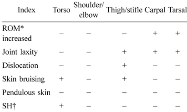
subcutaneous hematoma after stifle surgery because of bilateral medial patella luxation (grade II) and right cruciate ligament rupture. The owner informed that the dog had fragile skin and history of bruising, since 8 months of age. On physical examination, the dog revealed predominant periodontitis, alopecia, skin fragility and hypermorbility, and carpal and tarsal joint laxity.

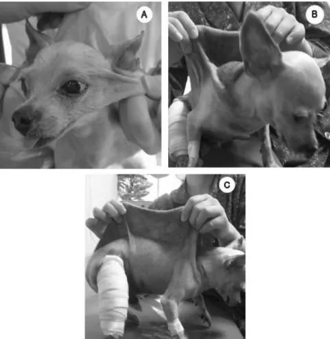
Especially, the skin showed marked stretchy, fragile and thinly over the trunk. Skin elasticity was also decrease, as well as the penlight penetrated thinly skin and the dermal vessels are exposed (Fig. 1, 2). Skin hyperextensibily appears over the torso and head except for extremities. Skin bruising was detected around neck and abdomen. Otherwise the dog appeared healthy, and no other abnormalities were found. There was no ophthalmic symptom and we don't obtain pedigree history.

To determine the skin laxity, skin extensibility index (SEI) was calculated as objective measurements. SEI was examined as follows: skin extensibility index (%)

= [height of fold of skin over lumbar region (cm)/

distance from occipital to base of tail (cm)]

×

100.

*Corresponding author: Okjin Kim

College of Life Science and Natural Resources, Wonkwang University, Iksan 570-749, Korea

[Tel: +82-63-850-6668, Fax: +82-63-850-7308, E-mail: kimoj@wonkwang.ac.kr]

(2)

92 Sung-Jin Cho, Okjin Kim

Vertical height of extensible skin (dorsolumbar skin fold) was 7.5 cm and body length (from the base of the tail to the occipital crest) was 32 cm, therefore calculated SEI was 23.4% (Table 1). On laboratory result, WBC, ALP, AST and ALP were mildly elevated (Table 2).

Skin biopsy was performed using a 4-mm skin biopsy punch and skin specimens obtained from the dorsum was fixed in 10% formalin-glutaraldehyde solution, and stained with hematoxylin-eosin. Histo- pathologically, there were a diffuse in the dermis layer and a loose in the hypodermis layer, and moreover there were decreased dense collagen fiber of dermis and number of hair follicles (Fig. 3). This case was diagnosed as EDS based on the SEI increased and collagen fiber decreased.

Fig. 1. A Yorkshire terrier with skin hyperextensibility by Ehlers-Danlos syndrome or cutaneous asthenia. This case showed excess cutaneous laxity on the head (A) and dorsum (B) with gentle traction. The hyperextensible skin was severe thinly and showing the dermal vessels through the other side. The copious bruising was noted around the right thigh and lower abdomen after stifle surgery (C).

Fig. 2. Histologic micrograph of dorsal dermis from Ehlers-

Danlos syndrome showing decreased dense collagen fiber

of skin. Note lack, shortened and fragmented dermal

collagen bundles (H&E stain,

×

200).

(3)

Ehlers-Danlos syndrome in a Yorkshire terrier 93 EDS or cutaneous asthenia is a rare disease in

veterinary and human medicine, which is a group of inheritable collagen mutation in connective tissue genes [4, 9]. Collagen, glue-like protein of connective tissue, defect reduce mechanical properties of skin, joints, ligaments, and blood vessels. Thus, EDS is characterized by skin hyperextensibility, joint hypermo- rbility and fragility of skin and blood vessels [6, 8].

The clinical and histopathological findings were consistent with the diagnosis of EDS [5, 6]. In human medicine, EDS have been divided ten subtypes based on mode of inheritance and biochemical defect and clinical signs [2, 4, 9]: Type I; gravis, Type II; mitis, Type III; benign familial hypermorbility, Type IV;

ecchymotic or arterial, Type V; x-linked, Type VI;

ocular, Type VII; arthochalasis multiplex congenita, Type VIII; periodontal form, Type IX; x-linked cutis laxa, skeletal, and Type X; fibronetic deficient. In veterinary medicine, EDS is described with cutaneous asthenia that is results from abnormalities in the biosynthesis of collagen and disturbance in connective tissue function [3, 7, 8]. According to lack of collagen bundle, these diseases are commonly manifested soft tissue hyperextensibility or fragility, for instance; skin

fragility (dermatosparaxia), skin laxity (cutaneous asthenia) or joint laxity, and periodontitis [3, 5, 6]. Skin laxity was principally produced also in this case. Cutaneous asthenia was first identified in cattle and then, reported many species, sheep, cat, dog, and mink. But classifi- cation of collagenopathies in veterinary medicine is insufficient because rare genetic, biochemical and ultrastructural studies have been carried out [6]. This case was evaluated lack of dense collagen fiber in the extensible and thin skin on microstructural and clinical studies. The skin extensibility index is used to quantitate the extensibility of affected dorsolumbar skin. SEI has been used as common criterion for clinical identification of EDS in dog and cat [3, 8].

Normally, SEI of non-affected dog is 8% to 15% [3, 5], but this case was 23.4%. More significantly, the skin of EDS-affected animal is fragile and easily laceration [1, 2]. Consequently, readily manifests subcu- taneous hematomas produced by minimal trauma and poor wound healing in EDS [2, 5, 9]. In this case, the copious bruising area of surgical stifle and ecchymosis around the abdomen are typical clinical features caused by fragile vessels. These signs are like EDS subtype I, II, III and IV [3, 6, 8]. Cutaneous asthenia resem- bling EDS type I-III is cause by a deficiency of type I procollagen-N-peptidase, which lead to accumulation of partially processed type I procollagen [1, 8].

Collagen fibers of affected animals were irregular arranged and lost mechanical strength [1, 3]. In light and electron microscopic study, collagen bundle were loosely integrated and separated on ground substance.

Collagen fibers appeared fragmented and isolated [6, 8]. The principal manifestation of ultrastructural finding was the disorganization in collagen fibril of dermis, for example; loosely fibril packing, variation in fiber diameter and disarray arrangement of collagen fiber [3, 6]. Lacked and fragmented dermal collagen Table 2. Results of hematological analysis

CBC count Differential count Chemistry

WBC (10

3

/ul) 25.45 Neutrophils seg 87 ALT (IU) 97

RBC (10

6

/ul) 3.73 Lymphocyte 7 AST (IU) 73

HB (g/dl) 9.2 Monocyte 6 ALP (IU) 318

PCV % 27.3 Eosinophil 0 BUN (mg/dl) 21

MCV fl 73 Basophil 0 Creatinine (mg/dl) 0.45

MCHC % 33.6

Table 1. Clinical appearance of this case

Index Torso Shoulder/ elbow Thigh/stifle Carpal Tarsal ROM*

increased

+ +

Joint laxity

− −

+ + +

Dislocation

− −

+

− −

Skin bruising +

+

− −

Pendulous skin

SH† +

*ROM (range of motion), † SH (skin hyperextensibility).

(4)

94 Sung-Jin Cho, Okjin Kim bundles were eminently observed also in this case. It

is clinical important to perceive cutaneous asthenia.

This abnormality are easily lacerated and presented large wound [2, 3]. In human and veterinary practical management, tape closure has been indicated to reduce large scar [3]. However, dog and cats suffer with cutaneous asthenia appear to have a normal life span, if ensure caution and free from trauma [3]. In this case, the bruising and wound were treated with carefully nursing and general post-operative treatment. On follow up after treatment, the surgical wound and bruising recovered but skin and joint laxity persisted.

This case is first report of cutaneous asthenia asso- ciated with EDS in a dog in Korea. Skin extensibility index and histological evaluation were useful for the diagnosis of cutaneous asthenia. The prominent signs of this rare disease were skin and joint laxity as also as poor wound healing that result in congenital abnormalities in the biosynthesis of collagen. Therefore, the affected animals were required carefully nursing and caution in trauma.

Acknowledgements

This paper was supported by Wonkwang University in 2007. The authors greatly appreciate Sangyun Lee, Tanwoo Park, Nanhee Bok, Sunhwa Hong, Namjin Sin and Bokyung Kim in Center for Animal Resources Development, Wonkwang University for their excellent technical assistance.

References

1. Benitah N, Matousek JL, Barnes RF, Lichtensteiger CA, Campbell KL. Diaphragmatic and perineal hernias

associated with cutaneous asthenia in a cat. J Am Vet Med Assoc 2004, 224 , 706-709, 698.

2. De Paepe A, Malfait F. Bleeding and bruising in patients with Ehlers-Danlos syndrome and other collagen vascular disorders. Br J Haematol 2004, 127 , 491-500.

3. Freeman LJ, Hegreberg GA, Robinette JD. Ehlers- Danlos syndrome in dogs and cats. Semin Vet Med Surg (Small Anim) 1987, 2 , 221-227.

4. Lawrence EJ. The clinical presentation of Ehlers- Danlos syndrome. Adv Neonatal Care 2005, 5 , 301- 314.

5. Minor RR, Lein DH, Patterson DF, Krook L, Porter TG, Kane AC. Defects in collagen fibrillogenesis causing hyperextensible, fragile skin in dogs. J Am Vet Med Assoc 1983, 182 , 142-148.

6. Paciello O, Lamagna F, Lamagna B, Papparella S.

Ehlers-Danlos-like syndrome in 2 dogs: clinical, histologic, and ultrastructural findings. Vet Clin Pathol 2003, 32 , 13-18.

7. Poulsen PH, Thomsen MK, Kristensen F. Cutaneous asthenia in the dog. A report of two cases. Nord Vet Med 1985, 37 , 291-297.

8. Sinke JD, van Dijk JE, Willemse T. A case of Ehlers- Danlos-like syndrome in a rabbit with a review of the disease in other species. Vet Q 1997, 19 , 182-185.

9. Yen JL, Lin SP, Chen MR, Niu DM. Clinical features

of Ehlers-Danlos syndrome. J Formos Med Assoc

2006, 105 , 475-480.

수치

Fig. 1.  A Yorkshire terrier with skin hyperextensibility by Ehlers-Danlos syndrome or cutaneous asthenia
Table 1.  Clinical appearance of this case

참조

관련 문서

The feed is commonly a solution in a solvent like ethanol or t-butanol, and the nonsolvent is water..

Serum uric acid levels and risk for vascular disease in patients with metabolic syndrome... Prevalence if the metabolic syndrome in a Turkish

Objective: The purpose of this study was to investigate the expression of syndecan-1, E-cadherin, and beta-catenin in tissue sections of normal sun-damaged skin,

In this group of high-risk patients with severe COVID-19 pneumonia, treatment with lenzilumab was associated with a significantly shorter time to clinical improvement compared

An investigation of adults with Asperger syndrome or high functioning autism, and normal sex differences.. The facilitation of social-emotional understanding

Actinic keratoses showed a trend towards increased expression in the basal layer compared with normal skin.. In actinic keratoses, Keratoacanthomas and seborrheic

A Study on the Meteorological Characteristics Associated with Heavy Rainfall Case of July 9, 2009 Using KLAPS Reanalysis Data..

environmental burdens associated with a product, process, or activity by identifying energy and materials used and. wastes released