KISEP Case Reports J Korean Neurosurg Soc 30::::211-216, 2001
반복적인 소뇌 출혈로 발현한 후두와 모세혈관 확장증
- 증 례 보 고 -
충남대학교 의과대학 신경외과학교실
이용묵·고현송·조준희·염진영·송시헌·김 윤
= Abstract =
Capillary Telangiectasia of the Posterior Fossa Presenting with Repeated Cerebellar Hemorrhage
- - -
- A Case Report --- -
Yong-Mook Lee, M.D., Hyeon-Song Koh, M.D., Jun-Hee Cho, M.D., Jin-Young Youm, M.D., Shi-Hun Song, M.D., Youn Kim, M.D.
Department of Neurosurgery, College of Medicine, Chungnam National University, Taejon, Korea
emorrhage due to capillary telangiectasia is rare. We report a case of capillary telangiectasia presenting with repeated cerebellar hemorrhage in a 38-year-old male. To our knowledge this is the first case of repeated cerebellar hemorrhage from the capillary telangiectasia. At the first operation, we removed hematoma only, but rebleeding occurred repeatedly in the same area. Finally, we evacuated the recurrent hematoma and vascular mass of capillary telangiectasia at the second operation under surgical microscope. Based on the findings of this case and a review of the literature, we conclude that capillary telangiectasia can be the cause of the massive repeated hemorrhage.
KEY WORDS:Capillary telangiectasia・Repeated cerebellar hemorrhage.
서 론
신경계의 혈관 기형은 일반적으로 동정맥 기형(Arterio- venous malformation), 해면상 혈관종(Cavernous hema- ngioma), 정맥 기형(Venous malformation), 모세혈관 확 장증(Capillary telangiectasia)으로 분류하며5)10), 이러한 병변들이 혈관 조영술에서 보이지 않는 경우를 혈관 조영 술상 잠재성 혈관 기형(Angiographically occult vascular malformation : AOVM)이라고 한다. 동정맥 기형, 해면상 혈관종, 정맥 기형은 자기공명 영상이나 혈관 조영술에서 각각의 독특한 형태를 가지고 있으며, 혈관 조영술에서 잠 재성인 혈관 기형은 일반적으로 조직병리학적 검사상 모세 혈관 확장증이나 해면상 혈관종으로 진단되는 경우가 대부 분이다1)5)10).
모세혈관 확장증은 혈관 조영술상 잠재성 혈관 기형이며 대부분 증상을 일으키지 않으므로 부검에서 우연히 발견되 는 경우가 대부분이고, 주로 뇌간이나 소뇌에 분포하여, 뇌 간 등에서 출혈을 일으킨다고 보고되지만, 다른 혈관 기형 의 동반 없이 순수한 모세혈관 확장증이 출혈을 일으킨다 는 보고는 매우 희소하다9)10). 본 교실에서는 젊은 남자 환 자에서 세차례의 반복적인 출혈을 일으킨 모세혈관 확장증 1례를 수술적 치험하고 방사선학적, 조직학적 검사를 시행 하여 문헌 고찰과 함께 보고하는 바이다.
증 례
환자는 38세 남자로 내원 10일전부터 현기증과 두통으 로 개인 의원에서 치료받았으나 호전 없이 지내던 중, 내 원 당일 증상이 더욱 악화되고 오심 및 구토 등이 발생하
HHHH
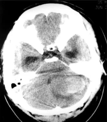
여 본원에 내원하였다. 이학적 검사상 유전성 모세혈관 확 장증의 소견은 발견할 수 없었으며, 신경학적 검사상 좌 측방 주시시 안구 진탕이 발생하였으나 소뇌 기능 검사상 특이 소견은 없었다. 요추천자 검사에서 뇌척수액 압력은 190mmH2O 이었으며 뇌척수액 색깔은 투명하였고, 혈구 는 관찰되지 않았다. 내원 다음날 시행한 자기공명 영상 에서 좌측 소뇌 반구에 T1 강조 영상에서 고신호 강도, T2 강조 영상에서 저신호 강도의 세포내 메트헤모글로빈 (methemoglobin) 상태의 혈종이 3×4.5cm 크기로 관찰 되었으며, 조영제 주입 후 조영 증강은 없었다(Fig. 1). 또 한 혈관 조영술에서는 혈관 기형을 의심할 만한 소견은 보 이지 않았다(Fig. 2).
입원 후 만니톨로 두개강내압을 조절하였으나 현기증 및 두통과 간헐적인 구토 증세가 지속되었으며, 내원 10일째 증상이 더욱 악화되고 뇌전산화 단층촬영상 수두증 소견이 보여(Fig. 3), 응급 수술을 시행하였다. 수술은 후두부에 방 정중 두피 절개 후, 좌측 후두골 절제술을 시행하고 혈종을 제거하였고, 혈종의 피막을 확인할 수 있었으며, 혈종 제거 부위에 유치 도관을 삽입하였다. 수술 후 1일째 시행한 추 적 뇌전산화 단층촬영상 혈종은 대부분 제거되었으며 수두 증 소견도 완화되었다(Fig. 4).
수술 후 11일째 환자는 다시 심한 두통 및 현기증을 호 소하였으며 추적 뇌전산화 단층촬영상 동일 위치에 재출혈 소견을 보였으며(Fig. 5), 추적 관찰 중이던 수술 후 19일
Fig. 1. A magnetic resonance imaging of the brain obtained on the first admission day. T1-weighted axial image showing the high signal intensity lesion in the left cerebellar hemisphere(A). T2-weighted axial image demonstrating the low signal intensity in center and the high signal intensity at periphery in the corresponding site(B).
AAA
A BB BB
Fig. 2. Anterio-posterior view of vertebral angiogram showing no definite vascular malformation.
이용묵·고현송·조준희·염진영·송시헌·김 윤
째 의식 저하와 함께 뇌전산화 단층촬영상 3차 출혈 소견 을 보여(Fig. 6), 재수술을 시행하였다. 2차 및 3차 출혈 당시 혈액학적 이상 소견은 발견할 수 없었다. 재수술은 이
전 두피 절개 부위를 다시 절개하고 후두골 절제술을 더욱 광범위하게 하였다. 현미경하에서 혈종을 모두 제거하자 혈 종의 전하측방에 밝은 갈색을 띠는 혈관 기형이 의심되는 부위가 있었으며, 이를 적출하여 조직 검사를 시행하였고,
Fig. 3. A computed tomography scan of the brain obtained on the tenth admission day showing a large hematoma and peripheral edema in the left cerebellar hemisphere with enlarged temporal horns of both lateral ventricles and deviation of the fourth ventricle.
Fig. 4. A computed tomography scan of the brain obtained on the first postoperative day showing the near total removal of the previous hematoma.
Fig. 6. A computed tomography scan of the brain obtained on the 19th postoperative day revealing the enlargement of hematoma and the change of shape.
Fig. 5. A computed tomography scan of the brain obtained on the 11th postoperative day demonstrating a reblee- ding in the previous operated site.
일부 잔유 병소는 수차례 전기 소작하였다. 조직 검사상 한 층의 내피 세포로 구성된 확장된 모세혈관 내에 충혈 소견 이 보였으며 모세혈관 사이에는 정상 신경조직 소견이 관찰 되었고, 헤모시데린 성분이 군데군데 있어 이전의 만성 출 혈 소견과 급성 출혈 소견이 같이 보였다(Fig. 7). Elastin 염색에서 elastin 성분은 보이지 않았다. 2차 수술 후 환자 는 의식 및 임상 증상의 급격한 호전을 보였으며, 2차 수술 13일째 촬영한 뇌전산화 단층촬영 소견상 혈종의 흔적은 완 전 소실되고 뇌실 크기도 완전 정상 소견을 보였다(Fig. 8).
내원 52일째 환자는 신경학적 이상 소견과 자각 증상 없이 상태로 퇴원하였으며, 현재까지 약 9개월간의 추적 관찰상 간헐적이고 경미한 두통과 현훈 이외에 특이 소견은 없었다.
고 찰
모세혈관 확장증은 임상적으로 증상이 없고, 방사선학적 검사도 발견하기 어려우며, 대개 부검에서 우연히 발견되는
신경계의 혈관 기형이다. 부검 례에서 약 0.04~0.15%의 빈도를 보이며8)9)10), 70% 정도가 뇌교 등의 후두와에 위치
하나5)9)10) 뇌기저핵이나 대뇌 피질에서도 발견할 수 있다6).
조직학적으로 정상 뇌실질에 의하여 나뉘어진 비정상적으로 확장된 모세혈관으로 이루어져 있으며, 모세혈관은 낭상 또 는 방추상의 확장을 보이기도 하며, 평활근이나 탄성 섬유, 석회화 등은 관찰되지 않고, 특징적으로 직접적인 동맥 유 입이 없으며, 유출 정맥도 없다2)4)5)10). 대부분의 혈관 기형 처럼 모세혈관 확장증도 선천적 원인으로 생각되며10), 발생 과정 중 혈관의 퇴화 과정의 이상으로 인하여 혈관 기형이 유발된다고 여겨지고 있다. 즉, 배아기 2개월에 발생하는 뇌실질내에는 많은 모세혈관이 존재하고, 동맥과 정맥이 점 진적으로 성숙됨에 따라 퇴화되는데, 이 과정 중에 혈관 형 성(angiogenesis)의 저해가 오면 결과적으로 모세혈관 확 장증이 나타나게 된다1).
Russel 등은 해면상 혈관종과 모세혈관 확장증이 서로 유 사한 면을 많이 갖고 있기 때문에 두 병변의 구분이 어려
Fig. 7. Photographs of biopsy specimen obtained in the second operation. A:There are many dilated capillaries with intervening parenchyma and intraparenchymal fresh hemorrhage(H & E, ×100). B:Capillaries consist with one layer of endothelial cells without the muscular layer, and the brownish hemosiderin component is shown around the ectatic capillaries (arrow)(H & E, ×200).
AAA
A BB BB
이용묵·고현송·조준희·염진영·송시헌·김 윤
운 경우가 때때로 있다고 보고하며, 둘 사이의 구분에 가장 중요한 구별점은 확장된 혈관 사이에 뇌실질의 존재 여부 라고 기술하였다9). 그러나 두 병변 사이의 이행(transitional) 형태의 존재에 대하여는 설명하지 못하였다. Rigamonti 등 은 20례의 해면상 혈관종을 분석한 결과, 해면상 혈관종과 모세혈관 확장증을 서로 다른 종류로 구분하기 애매모호하 다고 발표하며, 둘 사이의 이행 형태를 밝혀냈고 이들을 동 일한 혈관 기형 분류내의 양립하는 병변이라고 주장하였다7). McCormick 등은 모세혈관 확장증에서 해면상 혈관종으로 상당히 진행한 이행 형태의 병변은 그렇지 않은 병변보다 좀더 나쁜 자연 경과를 취할 수 있다고 보고하였다4). 이처 럼 몇몇 보고에서 모세혈관 확장증과 해면상 혈관종을 하 나의 질환 분류로 명명할 것을 주장하고 있지만, 현재까지 혈관 기형의 분류는 위에서 언급한 네가지의 분류가 일반 적이며, 본 증례의 경우 해면상 혈관종과 확연한 구분을 보 인 모세혈관 확장증 소견을 보였으며, 모세혈관 확장증 자 체로도 출혈의 원인이 될 수 있다는 것을 입증한 것으로 사 료된다. 모세혈관 확장증은 뇌혈관 조영술과 자기공명 영 상에서도 일반적으로 발견되지 않으나, Rigamonti 등은 부 검 례에서 사후에 시행한 자기공명 영상에서 저신호 강도 를 보인다고 보고하였으며7), Lee 등은 혈액내 산소 농도와 관련된 대조(blood oxygen-level-dependent contrast)
에 의하여 gradient-echo image(GRE)에서 저신호 강도를 나타나며, 이는 가장 민감도가 높은 검사라고 보고하였다3). 모세혈관 확장증은 동정맥 기형이나 해면상 혈관종과는 대 조적으로 대부분 출혈을 일으키지 않으며, 모세혈관 확장증 주변에 소량의 점상 출혈에 의하여 헤모시데린 침착이 있다 는 보고도 있으나 매우 희소하며, 부검 례에서도 흔하지 않 는 소견이다5)10). 모세혈관 확장증에서의 출혈은 극히 희소 하게 보고되는데, 1997년 Roost 등은 여러 보고를 정리하여 발표한 논문에서 지난 45년간 증상이 있는 순수한 모세혈관 확장증은 18례가 발표되었으며, 이들 중 11례에서 출혈에 의하여 증상이 발생하였다고 기술하고 있다8). 그러나 모세혈 관 확장증은 종종 해면상 혈관종과 동반되어 증상을 유발하 는 출혈을 일으킨다고 하며1)7), 또한 정맥 기형과 동반되어 출혈을 일으켰다는 보고도 1례 있었다4). 또한 출혈 부위도 뇌교나 연수 등의 뇌간부가 대부분으로 소뇌 출혈은 매우 드 물게 보고되고 있다. 본 저자들의 조사에 의하면 모세혈관 확장증이 원인이 되어 다량의 급성 소뇌 출혈을 일으킨 예 는 있으나2), 다량의 반복적 소뇌 출혈이 세차례 있었다는 지 상 보고는 현재까지 없었으며, 이는 국내뿐만 아니라 세계적 으로도 최초의 보고로 사료된다. 본 증례의 경우 1차 수술에 서는 병소의 완전 제거와 혈종 주변의 조직 검사가 시행되 지 못했으며, 이로 인해 남아 있던 소뇌의 모세혈관 확장증 병소로부터 2차 및 3차 출혈이 일어났을 것으로 판단된다.
결 론
젊은 환자에서 혈관 조영술과 자기공명 영상에서 출혈의 원인이 될 만한 병변이 없고, 고혈압이나 혈액 응고 장애, 뇌종양 등의 원인 질환 등이 없을 때 모세혈관 확장증의 가능성을 고려해야 하겠으며, 이러한 경우 혈종 및 혈종 주 변에 혈관 기형으로 의심되는 부위가 있다면 반드시 완전 제거하고, 만일 잔유 병소가 의심되면 전기 소작 등과 함께 병변 부위의 조직 검사가 필수적이라고 사료된다. 본 교실 에서는 반복적인 소뇌 출혈로 발현한 후두와 모세혈관 확 장증 1례를 수술 치험하고 좋은 결과를 얻었기에, 문헌 고 찰과 함께 보고하는 바이다.
•논문접수일:2000년 5월 9일
•심사완료일:2000년 7월 18일
•책임저자:이 용 묵
301-721 대전광역시 중구 대사동 640 충남대학교 의과대학 신경외과학교실
전화:042) 220-7361, 전송:042) 220-7364 E-mail:[email protected]
[email protected] Fig. 8. A computed tomography scan of the brain obtained
on the 13th postoperative day after the second operation showing the totally removed repeated hemorrhage in the left cerebellar hemisphere, and restoration of shape of fourth ventricle.
References
1) Awad IA, Robinson JR, Mohanty S, Estes ML:Mixed vas- cular malformations of the brain:clinical and pathogenetic considerations. Neurosurgery 33:179-188, 1993
2) Bland LI, Lapham LW, Ketonen L, Okawara SH:Acute cerebellar hemorrhage secondary to capillary telangiectasia in an infant. A case report. Arch Neurol 51:1151-1154, 1994 3) Lee RR, Becher MW, Benson ML, Rigamonti D:Brain capi-
llary telangiectasia:MR imaging appearance and clinicohis- topathologic findings. Radiology 205:797-805, 1997 4) McCormick PW, Spetzler RF, Johnson PC, Drayer BP:
Cerebellar hemorrhage associated with capillary telangiectasia and venous angioma:A case report. Surg Neurol 39:451- 457, 1993
5) McCormick WF:The pathology of vascular(“arteriove-
nous”) malformations. J Neurosurg 24:807-816, 1966 6) Milandre L, Pellisier JF, Boudouresques G, Bonnefoi B,
Cherif AA, Khalil R:Nonhereditary multiple telangiectasias of the central nervous system. Report of two clinicopatholo- gical cases. J Neurol Sci 82:291-304, 1987
7) Rigamonti D, Johnson PC, Spetzler RF, Hadley MN, Drayer BP:Cavernous malformations and capillary telangiectasia: A spectrum within a single pathological entity. Neurosurgery 28:60-64, 1991
8) Roost DV, Kristof R, Wolf HK, Keller E:Intracerebral capi- llary telangiectasia and venous malformation:A rare associ- ation. Surg Neurol 48:175-183, 1997
9) Russell DS, Rubinstein LJ:Pathology of tumours of the ner- vous system. 5th ed. Baltimore, Md:Williams & Wilkins, 1989, pp727-746
10) White RJ, Kernohan JW, Wood MW:A study of fifty intra- cranial vascular tumors found incidentally at necropsy. J Neuropathol Exp Neurol 17:392-398, 1958