보 문
Novel substrate specificity of a thermostable β-glucosidase from the hyperthermophilic archaeon, Thermococcus pacificus P-4
Yun Jae Kim
1,2, Jae Eun Lee
1, Hyun Sook Lee
1,2, Kae Kyoung Kwon
1,2, Sung Gyun Kang
1,2, and Jung-Hyun Lee
1,2*
1
Korea Institute of Ocean Science and Technology, Ansan 426-744, Republic of Korea
2
Department of Marine Biotechnology, University of Science and Technology, Daejeon 305-350, Republic of Korea
초고온 고세균 Thermococcus pacificus P-4로부터 내열성 β-glucosidase의 새로운 기질 특이성
김윤재
1,2・ 이재은
1・ 이현숙
1,2・ 권개경
1,2・ 강성균
1,2・ 이정현
1,2*
1
한국해양과학기술원,
2과학기술연합대학원대학교 해양생명공학부
(Received February 4, 2015; Accepted March 13, 2015)
ABSTRACT: Based on the genomic analysis of Thermococcus pacificus P-4, we identified a putative GH1 β-glucosidase-encoding gene (Tpa-glu). The gene revealed a 1,464 bp encoding 487 amino acid residues, and the deduced amino acid residues exhibited 77% identity with Pyrococcus furiosus β-glucosidase (accession no. NP_577802). The gene was cloned and expressed in Escherichia coli system. The recombinant protein was purified by metal affinity chromatography and characterized. Tpa-Glu showed optimum activity at pH 7.5 and 75°C, and thermostability with a half life of 6 h at 90°C. Tpa-Glu exhibited hydrolyzing activity against various pNP-glycopyranosides, with k
cat/K
mvalues in the order of pNP- β-glucopyranoside, pNP-β-galactopyranoside, pNP-β-mannopyranoside, and pNP-β-xylopyranoside.
In addition, the enzyme exhibited exo-hydrolyzing activity toward β-1,3-linked polysaccharide (laminarin) and β-1,3- and β-1,4-linked oligosaccharides. This is the first description of an enzyme from hyperthermophilic archaea that displays exo-hydrolyzing activity toward β-1,3-linked polysaccharides and could be applied in combination with β-1,3-endoglucanase for saccharification of laminarin.
Key words: β-1,3-linked polysaccharide, β-glucosidase, Thermococcus pacificus P-4, exo-hydrolyzing activity, laminarinase activity
*For correspondence. E-mail:
[email protected]
; Tel.: +82-31-400-6243; Fax: +82-31-406-2495The hyperthermophilic archaeon, Thermococcus pacificus P-4 was isolated from a geothermal vent in Whale Island, New Zealand and grows optimally at 80 to 88°C. The strain can grow on various substrates, such as peptides and starch (Miroshnichenko et al., 1998). Most proteins from these organisms are thermostable and thermoactive, and these features make these proteins attractive due to the biotechnological advantages offered by stabilized biocatalysts (Imanaka and Atomi, 2002; Schiraldi et al., 2002; de Miguel Bouzas et al., 2006; Atomi et al., 2011). The glycosyl hydrolases from hyperthermophilic archaea have recently been the focus of intense research in the fields of biotechnology due to their high
stability and reactivity. Among them, β-glucosidases are attractive targets for the saccharification of cellulosic materials through the hydrolytic activity and synthesis of glycosides with glycosylation activity.
β-Glucosidases are enzymes that catalyze β-1,4-linked
disaccharides or glucose-substituted molecules. At present,
many β-glucosidases have been isolated and characterized
from all domains, including bacteria, archaea, and eukarya
(Bhatia et al., 2002). These enzymes were classified by
nucleotide sequence identity or substrate specificity. Extremely
thermostable β-glucosidases from some hyperthermophilic
archaea were isolated and characterized, and it was revealed
that these enzymes form a distinct subfamily among the
glycosyl hydrolase family 1 by sequence identity and exhibit
distinct substrate specificities in comparison with their mesophilic counterparts (Ezaki et al., 1999). However, these compounds also show intrinsic substrate specificity. The β- glucosidase from Thermococcus kodakaraensis KOD1 shows bifunctional enzyme activity towards pNP-glucopyranoside and pNP-mannopyranoside (Bhatia et al., 2002). The β- glucosidases from Pyrococcus furiosus (Kengen et al., 1993;
Bauer et al., 1996; Kado et al., 2011) and Sulfolobus solfataricus (Nucci et al., 1993; Moracci et al., 1995; Aguilar et al., 1997) are well studied through biochemical characterizations and three-dimensional structure studies. The β-glucosidase from P.
furiosus exhibits broad substrate specificities toward various pNP-glycopyranosides with the highest specific activity toward pNP-glucopyranoside, whereas it did not present hydrolyzing activities toward polysaccharides and only very little activity toward β-1,3-glucans such as laminarin (Kengen et al., 1993; Bauer et al., 1996). These features were similar to those of β-glycosidase from S. solfataricus. To date, only the β- glucosidase from the mesophilic bacterium Cellvibrio mixtus has been reported to hydrolyze laminarin as a substrate (Sakellaris et al., 1997). However, no report has revealed a β- glucosidase from hyperthermophilic archaea that can actively hydrolyze β-1,3-glucan.
In this study, we identified a novel GH1 β-glucosidase- encoding gene (Tpa_orf01364) from T. pacificus genome, cloned and expressed of it in the Escherichia coli system, and examined the enzymatic properties of the purified recombinant enzyme toward various pNP-glycopyranosides and polysaccharides.
Materials and Methods
Strains and growth conditions
Thermococcus pacificus P-4 (=DSM 10394
T) was obtained from the DSMZ (German Collection of Microorganisms and Cell Cultures). The strain was routinely cultured on YPS (yeast extract-peptone-sulfur) medium (Bae et al., 2006) at 85°C. E.
coli DH5 α and E. coli BL21-Rosetta
TM(DE3) pLysS (Stratagene) strains were used for the cloning and overexpression of the Tpa-glu encoding gene, respectively. E. coli strains were cultured in Luria-Bertani (LB) medium with 50 mg/L
kanamycin at 37°C.
Cloning of the Tpa-glu gene
The draft genome sequence of T. pacificus was analyzed by Illumina flatform (not published). The genomic DNA from T.
pacificus was prepared using a standard procedure (Robb et al., 1995). The Tpa-glu gene was amplified from the genomic DNA by PCR using the following primers: 5′-TAAGAAGGA GATATACATATGTATAAGTTTCCTAAAGATTT-3′
(sense primer, containing an NdeI site as underlined); 5 ′- CTCGAGTGCGGCCGCAAGCTT CCTCCTCACGAGGAA GTCTA-3′ (antisense primer, containing a HindIII site as underlined). The PCR product was digested with NdeI/HindIII, ligated into the NdeI/HindIII-digested pET-24a(+) vector (Novagen Inc.), and transformed into E. coli DH5 cells.
Positive transformants were selected using restriction enzyme digestion, and the clones were confirmed by DNA sequencing.
Expression and purification of Tpa-glu
The recombinant plasmid harboring the Tpa-glu gene was retransformed into E. coli BL21-Rosetta
TM(DE3) pLysS cells.
The overexpression of the Tpa-glu gene was induced by the
addition of IPTG (0.5 mM) to an optical density of 0.5 at OD
600,
and the cells were further incubated for 16 h at 10°C. The cells
were harvested by centrifugation and resuspended in lysis
buffer (50 mM Tris-HCl, 0.1 M KCl, and 10% glycerol, pH
7.0). The cells were disrupted by sonication, the cellular debris
was removed by centrifugation at 20,000 × g for 1 h, and a
crude enzyme sample was prepared by heat treatment at 80°C
for 20 min. The supernatant was applied to a column of
TALON
TMmetal affinity resin (BD Biosciences Clontech)
washed with 10 mM imidazole (Sigma) in 50 mM Tris-HCl
buffer (pH 7.0) containing 0.1 M KCl and 10% glycerol. The
proteins were eluted in 50 mM Tris-HCl buffer (pH 7.0)
containing 300 mM imidazole, 0.1 M KCl and 10% glycerol,
and the eluted proteins were desalted with 50 mM sodium
phosphate buffer (pH 7.0), using a Centricon YM-10 column
(Millipore). The protein concentration was determined through
the Bradford assay (Bradford, 1976) and the purity was
examined by SDS-PAGE according to the standard procedure.
Enzyme assay
The optimal pH and temperature of Tpa-Glu were deter- mined using a p-nitrophenyl- glucopyranoside (pNP-glu) as the substrate. The pH for the enzyme was determined to be 80°C at a pH range of 4.0 to 8.5 using the following buffers: 50 mM sodium citrate (pH 4.0 –6.0), sodium phosphate (pH 6.0–
8.0), and Tris-HCl (pH 8.0–8.5). The temperature range for the enzyme was determined under the standard condition to be 60–
100°C. All of the reaction mixtures (200 μl) containing 0.5 μg of enzyme and 1 mM pNP-glu, were incubated for 10 min. The absorbance of released p-nitrophenol was measured at 405 nm using an Asys UVM 340 microplate reader (Biochrom). All of the reactions were performed in triplicate. The thermostability of Tpa-glu was assessed by incubating the enzyme at 75°C and 90°C for various periods, and the remaining activities were measured using a pNP-glu as a substrate.
To determine the kinetic properties of Tpa-Glu against various pNP-glycopyranosides (ε of pNP at 405 nm = 8.4 mM
-1․ cm
-1), the standard reaction mixture contained 50 mM sodium phosphate (pH 7.0), 0.25–5 mM pNP-glycopyranosides, and H
2O in a final volume of 1 ml. The reaction mixture was preheated at 75°C for 10 min before addition of the enzymes (1–
3 μg). The reaction was conducted at 70°C for 2 min using a UV-2600 spectrophotometer (Shimadzu) at 405 nm. The initial velocities were obtained directly from the initial slopes of the time course plots. The K
mand k
catvalues were calculated using the Michaelis-Menten equation. To determine the kinetic properties of Tpa-Glu against cellobiose and laminaribiose, the standard reaction mixture contained 50 mM sodium phosphate (pH 7.0), 0.25–7 mM substrates, and H
2O in a final volume of 200 μl. The reaction mixture was preheated at 75°C for 10 min before addition of the enzymes (2 μg). The reaction was carried out at 75°C for 1–10 min, and the reaction was stopped by the addition of 800 μl of 0.1% dinitrosalicylic acid (DNS). The reaction mixture was incubated at 100°C for 5 min. The absorbance was measured at 540 nm using a UV-2600 spectrophotometer, and then converted into the amount of glucose released using a standard curve.
TLC assay
To confirm the hydrolytic activity toward oligosaccharides
and polysaccharides, we used cellooligosaccharides (2 –6 mers), laminarioligosaccharides (2–6 mers), and laminarin.
The reaction mixture (100 μl) contained 50 mM sodium phosphate (pH 7.0), 1 mM substrates, and 1 μg enzyme. The products were analyzed by thin-layer chromatography using silica gel 60 F
254(Merck) and butanol : acetate : water (2 : 1 : 1).
The plate was dried in a hood and stained with 0.03%
N-(1-naphthly)-ethylendiamine and 5% H
2SO
4in methanol.
The plate was then dried in a hood and incubated at 110°C for 10 min.
Nucleotide sequence accession number
The nucleotide sequence of Tpa-glu can be accessed through the NCBI Database under the accession number KM583848.
Results and Discussion
The organization of gene cluster and sequence analysis of Tpa-glu
The hyperthermophilic archaeon, T. pacificus (T
optof 80–
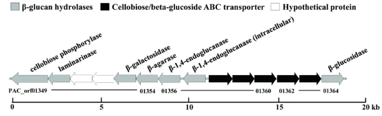
88°C) can grow on various substrates, such as peptides and starch (Miroshnichenko et al., 1998). Based on the genomic analysis of the strain, we identified a novel GH1 β-glucosidase- encoding gene located in a novel glycoside hydrolase gene cluster consisting of cellulose-, laminarin-, and agarose- degrading enzymes (Fig. 1). The gene revealed an ORF of 1,464 bp encoding 487 amino acid residues.
The deduced amino acid sequences of Tpa-glu showed 77%
identity with the β-glucosidase from P. furiosus (Bauer et al., 1996), 54% identity with the β-glycosidase from S. solfataricus (Cubellis et al., 1990), 40% identity with the β-glycosidase from T. kodakaraensis (Ezaki et al., 1999), and 33% identity with the β-glucosidase from P. horikoshii (Matsui et al., 2000).
We analyzed the phylogenetic tree among the family 1 glycosyl hydrolases from hyperthermophilic archaea. As shown in Fig.
2A, the tree has three branches. One branch includes the β-
glucosidase from P. horikoshii, which is known to be a
membrane protein and approximately 13% shorter than the
others in polypeptide length (Matsui et al., 2000). The second
branch includes the β-glycosidase from T. kodakaraensis and
β-mannosidase from P. furiosus. According to previously
Fig. 3. SDS-PAGE analysis of Tpa-Glu. M, molecular weight marker.
Fig. 1. Novel β-glycosyl hydrolase gene cluster in T. pacificus.
Fig. 2. Phylogenetic relationships of various family 1 glycosyl hydrolases from hyperthermophilic archaea (A) and alignments of Tpa-glu and Pfu-glu (B). The abbreviations are as follows: Tpa-glu, β-glucosidase from T. pacificus; Pfu-glu, β-glucosidase from P. furiosus; Sso-gly, β- glycosidase from S. solfataricus; Pho-glu, β-glucosidase from P.
horikoshii; Tko-gly, β-glycosidase from T. kodakaraensis; Pfu-man, β- mannosidase from P. furiosus.
reported results, the β-glycosidase from T. kodakaraensis exhibits bifunctional β-glycosidase and β-mannosidase activity, which is related to a conserved region (Ezaki et al., 1999).
Tpa-glu was located in the third branch, which includes β- glucosidase from P. furiosus and β-glycosidase from S.
solfataricus.
When pair-wise structure-based alignment with the β- glucosidase from P. furiosus, Tpa-glu showed an insertion sequence of 12 amino residues (299 to 310) in the loop region between α-helix 12 and β-strand 9 of the β-glucosidase from P.
furiosus (Fig. 2B). This extended loop was also observed in the
β-glucosidases from other hyperthermophilic archaea and exhibited variable size and sequence (data not shown).
However, the function of this loop region is unclear.
Expression and characterization of Tpa-glu
Tpa-glu in pET-24a(+) was expressed well in E. coli and was
purified using TALON
TMmetal affinity chromatography. The
purified Tpa-Glu revealed a major protein band with a
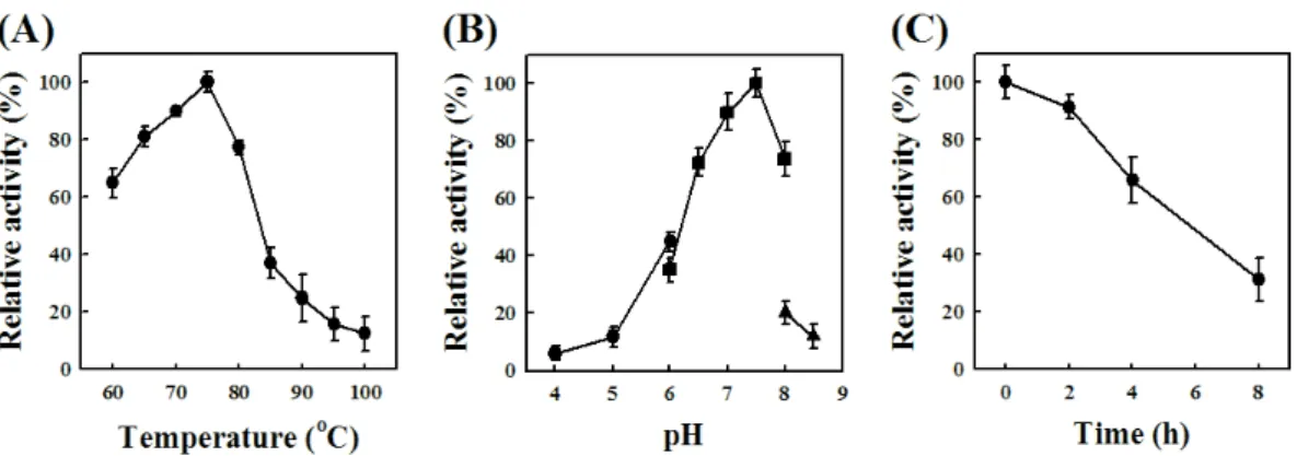
molecular mass of 58 kDa (Fig. 3). The effect of the pH on
hydrolyzing activity of Tpa-Glu was examined in the pH range
of 4.0 to 8.5. The optimum activity occurred at pH 7.5 in an
assay using pNP-glu as a substrate (Fig. 4A). The effect of
temperature on the hydrolyzing activity was also determined in
the range of 60 to 100°C, and the optimum polymerase activity
of Tpa-Glu was found to occur at 75°C (Fig. 4B). The
thermostability of Tpa-Glu was tested by measuring the
decrease in hydrolyzing activity after pre-incubation at 75°C
and 90°C. The half-life (t
1/2) of the enzyme at 75°C and 90°C
was 16.0 h and 6.0 h, respectively (Fig. 4C).
Fig. 4. Effects of temperature (A) and pH (B) on the activity of Tpa-Glu and measurement of the thermostability of Tpa-Glu (C). The activity assays were performed under standard conditions with the following buffers (each at 50 mM): sodium citrate, pH 4.0–6.0 (●); sodium phosphate, pH 6.0–8.0 (■);
Tris-HCl, pH 8.0–8.5 (▲). Tpa-glu was pre-incubated at 90°C, and the remaining activities were assayed at 75°C.
Table 1. Kinetic constants for Tpa-Glu
Substrates Km (mM) kcat (s-1) kcat/Km (s-1 mM-1)
pNP-β-glucopyranoside 0.58 64 110.34
pNP-β-galactopyranoside 10.36 58 5.62
pNP-β-mannopyranoside 5.06 6 1.18
pNP-β-xylopyranoside 6.47 1.18 0.18
Cellobiose 22.48 89 3.96
Laminaribiose 2.61 41 15.71
Kinetic properties of Tpa-Glu
The hydrolyzing activity of Tpa-Glu toward various pNP-glycopyranosides, such as pNP-β-glucopyranoside (pNP-glu), pNP-β-galactopyranoside (pNP-gal), pNP-β- mannopyranoside (pNP-man), and pNP- β-xylopyranosie (pNP-xyl), was tested. Tpa-Glu exhibited hydrolyzing activity against various pNP-glycopyranosides and showed the highest affinity with pNP-glu, with a K
mvalue of 0.58 mM. The highest catalytic efficiency (k
cat/K
mvalue) of Tpa-Glu was also observed for pNP-glu as the substrate, followed by pNP-gal, pNP-man, and pNP-xyl (Table 1). Additionally, the hydrolyzing activity of Tpa-Glu toward β-disaccharides, including cellobiose and laminaribiose, was examined. Tpa-Glu showed highest affinity with laminaribiose, with a K
mvalue of 2.61 mM, which is lower than the value of 22.48 mM found in the case of cellobiose. The k
cat/K
mvalue of Tpa-Glu against laminaribiose was 3.97-fold higher than that of cellobiose and 7.02-fold lower than that of pNP-glu (Table 1). The characteristics of these broad substrate specificities of Tpa-Glu were similar to those of previously characterized and reported β-glucosidases
and β-glycosidases from hyperthermophilic archaea (Sakellaris et al., 1997; Schiraldi et al., 2002; de Miguel Bouzas et al., 2006).
Substrate specificity of Tpa-Glu
The hydrolytic activity of Tpa-Glu toward various polysaccharides including avicel, cellulose, mannan, laminarin, and xylan, was tested. Interestingly, Tpa-Glu exhibits strong exo-hydrolyzing activity toward laminarin among poly- saccharides, however, unable to hydrolyze avicel, cellulose, mannan, and xylan (data not shown). The β-glucosidase from P. furiosus displays very little activity toward laminarin.
According to previously reported results, the β-glucosidase from the mesophilic bacterium, Cellovibrio mixtus was found to hydrolyze laminarin as a substrate (Sakellaris et al., 1997).
This substrate specificity of Tpa-Glu was different that of β- glucosidases or β-glycosidases from hyperthermophilic archaea (Cubellis et al., 1990; Bauer et al., 1996; Matsui et al., 2000).
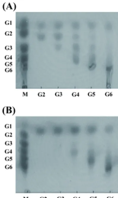
To address this question, we further tested the hydrolytic activity of Tpa-Glu toward β-1,3- and β-1,4-linked oligo- saccharides and laminarin through TLC analysis. Tpa-Glu exhibited hydrolyzing activity toward all of the tested oligosaccharides and released monomeric glucose as the final end-products (Fig. 5A and B). The results suggest that Tpa-Glu is an exo-type hydrolase. Additionally, Tpa-Glu showed substrate specificity with higher efficiency against β-1,3- linked oligosaccharides than β-1,4-linked oligosaccharides, and these results are consistent with the kinetics data.
Furthermore, as shown in Fig. 6, Tpa-Glu showed efficient
Fig. 5. The hydrolytic activities of Tpa-Glu toward β-1,4- (A) and β- 1,3-linked (B) oligosaccharides were measured by thin-layer chromato- graphy. The reaction was performed in 50 mM sodium phosphate buffer (pH 7.0) containing 1 mM substrates and 1 μg of enzyme at 75°C for 4 h.
The products were analyzed by thin-layer chromatography. Each substrate is indicated on the bottom line. The abbreviations are as follows: M, size marker; G1, glucose; G2, glucose dimer; G3, glucose trimer, G4, glucose tetramer; G5, glucose pentamer; G6, glucose hexamer.
Fig. 6. The hydrolytic activities of Tpa-Glu toward laminarin were measured by thin-layer chromatography. The reaction was performed in 50 mM sodium phosphate buffer (pH 7.0) containing 0.25% laminarin and 1 μg of enzyme at 75°C for 0.5, 1, 2, 3, and 4 h. The products were analyzed by thin-layer chromatography. The reaction time is indicated on the bottom line. The abbreviations are as follows: M, size marker; G1, glucose; G2, glucose dimer; G3, glucose trimer, G4, glucose tetramer;
G5, glucose pentamer; G6, glucose hexamer.
hydrolytic activity against laminarin and also produced monomeric glucose as the major end-products depending on the reaction time. The characteristics of Tpa-Glu, which hydrolyzes the polysaccharide laminarin, were distinct from those of previously reported β-glucosidases or β-glycosidases from hyperthermophilic archaea. Although Tpa-Glu is very
similar to the β-glucosidase of P. furiosus in terms of primary structure, it is unclear why Tpa-Glu has unique substrate specificity. It was postulated that the loop region of Tpa-Glu may be important for the substrate specificity of the enzyme.
As mentioned above, the structure of the loop region was located in A-C dimers of the tetrameric structure of β- glucosidase from P. furiosus (Kado et al., 2011). Moreover, in the β-glycosidase from S. solfataricus, it was reported to be involved in ion-pair networks with connecting sequences from the second and fifth βα units, and the network occurs at the tetrameric interface between the carboxy termini and involves 16 individual ion-pair interactions (Aguilar et al., 1997). We predicted that the substrate specificity of Tpa-Glu was associated with the ionic interaction between the loop region and the fourth barrel helix at the tetrameric interface. To understand the characteristics of the substrate specificity of Tpa-Glu, additional investigations, such as structural and/or mutational analyses of protein, are required.
In this paper, we described the cloning and characterization of a gene encoding a thermostable β-glucosidase from T. pacificus.
The purified Tpa-Glu showed hydrolyzing activity toward not only various pNP-glycopyranosides and oligosaccharides but also laminarin. The feature of the exo-hydrolyzing activity of Tpa-Glu toward laminarin suggests that it may be applied in combination with β-1,3-endoglucanase for the saccharification of brown algae containing laminarin as a cell wall component.
적 요
Thermococcus pacificus P-4의 유전체 서열 분석을 통하여 예 측되는 GH1 β-glucosidase를 암호화하는 유전자를 동정하였다.
그 유전자는 487 아미노산들을 암호화하는 1,464 bp 나타내었 으며, 그 아미노산 서열은 Pyrococcus furiosus β-glucosidase와 77% 상동성을 나타내었다. 그 유전자는 Escherichia coli 시스 템 내에서 복제 및 발현하였다. 재조합 된 단백질은 금속 친화 크로마토그래피를 통하여 정제하고 특성을 분석하였다. 정제 된 단백질(Tpa-Glu)은 pH 7.5와 75°C에서 최적활성을 나타내 었으며, 열안정성은 90°C에서 약 6시간의 반감기를 보였다.
Tpa-Glu는 pNP-β-glucopyranoside, pNP-β-galactopyranoside,
pNP-β-mannopyranoside, 그리고 pNP-β-xylopyranoside 순
으로 우수한 k
cat/K
m값을 나타내었다. 또한, Tpa-Glu는 β-
1,3-linked polysaccharide (laminarin) 그리고 β-1,3-와 β- 1,4-linked oligosaccharides에 대하여 exo-hydrolyzing 활성을 보였다. 본 연구는 초고온 고세균으로터 β-glucosidase가 exo- hydrolyzing 활성을 처음 확인한 것으로 이 효소는 laminarin 의 당화공정에 β-1,3-endoglucanase와 함께 적용할 수 있을 것 으로 기대된다.
Acknowledgements
This work was supported by the KIOST In-house Program (PE99314), the Development of Biohydrogen Production Technology using Hyperthermophilic Archaea Program of the Ministry of Ocean and Fisheries of the Republic of Korea.
References
Aguilar, C.F., Sanderson, I., Moracci, M., Ciaramella, M., Nucci, R., Rossi, M., and Pearl, L.H. 1997. Crystal structure of the beta-glycosidase from the hyperthermophilic archaeon Sulfolobus solfataricus: resilience as a key factor in thermostability. J. Mol.
Biol. 271, 789–802.
Atomi, H., Sato, T., and Kanai, T. 2011. Application of hyper- thermophiles and their enzymes. Curr. Opin. Biotechnol. 22, 618–626.
Bae, S.S., Kim, Y.J., Yang, S.H., Lim, J.K., Jeon, J.H., Lee, H.S., Kang, S.G., Kim, S.J., and Lee, J.H. 2006. Thermococcus onnurineus sp. nov., a hyperthermophilic archaeon isolated from a deep-sea hydrothermal vent area at the PACMANUS field. J.
Microbiol. Biotechnol. 16, 1826–1831.
Bauer, M.W., Bylina, E.J., Swanson, R.V., and Kelly, R.M. 1996.
Comparison of a beta-glucosidase and a beta-mannosidase from the hyperthermophilic archaeon Pyrococcus furiosus. Purification, characterization, gene cloning, and sequence analysis. J. Biol.
Chem. 271, 23749–23755.
Bhatia, Y., Mishra, S., and Bisaria, V.S. 2002. Microbial β- glucosidases: cloning, properties, and applications. Crit. Rev.
Biotechnol. 22, 375–407.
Bradford, M.M. 1976. A rapid and sensitive method for the quantitation of microgram quantities of protein utilizing the principle of protein-dye binding. Anal. Biochem. 72, 248–254.
Cubellis, M.V., Rozzo, C., Montecucchi, P., and Rosssi, M. 1990.
Isolation and sequencing of a new beta-galactosidase-encoding
archaebacterial gene. Gene 94, 89–94.
de Miguel Bouzas, T., Barros-Velázquez, J., and Villa, T.G. 2006.
Industrial applications of hyperthermophilic enzymes: a review.
Protein Pept. Lett. 13, 645–651.
Ezaki, S., Miyaoku, K., Nishi, K., Tanaka, T., Fujiwara, S., Takagi, M., Atomi, H., and Imanaka, T. 1999. Gene analysis and enzymatic properties of thermostable beta-glycosidase from Pyrococcus kodakaraensis KOD1. J. Biosci. Bioeng. 88, 130–135.
Imanaka, T. and Atomi, H. 2002. Catalyzing “hot” reactions: enzymes from hyperthermophilic archaea. Chem. Rec. 2, 149–163.
Kado, Y., Inoue, T., and Ishikawa, K. 2011. Structure of hyper- thermophilic β-glucosidase from Pyrococcus furiosus. Acta Crystallogr. Sect. F Struct. Biol. Cryst. Commun. 67, 1473– 1479.
Kengen, S.W., Luesink, E.J., Stams, A.J., and Zehnder, A.J. 1993.
Purification and characterization of an extremely thermostable β-glucosidase from the hyperthermophilic archaeon Pyrococcus furiosus. Eur. J. Biochem. 213, 305–312.
Matsui, I., Sakai, Y., Matsui, E., Kikuchi, H., Kawarabayasi, Y., and Honda, K. 2000. Novel substrate specificity of a membrane- bound β-glycosidase from the hyperthermophilic archaeon Pyrococcus horikoshii. FEBS Lett. 467, 195–200.
Miroshnichenko, M.L., Gongadze, G.M., Rainey, F.A., Kostyukova, A.S., Lysenko, A.M., Chernyh, N.A., and Bonch-Osmolovskaya, E.A. 1998. Thermococcus gorgonarius sp. nov. and Thermococcus pacificus sp. nov.: heterotrophic extremely thermophilic archaea from New Zealand submarine hot vents. Int. J. Syst. Bacteriol.
24, 23–29.
Moracci, M., Nucci, R., Febbraio, F., Vaccaro, C., Vespa, N., La Cara, F., and Rossi, M. 1995. Expression and extensive characterization of a β-glycosidase from the extreme thermoacidophilic archaeon Sulfolobus solfataricus in Escherichia coli: authenticity of the recombinant enzyme. Enzyme Microb. Technol. 17, 992–997.
Nucci, R., Moracci, M., Vaccaro, C., Vespa, N., and Rossi, M. 1993.
Exo-glucosidase activity and substrate specificity of the β- glycosidase isolated from the extreme thermophile Sulfolobus solfataricus. Biotechnol. Appl. Biochem. 17, 239–250.
Robb, F.T., Place, A.R., Sowers, K.R., Schreier, H.J., DasSarma, S., and Fleischmann, E.M. 1995. Archaea: A Laboratory Manual, pp. 3–29. Cold Spring Harbor Laboratory Press, Cold Spring Harbor, New York.
Sakellaris, H., Manners, J.M., and Pemberton, J.M. 1997. A gene encoding an exo-β-glucosidase from Cellvibrio mixtus. Curr.
Microbiol. 35, 228–232.
Schiraldi, C., Giuliano, M., and De Rosa, M. 2002. Perspectives on biotechnological applications of archaea. Archaea 1, 75–86.