• 검색 결과가 없습니다.

Implant Fracture After Screw Fracture in a TissueLevel Implant: A Case Report

N/A
N/A
Protected

Academic year: 2021

Share "Implant Fracture After Screw Fracture in a TissueLevel Implant: A Case Report"

Copied!
6
0
0

로드 중.... (전체 텍스트 보기)

전체 글

(1)

pISSN 1229-5418 eISSN 2671-6623

Implantology 2019; 23(4): 229-234 https://doi.org/10.32542/implantology.2019020

Received: December 24, 2019 Revised: December 27, 2019 Accepted: December 27, 2019 ORCID

Kyehee Wee

https://orcid.org/0000-0003-3185-7835 Bo-Ah Lee

https://orcid.org/0000-0003-3586-4928 Copyright © 2019. The Korean Academy of Oral &

Maxillofacial Implantology

This is an Open Access article distributed under the terms of the Creative Commons Attribution Non-Commercial License (http://creativecommons.

org/licenses/by-nc/4.0/) which permits unrestricted non-commercial use, distribution, and reproduction in any medium, provided the original work is properly cited.

OPEN ACCESS

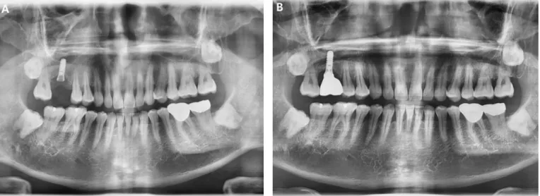
Dental implants may be a valid prosthetic treatment to replace a single tooth, but there are some complications. One complication of dental implants is a fracture of the implant. This case report describes the implant fracture of a tissue-level implant. A patient with a tissue-level implant that was placed on the right maxillary first molar ten years ago visited the clinic because the implant prosthesis was fallen out with the fractured abutment screw. Panoramic radiograph confirmed the fracture of the abutment screw. After removing the screw fragment, a new restoration was applied.

After two years, not only the screw but also the fixture was broken. The fixture fragment was surgically removed. This case implies that screw fractures can be a precursor to fixture fractures and regular check-up is necessary for preventing implant fractures.

Keywords: Dental implants, Fatigue fractures, Marginal bone loss, Peri-implant fractures

Abstract

Level Implant: A Case Report

Kyehee Wee, MS

1

, Bo-Ah Lee, DDS, PhD

2*

1

Specialist, Department of Dentistry, National Police Hospital, Seoul, Korea

2

Fellow, Department of Periodontology, National Health Insurance Service Ilsan Hospital, Goyang, Korea

* Corresponding author: Bo-Ah Lee, Department of Periodontology, National Health Insurance Service Ilsan Hospital, 100, Ilsan-ro, Ilsandong-gu, Goyang, Gyeonggi-do, 10444, Korea.

Tel:+82-31-900-0625. Fax: +82-0303-3448-7138. E-mail: balee@nhimc.or.kr

Ⅰ. Introduction

Dental implants can be a valid prosthetic treatment to replace single tooth.

1

Although the 10-year survival rates of tissue-level dental implants has been reported to be greater than 98%,

2

complications are another issue. Complications of the implant include osseointegration failure of implant fixture, peri-implantitis, abutment screw loosening, screw fracture, loss of implant prostheses and implant fracture.

3–5

Marcelo et al. (2014)

6

suggested implant failures can be classified as early failures and late failures. Early failure occurs during the stage of osseointegration, while late failure occurs after the occlusal load is applied.

Fractures of the implant, one of the late failures, are uncommon but once they occur, they are difficult to treat.

7

Tissue-level implants are implant types designed to reduce the marginal bone loss after

implant placement by placing the platform 1.5 to 3 mm above the bone level. Some studies

(2)

using finite element analysis have reported that marginal bone loss may be associated with screw fractures and fixture fractures.

8, 9

In addition, the ITI standard diameter solid screw fixtures have a thick wall with excellent strength, which can withstand heavy occlusal force.

10

In this regard, some clinicians may expect that tissue-level implants are resistant to fractures. Indeed, the reported prevalence of fracture in tissue-level implants is very low.

11, 12

However, there was no study to compare the prevalence of fractures between tissue-level and bone-level implants.

Risk factors for implant fractures are the type of prosthetic structure (single crown or fixed partial denture), parafunction, inappropriate occlusion, crestal bone resorption and screw loosening.

13-15

This case report shows fixture fracture that occurred two years after screw fracture in a tissue-level implant.

Ⅱ. Case Report

A 41-year-old male patient visited the clinic with a chief complaint of tooth mobility, and the maxillary left first molar was extracted. Six months later, a tissue-level implant (Straumann

®

Tissue Level Implant 4×10 mm, Straumann, Basel, Switzerland) was placed with maxillary sinus elevation.

The screw-retained prosthesis was applied 5 months after implant placement.

About 10 years later, the patient visited the clinic again with the fractured upper part of the implant.

Panoramic radiograph confirmed the fracture of abutment screw (Fig. 1A), but the screw fragment was

Fig. 1. (A) Panoramic radiograph revealed the fracture of abutment screw on the maxillary right first molar implant, (B) A

new prosthesis was applied.

(3)

not visible due to overgrown soft tissue. The screw fragment remaining inside the fixture was removed using a screw retrieval kit (HELP kit

®

, Dentium, Seoul, Korea) under flap elevation, and a healing abutment was placed. After soft tissue healing, a conventional impression process was performed and the newly made cement type prosthesis with a solid abutment was applied (Fig. 1B).

However, only about 1.5 years later, he returned to the clinic with a fractured implant. In addition to abutment and screw fracture, fixture fracture was also observed at the interface between the machined collar and the rough surface (Fig. 2A). The fractured implant was surgically removed using a trephine bur under flap elevation (Fig. 2B). The placement of a new implant wider than the previous one was planned.

Ⅲ. Discussion

Various causes of implant fractures have been reported.

13–17

Chrcanovic et al. (2018)

16

and Goiato et

al. (2019)

17

suggested that implant diameter can influence the incidence of implant fractures. Small

diameter implants have a reduced surface area and are mechanically weak.

17

Chrcanovic et al. (2018)

16

also reported that fractures of the ITI tissue-level implants were more frequent than other implants, but

were limited to narrow implants with a diameter of less than 3.3 mm. In this case, the diameter of the

fixture was 4.0 mm. However, it might not be sufficient for the posterior teeth, since they are subjected

to greater occlusal load than the anterior teeth. In addition, the occlusal surface of molar teeth is greater

Fig. 2. (A) The fracture of the fixture occurred at the interface between the machined collar and the rough surface, (B) Post-

operative radiograph shows alveolar bone defect after removal of the fixture using a trephine bur.

(4)

than that of premolar or anterior teeth. Therefore, it is recommended to use wide implant with a diameter of 5 mm or more in the posterior area. Chrcanovic et al. (2018)

16

reported small diameter implants were more fractured, especially in the posterior area.

Another cause of implant fracture is excessive bite force.

18

The parafunctional habits (bruxism, clenching etc.) and the excessive occlusal forces can cause not only implant failure, but also many other complications. However, no traumatic occlusion or parafunctional habits were observed in this study and tooth wear in all dentition was not severe enough to consider abnormal function. On the contrary, Esquivel-Upshaw et al. (2014)

19

reported no association between occlusal force and implant fracture.

The fracture can be resulted from mechanical fatigue failure due to prolonged use. Goiato et al. (2019)

17

reported implant fractures usually occur 3-4 years after placement. In this study, a screw fracture occurred 10 years after implant placement and a fixture fracture occurred 1.5 years later. Yokoyama et al. (2002)

20

concluded that titanium in a biological environment absorbs hydrogen, which may be the cause of fractures of titanium implants.

It should be noted that the screw fracture occurred before the fixture fracture in this case. In screw- retained implants, the stress produced by the micromovement of screws can apply constant tension on the implant fixtures.

14

Therefore, screw loosening often causes implant fracture and can be considered as a warning signal.

10, 18

This implies the importance of periodic maintenance. If screw loosening is found during the recall, clinicians should retighten the screw using an appropriate torque.

When screw fracture occurs, it is necessary to remove the fractured screw without damaging the internal threads. Various techniques have been introduced to manage fractured abutment screws, including hand instruments, ultrasonic scalers, and rotary instruments.

21

Mizumoto et al. (2018)

21

suggested that if the fractured fragment was removed and the fixture was not damaged, a new restoration could be applied. In this case, the remaining fragment was removed and a new restoration was applied.

After 1.5 years, however, the screw and the fixture fractured. One possible reason may be that the damage of the internal threads was not recognized. If the internal threads were damaged, the new abutment screw would not fit properly. Non-passive fit of the prosthetic structure is one of the causes of implant fracture.

22

In addition, repeated instrumentation to remove the fragment may have applied fatigue stress on the fixture. The fact that the location of fixture fracture was similar to that of screw fracture supports the above explanation.

The location of the fixture fracture was at the crestal bone level, where the interface between the

smooth collar and the rough surface was located. The thickness of tissue level implants is usually thinnest

at the bone crest level and the lateral bending force applied on the fixture causes fatigue stress.

16

(5)

Abutment type can also affect the fracture of implants.

23

Literatures reported that internal connection types provide better joint stability than external connection types and among the internal type, the conical type provides enhanced resistance to horizontal and oblique forces than the hexagon type. However, there are studies reporting no significant differences between two types.

13

In this case, an internal conical abutment was used. It seems that the misfit between abutments and fixtures is more important to implant fractures than the connection type of abutments and implants.

There are some treatment options for managing implant fractures.

6, 24

One is to remove the coronal portion of the fractured implant, leaving the remaining part integrated in the bone. However, this approach does not allow the placement of a new implant. In most cases, complete removal of the fractured implants can be the treatment of choice. In the present case, the fractured implant was removed using a trephine bur.

Ⅳ. Conclusion

The most important thing about implant failure is prevention. In the posterior area, it is recommended to choose a wide diameter implant and place it in the center of the occlusion if possible. Regular maintenance is also important. The occlusal adjustment should be necessary to avoid excessive occlusal force and the loosened screw should be retightened. The presence of parafunctional habits should also be examined.

References

1. Creugers NH, Kreulen CM, Snoek PA, de Kanter R.J.A.M. A systematic review of single-tooth restorations supported by implants. J Dent. 2000;28:209-17.

2. Kim S, Jung UW, Cho KS, Lee JS. Retrospective radiographic observational study of 1692 Straumann tissue-level dental implants over 10 years: I. Implant survival and loss pattern. Clin Implant Dent Relat Res 2018;20:860-6.

3. Donati M, Ekestubbe A, Lindhe J, Wennström JL. Implant-supported single-tooth restorations. A 12-year prospective study. Clin Oral Implants Res 2016;27:1207-11.

4. Pjetursson BE, Tan K, Lang NP, Brägger U, Egger M, Zwahlen M. A systematic review of the survival and complication rates of fixed partial dentures (FPDs) after an observation period of at least 5 years. Clin Oral Impl Res 2004; 15:625-42.

5. Jung RE, Pjetursson BE, Glauser R, Zembic A, Zwahlen M, Lang NP. A systematic review of the 5-year survival and complication rates of implant-supported single crowns. Clin Oral Impl Res 2008;19:119-30.

6. Marcelo CG, Filié Haddad M, Gennari Filho H, Luiz Marcelo RV, Daniela Micheline DS, Aldiéris

(6)

AP. Dental implant fractures - aetiology, treatment and case report. J Clin Diagn Res 2014;8:300-4.

7. Lang NP, Mombelli A, Tonetti MS, Brägger U, Hämmerle CH.F. Clinical trials on therapies for peri- implant infections. Ann Periodontol 1997;2:343-56.

8. Romeed SA, Malik R, Dunne SM. Marginal bone loss influence on the biomechanics of single implant crowns. J Craniofac Surg 2013;24:1459-65.

9. Jimbo R, Halldin A, Janda M, Wennerberg A, Vandeweghe S. Vertical fracture and marginal bone loss of internal-connection implants: a finite element analysis. Int J Oral Maxillofac Implants 2013;28:e171-6.

10. Schwarz MS. Mechanical complications of dental implants. Clin Oral Implants Res 2000;11:156-8.

11. Weber HP, Crohin CC, Fiorellini JP. A 5-year prospective clinical and radiographic study of non- submerged dental implants. Clin Oral Implants Res 2000;11:144-153.

12. Gokcen-Rohlig B, Yaltirik M, Ozer S, Tuncer ED, Evlioglua G. Survival and Success of ITI Implants and Prostheses: Retrospective Study of Cases with 5-Year Follow-Up. Eur J Dent 2009;3:42-9.

13. Lee DW, Kim NH, Lee Y, Oh YA, Lee JH, You HK. Implant fracture failure rate and potential associated risk indicators: an up to 12-year retrospective study of implants in 5,124 patients. Clin Oral Implants Res 2019;30:206-17.

14. Tabrizi R, Behnia H, Taherian S, Hesami N. What are the incidence and factors associated with implant fracture? J Oral Maxillofac Surg 2017;75:1866-72.

15. Stoichkov B, Kirov D. Analysis of the causes of dental implant fracture: A retrospective clinical study. Quintessence Int 2018;49:279-86.

16. Chrcanovic BR, Kisch J, Albrektsson T, Wennerberg A. Factors influencing the fracture of dental implants. Clin Implant Dent Relat Res 2018;20:58-67.

17. Goiato MC, Andreotti AM, Dos Santos DM, Nobrega AS, de Caxias FP, Bannwart LC. Influence of length, diameter and position of the implant in its fracture incidence: a systematic review. J Dent Res Dent Clin Dent Prospects 2019;13:109-16.

18. Eckert SE, Meraw SJ, Cal E, Richard K O. Analysis of incidence and associated factors with fractured implants: a retrospective study. Int J Oral Maxillofac Implants 2000;15:662-7.

19. Esquivel-Upshaw JF, Mehler A, Clark AE, Neal D, Anusavicea KJ. Fracture analysis of randomized implant-supported fixed dental prostheses. J Dent 2014;42:1335-42.

20. Yokoyama K, Ichikawa T, Murakami H, Miyamotod Y, Asaokaa K. Fracture mechanisms of retrieved titanium screw thread in dental implant. Biomaterials 2002;23:2459-65.

21. Mizumoto RM, Jamjoom FZ, Yilmaz B. A risk-based decision making tree for managing fractured abutment and prosthetic screws: a systematic review. J Prosthet Dent 2018;119:552-9.

22. Balshi TJ. An analysis and management of fracture implants: a clinical report. Int J Oral Maxillofac Implants 1996;11:660-6.

23. Bordin D, Witek L, Fardin VP, Bonfante EA, Coelho PG. Fatigue failure of narrow implants with different implant-abutment connection designs. J Prosthodont 2018;27:659-64.

24. Gargallo Albiol J, Satorres-Nieto M, Puyuelo Capablo JL, Sánchez Garcés MÁ, Pi Urgell J, Gay

Escoda C. Endosseous dental implant fractures: an analysis of 21 cases. Med Oral Patol Oral Cir

Bucal 2008;13:E124-8.

수치

Fig. 1. (A) Panoramic radiograph revealed the fracture of abutment screw on the maxillary right first molar implant, (B) A  new prosthesis was applied.

참조

관련 문서

Methods to overcome insufficient bone due to poor bone quality, the pneumatization of a maxillary sinus and other anatomical limitations of implant placement

A clinical study of the efficacy of gold-tite square abutment screws in cement-retained implant restorations.. An abutment screw loosening study of a Diamond

Histopathologic findings of control group at 4 weeks show little bone- implant contact (BIC) around the implant (asterisks) and new-bone formation in the defect

Measurement of proximal contact tightness between the left first molar and second molar(dental implant) in the mandible... Clinical dental anatomy, histology,

Evaluating parameters of osseointegrated dental implants using finite element analysis-a two- dimentional comparative study examining the effects of implant

Experimental alveolar ridge augmentation by distraction osteogenesis using a simple device that permits secondary implant placement. Anteroinferior distraction of

As the number of clinical cases requiring supracrestal apical-coronal positioning is increasing, we examined the utility of FEA in such cases. In this study,

Determining optimal surface roughness of TiO₂blasted titanium implant material for attachment, proliferation and differentiation of cells derived from