• 검색 결과가 없습니다.

Isolated Cervical Lymph Node Sarcoidosis Presenting in an Asymptomatic Neck Mass: A Case Report

N/A
N/A
Protected

Academic year: 2021

Share "Isolated Cervical Lymph Node Sarcoidosis Presenting in an Asymptomatic Neck Mass: A Case Report"

Copied!
4
0
0

로드 중.... (전체 텍스트 보기)

전체 글

(1)

Isolated Cervical Lymph Node Sarcoidosis Presenting in an Asymptomatic Neck Mass: A Case Report

Yong Shik Kwon, M.D.

1

, Hye In Jung, M.D.

1

, Hyun Jung Kim, M.D.

1

, Jin Wook Lee, M.D.

1

, Won-Il Choi, M.D., Ph.D.

1

, Jin Young Kim, M.D.

2

, Byung Hak Rho, M.D., Ph.D.

2

, Hye Won Lee, M.D.

3

and Kun Young Kwon, M.D., Ph.D.

3

Departments of

1

Internal Medicine,

2

Radiology, and

3

Pathology, Keimyung University Dongsan Medical Center, Keimyung University School of Medicine, Daegu, Korea

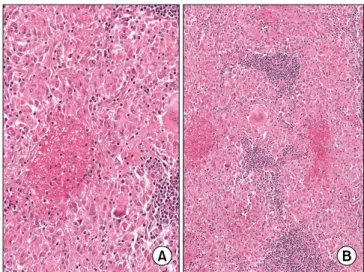
Sarcoidosis, a systemic granulomatous disease of unknown etiology. The presentation of sarcoidal granuloma in neck nodes without typical manifestations of systemic sarcoidosis is difficult to diagnose. We describe the case of a 37-year-old woman with an increasing mass on the right side of neck. The excisional biopsy from the neck mass showed noncaseating epithelioid cell granuloma of the lymph nodes. No evidence of mycobacterial or fungal infection was noted. Thoracic evaluations did not show enlargement of mediastinal lymph nodes or parenchymal abnormalities. Immunohistochemistry showed abundant expression of tumor necrosis factor-α in the granuloma.

However, transforming growth factor-β was not expressed, although interleukin-1β was focally expressed. These immunohistochemical findings supported characterization of the granuloma and the diagnosis of sarcoidosis. Sarcoidosis can present with cervical lymph node enlargement without mediastinal or lung abnormality. Immunohistochemistry may support the diagnosis of sarcoidosis and characterization of granuloma.

Keywords: Sarcoidosis; Lymphatic Diseases; Neck; Immunohistochemistry

hilar lymphadenopathy, pulmonary infiltration, and skin and ocular lesions. Sarcoidosis is characterized by noncaseating epithelioid cell granulomas in affected organs, particularly in the lung, hilar lymph nodes (LNs), skin, and eyes

1

. Sarcoidosis can affect individuals of any race, sex, and age, but it common- ly affects young to middle-aged adults. Thoracic radiologic abnormalities are common, and most of the morbidity and mortality associated with sarcoidosis involves the lung.

The diagnosis of sarcoidosis is established on the basis of compatible clinical and radiologic findings, supported by histologic evidence of noncaseating epithelioid-cell granu- lomas in one or more organs in the absence of organisms or particles

2

. Biopsy is recommended for all patients presumed to have sarcoidosis, except for those with Lofgren’s syndrome

2

. Pathologists can identify granulomas, but the diagnosis should not be based on pathological findings alone.

Clinically, many conditions result in sarcoid-like granulo- mas that may be interpreted as a local reaction to a malig- nancy, a noncaseating reaction to a focus of caseating tuber- Copyright © 2013

The Korean Academy of Tuberculosis and Respiratory Diseases.

All rights reserved.

Introduction

Sarcoidosis is a multi-system chronic inflammatory condi- tion of unknown etiology. It usually presents with bilateral

CASE REPORT

http://dx.doi.org/10.4046/trd.2013.75.3.116

ISSN: 1738-3536(Print)/2005-6184(Online) • Tuberc Respir Dis 2013;75:116-119

116

Address for correspondence: Won-Il Choi, M.D., Ph.D.

Department of Internal Medicine, Keimyung University Dongsan Medical Center, Keimyung Universiy School of Medicine, 56 Dalseong- ro, Jung-gu, Daegu 700-712, Korea

Phone: 82-53-250-7527, Fax: 82-53-250-7434 E-mail: [email protected]

Received: Jan. 23, 2013 Revised: May 14, 2013 Accepted: Jun. 17, 2013

cc

It is identical to the Creative Commons Attribution Non-Commercial

License (http://creativecommons.org/licenses/by-nc/3.0/).

(2)

Immunohistochemical features of neck sarcoidosis

http://dx.doi.org/10.4046/trd.2013.75.3.116 117

www.e-trd.org

culosis, or as another inflammatory disease

3

. These known granulomatous diseases should be excluded in patients with- out typical symptoms of sarcoidosis. The most common clini- cal features are respiratory symptoms, fatigue, night sweats, weight loss, erythema nodosum

2

. However, 50% of cases of sarcoidosis are asymptomatic, with abnormalities detected incidentally during chest radiography. Bilateral hilar lymph- adenopathy is the earliest and most common manifestation of sarcoidosis. Peripheral lymphadenopathy is less likely to pres- ent first

4

.

Currently, the presentation of sarcoidal granuloma in neck nodes without typical manifestation of systemic sarcoidosis remains a diagnostic challenge. We present a case of cervical LN enlargement without mediastinal or lung parenchymal abnormality. Immunohistochemistry supported a diagnosis of sarcoidosis and excluded other granulomatous disorders.

Case Report

A 37-year-old female patient presented with a palpable non- tender mass on the right side of her neck for 1 month. She had no other symptoms such as coughing, sputum production, rhinorrhea, arthralgia, fever, body weight loss, or dyspnea.

Physical examination revealed a palpable non-tender mass on the right side of her neck but no splenomegaly or hepato- megaly. She had no uveitis. The erythrocyte sedimentation rate was 11 mm/hr (normal range for female individuals, <25 mm/hr), with a C-reactive protein level of 0.14 mg/dL (nor- mal range, 0−0.5 mg/dL) and a serum angiotensin converting enzyme level of 23.9 U/L (normal range, 9.0−47.0 U/L). Neck computed tomography showed an approximately 50-mm abnormal LN at level V on the right and an approximately 28- mm abnormal LN at level II on the right (Figure 1). Multiple enlarged LNs were noted at levels II, III, IV, and V on the right.

Chest computed tomography showed no abnormal nodular infiltration at either lung, but showed enlarged LNs in the left

supraclavicular and left axillar areas.

Fine needle aspiration from the right cervical LN revealed granulomatous inflammation with focal and central necrosis.

Acid fast bacilli (AFB) stain was negative, and polymerase chain reaction for mycobacterium was also negative. Exci- sional biopsy of right neck LN (level V) was performed. A 4×2.5-cm pink ovoid granulomatous mass was observed at the posterolateral side of the sternocleidomastoid muscle. The mass was well encapsulated, with mild adhesion to the sur- rounding soft tissue. Excisional LN biopsy from the right side of the neck revealed granulomatous inflammation with focal and central necrosis (Figure 2). AFB stain showed no AFB positive bacilli in the excisional LN. Polymerase chain reaction for mycobacterium tuberculosis and non-tuberculous myco- bacterium were again negative in the excisional LN. Clinically,

Figure 1. Sagittal (A) and axial (B) images of enhanced neck computed tomography (CT). (A) Sagittal images of enhanced neck CT show multiple enlarged lymph nodes at levels II, III, IV, and V. (B) Axial images of enhanced neck CT show mul- tiple enlarged lymph nodes at levels II, III, IV, and V. The short diameter of the larg- est lymph node is approximately 3 cm (arrow).

Figure 2. Microscopic findings of the case. (A) An epithelioid granu-

lomatous lesion with central necrosis and multinucleated giant

cells is present (H&E stain, ×100). (B) High magnification of (A)

shows hyalinized necrosis and surrounding epithelioid cells (H&E

stain, ×200).

(3)

YS Kwon et al.

118 Tuberc Respir Dis 2013;75:116-119 www.e-trd.org

we excluded all other exogenous conditions that may cause granulomatous inflammation in the lung, and sarcoidosis was diagnosed.

Immunohistochemistry showed diffuse high expression of tumor necrosis factor-α (TNF-α) in epithelioid cells and multi- nucleated giant cells. Interleukin (IL)-1β was focally expressed in the granuloma. However, transforming growth factor-β (TGF-β) was not expressed in the granulomatous lesion (Fig- ure 3). We administered 10 mg/day prednisolone. Follow-up chest computed tomography after 3 months showed no inter- val change from baseline. The patient had no symptoms and no palpable LNs at the 6-month follow-up visit.

Discussion

Sarcoidosis is an inflammatory systemic disease character- ized by noncaseating granulomas composed of epithelioid giant cells, macrophages, and lymphocytes

5

.

Cervical lymphadenopathy as the sole and initial presenta- tion of sarcoidosis is rare, thereby adding to the difficulty in diagnosis. Furthermore, sarcoidosis presenting as isolated as- ymptomatic neck mass was not reported yet. In this case, she did not recognize her neck mass until growing about 3 cm in diameter. From November 1996 to June 1999, 10 medical cen- ters (in the US) and 1 coordinating center conducted A Case Control Etiologic Study of Sarcoidosis (ACCESS) including 736 patients. In total, 699 patients showed thoracic involve- ment, 368 had concomitant extra-thoracic disease, and 6 had isolated head and neck sarcoidosis

6

.

A diagnosis of sarcoidosis is established on the basis of compatible clinical and radiologic findings and is supported by histologic evidence of noncaseating epithelioid-cell granu- lomas in one or more organs in the absence of organisms or particles. Sarcoidal granulomas have no unique histologic fea- tures to differentiate them from other granulomas

2

. Chronic

exposure to inflammatory stimuli and/or foreign bodies may contribute to the development of granulomatous disease

7

. Therefore, other important differential diagnoses of granu- lomatous disease (e.g., Crohn’s disease, toxoplasmosis, lym- phoma, and vasculitis) must be excluded. The pathological findings of sarcoidosis are granulomas, which are usually non- necrotizing, but occasionally, necrosis can be observed. Diag- nosis of sarcoidosis involves exclusion of the diagnoses of the other granulomatous diseases, and in particular, tuberculosis.

Immunohistochemical staining is helpful in the diagnosis of granulomatous disease. The presence of CD4 T cells that interact with antigen-presenting cells indicates initiation of the formation and maintenance of granulomas. The trigger- ing antigens activate selective T-cell clones that differentiate into type 1 helper T cells. Interferon-α and IL-2 are mainly secreted, and production of TNF-α is increased through mac- rophage activation. Pulmonary fibrosis occurs after a shift in content from Th1 cytokines to Th2 cytokines (mainly IL-4, IL- 10, and IL-13).

TNF-α is a key factor in sarcoidosis, both for initiation and at the chronic stage

8

. Cervical lymphadenopathy is dif- ficult to distinguish from other granulomatous diseases and malignancy. Therefore, TNF-α can be an adjuvant indicator of sarcoidosis. In the present case, abundant expression of TNF-α was noted in the granuloma, supporting the diagnosis of sarcoidosis. TNF-α is also thought to play a major role in the proliferation and spontaneous activity of macrophages and T- lymphocytes at the sites of inflammation and in the formation of noncaseating granuloma

9

. Therefore, TNF-α inhibitors may have a therapeutic effect on granulomatosis, such as that in the present case

8

.

In patients with pulmonary sarcoidosis, TGF-β is abundant- ly expressed in lung tissues and is localized to the epithelioid histiocytes within non-necrotizing granulomas

10

. However, TGF-β was not expressed in the granuloma in the present case. It is uncertain, therefore, whether TGF-β contributes Figure 3. Immunohistochemical stains.

(A) Diffuse positive expression of tumor

necrosis factor-α in epithelioid cells and

multinucleated giant cells. (B) Focal ex-

pression of interleukin-1β. (C) No expres-

sion of transforming growth factor-β in

the granulomatous lesion (A−C, ×200).

(4)

Immunohistochemical features of neck sarcoidosis

http://dx.doi.org/10.4046/trd.2013.75.3.116 119

www.e-trd.org

only to the formation of granuloma in the lung parenchyma.

IL-1β released from alveolar macrophages in pulmonary tu- berculosis is important in host defense against mycobacterial infection. The release of IL-1β was greater from alveolar mac- rophages of tuberculosis patients than from those of normal subjects

11

.

However, IL-1β was not expressed in the alveolar macro- phages of patients with sarcoidosis

12

. In the present case, IL-β was focally expressed in the granuloma, thereby supporting a diagnosis of sarcoidosis rather than tuberculosis.

Sarcoidosis can present with cervical LN enlargement with- out mediastinal or lung abnormality. Immunohistochemistry may support the diagnosis of sarcoidosis and characterization of granulomas. Furthermore, TNF-α inhibitors could be an option for treating sarcoidosis, as in the present case.

References

1. Baughman RP, Lower EE, du Bois RM. Sarcoidosis. Lancet 2003;361:1111-8.

2. Iannuzzi MC, Rybicki BA, Teirstein AS. Sarcoidosis. N Engl J Med 2007;357:2153-65.

3. Giuffrida TJ, Kerdel FA. Sarcoidosis. Dermatol Clin 2002;20:435-47.

4. Handa R, Aggarwal P, Wali JP, Wig N, Dinda AK, Biswas A.

Sarcoidosis presenting with peripheral lymphadenopathy.

Sarcoidosis Vasc Diffuse Lung Dis 1998;15:192.

5. Saltini C, Crystal RG. Pulmonary sarcoidosis: pathogenesis, staging and therapy. Int Arch Allergy Appl Immunol 1985;76 Suppl 1:92-100.

6. Newman LS, Rose CS, Bresnitz EA, Rossman MD, Barnard J, Frederick M, et al. A case control etiologic study of sar- coidosis: environmental and occupational risk factors. Am J Respir Crit Care Med 2004;170:1324-30.

7. Knopf A, Lahmer T, Chaker A, Stark T, Hofauer B, Pickhard A, et al. Head and neck sarcoidosis, from wait and see to tu- mor necrosis factor alpha therapy: a pilot study. Head Neck 2013;35:715-9.

8. Daien CI, Monnier A, Claudepierre P, Constantin A, Eschard JP, Houvenagel E, et al. Sarcoid-like granulomatosis in pa- tients treated with tumor necrosis factor blockers: 10 cases.

Rheumatology (Oxford) 2009;48:883-6.

9. Fehrenbach H, Zissel G, Goldmann T, Tschernig T, Vollmer E, Pabst R, et al. Alveolar macrophages are the main source for tumour necrosis factor-alpha in patients with sarcoidosis. Eur Respir J 2003;21:421-8.

10. Limper AH, Colby TV, Sanders MS, Asakura S, Roche PC, DeRemee RA. Immunohistochemical localization of trans- forming growth factor-beta 1 in the nonnecrotizing granu- lomas of pulmonary sarcoidosis. Am J Respir Crit Care Med 1994;149:197-204.

11. Kuo HP, Wang CH, Huang KS, Lin HC, Yu CT, Liu CY, et al.

Nitric oxide modulates interleukin-1beta and tumor necrosis factor-alpha synthesis by alveolar macrophages in pulmo- nary tuberculosis. Am J Respir Crit Care Med 2000;161:192-9.

12. Bost TW, Riches DW, Schumacher B, Carre PC, Khan TZ,

Martinez JA, et al. Alveolar macrophages from patients with

beryllium disease and sarcoidosis express increased levels of

mRNA for tumor necrosis factor-alpha and interleukin-6 but

not interleukin-1 beta. Am J Respir Cell Mol Biol 1994;10:506-

13.

수치

Figure 1. Sagittal (A) and axial (B) images  of enhanced neck computed tomography  (CT)

참조

관련 문서

Objective: We wanted to evaluate the outcomes of cervical cancer patients with supraclavicular lymph node (SCLN) involvement and who received radiation therapy (RT) combined

Therefore, we describe a case with minimal USC showing an unusual spread pattern, with cervical lymph node metastasis through the pelvic and para- aortic lymph nodes..

We managed a case of postoperative chylous ascites associa- ted with a robotic-assisted laparoscopic pelvic and para- aortic lymph node dissection for a cervical carcinoma that was

The T2-weighted axial image shows a soft tissue mass of low signal intensi- ty in the subcutaneous fat layer of the posterior neck, and it shows the multi- ple, variable sized,

In the case of a patient with lymph node enlarge- ment, as in the present case, the possibility of an infectious disease by viral or bacterial infection, an autoimmune disease, and

The case for branchiogenic cancer (malignant branchioma). Incidence of unsuspected metastases in lateral cervical cysts. Lateral cervical cysts and fistulas. Cystic

Tumor size(p=0.000), ETE(p=0.001), multifocality(p=0.014), and bilaterality(p=0.001) were significantly related factors for cervical lymph node metastasis clinically

Characteristics No.. Disease-free survival curves of PMC according to cervical lymph nodes involvement. LNP:lymph node positive group, LNN:lymph node negative group..