online©ML Comm
제 26 권 제 1 호 2010
갑상선 미세유두암에서 경부림프절 전이의 예측인자에 대한 연구
인제대학교 의과대학 부산백병원 외과학교실
유혜미·하태권·유성목·김운원·김상효
= Abstract =
A Study of Predictive Factors of Cervical Lymph Node Metastasis in Papillary Microcarcinoma(PMC) of Thyroid Gland
Hye-Mi Yu, MD, Tae-Kwun Ha, MD, Sung-Mock Ryu, MD, Woon-Won Kim, MD, Sang-Hyo Kim, MD
Department of Surgery, Busan Paik Hospital, Inje University College of Medicine, Busan, Korea
Background:Though papillary microcarcinoma(PMC) of thyroid gland is known to have very favorable long-term prognosis, the recurrence in the neck and distant metastasis have been often reported. The predictive factors of node metastasis and tumor recurrence in clinical course were investigated to define surgical decision or guidelines in surgery of papillary microcarcinoma. Methods:The authors conducted a retrospective an- alysis of 216 patients of PMC treated with surgery at Department of Surgery, Busan Paik Hospital for the period from 1997 to 2007. Of these patients, 58 cases showing cervical lymph node metastasis at initial sur- gery were studied. Results:In overall 216 patients, the sex ratio of male to female was 1:9.3(male 21, female 195 cases), the mean age at the time of diagnosis was 44.7 years and the median tumor size was 6.61mm. Neck lymph node metastasis was found in 58 patients(26.9%), thyroid capsular invasion was 56 cases(25.9%), multifocality and bilaterality were found in 32(14.8%) and 29 cases(13.4%), respectively. Through statistical analysis, sex, capsular invasion, ETE, and tumor size(>5mm) were considered to be predictive factors of cer- vical lymph nodes metastasis. Of them, capsular invasion was the most predictive indicator of cervical lymph node metastasis on multivariate analysis. Nodal recurrence was observed in 6 of 58 patients of node positive at initial surgery. Conclusion:The cervical lymph node metastasis is known to be a risk factor of prognosis in PMC of thyroid gland. The results of this study showed four statistically significant independent predictive factors of cervical lymph node metastasis in PMC:capsular invasion, tumor size(>5mm), ETE, and sex. On multivariate analysis, capsular invasion was a great influencing factor in prediction of lymph node metastasis.
Basically, patients who has predictive factors of cervical lymph node metastasis should have a thorough investigation, and close surveillance for nodal status is required in follow-up.
KEY WORDS:Papillary microcarcinoma(PMC) of thyroid gland·Lymph node metastasis·Predictive factor.
서 론
갑상선 유두상암은 갑상선암 중에서 대부분을 차지하며
일반적으로 서서히 진행하고 국소재발과 원격전이가 늦게 나타나는 특징과 수술에 의한 치료효과가 좋아 예후가 좋 은 암으로 알려져 있다.1) 이 중 약 30%는 직경 1cm 이하 의 미세 유두상암으로 분류하고 있으나 1cm 이상의 유두 상암과 마찬가지로 림프절 전이 및 다발성 원격전이의 소 견을 보이는 경우도 있어 종양크기에 의한 분류에 대해 의 문이 제기되기도 한다.2) 갑상선암에서 경부림프절 전이는 국 소 재발과 무병생존에 불량한 예측인자로 알려져 있으며,3) 교신저자:김상효, 614-735 부산광역시 진구 개금동 633-165
인제대학교 의과대학 부산백병원 외과학교실 전화:(051) 890-6347·전송:(051) 898-9427 E-mail:[email protected]
미세유두상암이라 할지라도 경부 림프절로의 전이가 보고 되고 있고, 특히 다발성 병변의 경우와 술 전 경부초음파 검 사에서 전이로 의심되는 림프절이 있는 경우에는 림프절 전 이 및 국소재발, 원격전이의 가능성이 높으나, 생존율에는 영향을 미치지 않지만, 무병생존율을 감소시킨다는 보고가 있어 유두상암과 같이 림프절 수술을 해야 한다는 주장이 많은 상황이다.4,5) 이에 대한 갑상선 미세 유두상암의 적절 한 치료로써 경부 림프절 곽청술의 범위 설정에 대해서는 아직도 이견이 많다.6)
이에 저자들은 경부림프절 전이가 진단된 갑상선 미세유 두상암 환자에서 림프절 전이에 영향을 미치는 위험 인자 들을 비교, 조사하여 경부 림프절 전이를 예측 할 수 있는 인자들의 분석을 통하여 적절한 갑상선 절제술 및 림프절 곽청술의 범위 선택과 향후 치료 방침의 결정에 보탬이 되 고자 본 연구를 시행하였다.
대상 및 방법
1997년 1월부터 2007년 12월까지 인제대학교 부산백 병원 외과에서 갑상선암으로 진단되거나 의심되어 수술을 시행 받고 수술 후 조직검사가 미세 유두상암으로 진단된 216명과 그 중 경부 림프절 전이가 진단된 58명의 환자를 대상으로 하였다. 미세 유두상암과 경부림프절 전이는 술 전 세침흡인세포 검사와 술중 동결절편 검사, 병리조직검 사로 진단되었으며, 술 후 국소 재발에 대한 진단은 경부림 프절의 영상학적 검사나 이학적 검사에서 증가되거나 촉지 되는 림프절에 대하여 세침 흡인 세포 검사를 시행하여 전 이가 진단되었을 경우로 하였다. 이들의 남녀 성비, 연령, 종 양 크기(0.5cm 이상 혹은 이하), 피막 침범, 갑상선외 침범, 다발성, 양측성, 혈관침범 등을 경부림프절 전이그룹과 비 전이그룹으로 분류하여 각각의 그룹과 관련된 경부림프절 전이 위험인자들을 분석, 예측 하였다. 추적관찰기간의 중 앙값은 58개월(범위:6~155)이었고, 진단 시 연령은 AJCC (American Joint Committee on Cancer)의 TNM(6th edi- tion)분류에 따라 45세를 기준으로 구분하였다. 통계분석은 단변량 분석 시 Chi-square test를, 다변량 분석은 multiple logistic regression analysis를 시행하였으며, 무병 생존율 을 계산하기 위해서 Kaplan-Meier법을 사용하였다. 통계 적인 유의성은 P값으로 표현하였으며 양측으로 0.05 미만 인 경우에 유의한 차이를 보이는 것으로 판정하였다.
결 과
1. 대상 환자의 임상병리학적인 특징
미세유두상 갑상선암 환자 216명의 연령 및 성별 분포
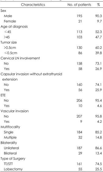
는 남자는 21례, 여자는 195례이었고, 남녀비는 1:9.3로 여자가 많았으며, 40대 81례, 30대 59례, 50대 46례 순으 로 호발 하였고, 나이구분에 따라 45세 미만이 113례, 45 세 이상은 103례이었으며 평균연령은 44.7세였다. 종양의 크기를 5mm을 기준으로 분류하였을 때, 5mm 이상은 130 례, 5mm 미만은 86례이었으며 종양의 평균크기는 6.61mm 였다. 경부림프절 전이는 58례(26.9%), 갑상선 피막침범은 56례(25.9%), 갑상선외 침범은 10례(4.6%), 혈관침범은 9 례(4.2%)였다. 다발성 병소는 32례(14.8%), 양측성은 29 례(13.4%)였다. 아전절제술을 포함한 전절제술은 161례 (74.5%), 완결 갑상선절제술을 포함한 일엽절제술은 55례 (25.5%)였다(Table 1).
2. 경부 림프절전이의 예측인자
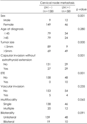
경부림프절 전이가 있는 환자 58명 중 46명(79.3%)이 여성이었고, 29명(50%)에서 피막침범이 있었으며, 49명 (84.5%)에서 5mm 이상의 종양크기를 나타내었고, 10명 (17.2%)에서 갑상선외 확장을 보였다. 경부 림프절 전이에
Table 1. Clinicopathologic characteristics of 216 patients with papillary microcarcinoma of thyroid gland
Characteristics No. of patients % Sex
Male Female Age at diagnosis
≤45
>45 Tumor size
>0.5cm
≤0.5cm
Cervical LN involvement No
Yes
Capsular invasion without extrathyroid extension
No Yes ETE
No Yes
Vascular invasion No
Yes Multifocality
Single Multiple Bilaterality Unilateral Bilateral Type of Surgery
TT/STT Lobectomy
195 021 113 103
130 086 158 058
160 056 206 010 207 009 184 032 187 029 161 055
90.3 09.7 52.3 47.7
60.2 39.8
73.1 26.9
74.1 25.9
95.4 04.6 95.8 04.2 85.2 14.8
86.6 13.4
74.5 25.5
대한 위험인자의 단변량분석에서 성별(p=0.001), 종양크기 (p=0.000), 피막침범(p=0.001), 갑상선외 확장(p=0.001) 등이 경부 림프절 전이와 연관이 있는 것으로 조사되었다.
연령, 다발성, 양측성, 혈관침범 등은 통계적으로 유의성을 가지는 인자가 아니었다(Table 2). 경부 림파절 전이에 영 향을 미치는 예측인자들간의 독립적인 상관관계 여부를 분 석하기 위하여 단변량 분석에서 p값이 0.05 미만인 4가지 변수를 가지고 다변량분석을 시행한 결과, 피막 침범이 경 부 림프절 전이에 가장 큰 영향을 미치는 위험인자이었으 며(OR=3.957, p=0.001), 종양크기(5mm 이상, OR=2.160, p=0.001)와 갑상선외 확장(OR=1.651, p=0.018)도 유용 한 예측인자였다(Table 3).
3. 경부 림프절전이 환자의 재발율
216명의 미세유두상암 환자에서 16명이 국소 재발 하
여, 조사대상 환자 전체의 10년 무병생존율은 97.2%였다.
추적 관찰기간 중 원격전이 및 사망은 발생하지 않았다. 진 단 및 첫 수술시 경부 림프절 전이 유무에 따른 경부 림프 절 재발까지의 10년 무병 생존율의 비교에서는 림프절 전 이가 없는 그룹에서 10명(10/148, 6.8%), 림프절 전이가 있는 그룹에서 6명(6/58, 10.3%)이 재발하였으며 중앙 림 프절 절제술을 포함하여 일엽절제술을 시행한 그룹에서 3 명(3/55, 5.5%), 아전절제술을 포함한 전절제술 및 중앙 림프절 절제술을 시행한 그룹에서 13명(13/161, 8.1%)에 서 재발하였다. 림프절 전이가 없는 미세유두상암에서 무 병생존율이 높았으나 이는 통계적으로 유의한 차이는 아니 었다(p=0.52)(Fig. 1).
고 찰
갑상선암은 예후가 비교적 양호하고 수술적 치료로 좋은 결과를 얻을 수 있다고 알려져 있다. 갑상선암 중 유두상암 은 가장 흔한 암으로 이 중 종양크기가 1cm 미만인 유두상 암을 미세 유두상암으로 정의하고 있다. 미세유두상암은 유 두상암중 약 30%를 차지하며, 유두상암에 비해 덜 침습적 이며 질병관련 사망률, 국소재발, 원격전이가 매우 드물어 다른 갑상선 종양에 비해 예후가 더 좋은 것으로 그 특징이 알려졌으나, 국소 질병 재발율 5%, 사망률 1%, 경부 림프절 전이율 22~64%에 이르는 등 유두상암과 동일한 공격성 을 가지는 것으로 보고되고 있어, 유사한 특징을 나타내는 유두상암과 미세유두상암을 단지 크기를 기준으로 분류하 는 것은 임상적으로 관련성이 적은 것으로 보여지며,7) 완치 를 목적으로는 T2 유두상암과 동일하게 적절한 치료가 필 요한 것으로 보고된다.4)
갑상선암의 경부 림프절 전이에 관여하는 인자들에 대해 서도 다양한 의견들이 제시되고 있는데, Byar 등8)은 환자
Table 2. Relationship of clinicopathologic factors in 58 PMC pa- tients with cervical lymph node metastasis
Cervical node metastasis LN(-)
(n=158)
LN(+)
(n=58) p value Sex
Male Female Age at diagnosis
≤45
>45 Tumor size
≤5mm
>5mm
Capsular invasion without extrathyroid extension
No Yes ETE
No Yes
Vascular invasion No
Yes Multifocality
Single Multiple Bilaterality Unilateral Bilateral
009 149
079 079
089 069
131 027 158 000 153 005 138 020 1390
19
12 46
34 24
09 49
29 29
48 10
54 04 46 12
48 10
0.001
0.280
0.000
0.001
0.001
0.235
0.065
0.091
Table 3. Multivariate analysis of influencing factors in cervical lymph node metastasis
Odds ratio p value
Sex 3.588 0.062
Capsular invasion without ETE 3.957 0.001
ETE 1.651 0.018
Tumor size 2.160 0.001
Fig. 1. Disease-free survival curves of PMC according to cervical lymph nodes involvement. LNP:lymph node positive group, LNN:lymph node negative group.
Disease-free surviva
1.0
0.8
0.6
0.4
0.2
0
0 24 48 72 96 120 144 Time(months after surgery)
LNP LNN p=0.52
의 연령, 성별 그리고 조직학적 분류를 중요시 했고, Cady 등9)은 AMES 즉 연령, 종양의 전이여부, 종양의 침범범위, 종양의 크기가 예후 결정의 기준이라 평가했다. 경부 림프 절 전이에 영향을 미치는 예후인자 중 논란의 대상은 종양 의 크기이다. 여러 연구에서 종양의 크기는 재발과 사망률 에 연관된 인자로 대표 되어 왔으며 종양의 공격성(다발성, 양측성, 갑상선외 침범, 림프절 침범) 등은 종양의 크기가 커 짐에 따라 증가한다.10) 또한 Kasai 등은 갑상선 미세 유두 암종을 Minute(5mm 미만), Tiny(5~10mm)로 더욱 세분 화하여 종양크기에 따라 공격성 정도의 차이를 제시하며, 경 부임파선 전이의 예측요인으로 주장하였다.11) Machens 등12) 과 Roti 등13)은 5mm 이상의 미세유두상암은 5mm 미만 의 종양과 비교하여 불량한 예후와 중요한 연관을 가진다 고 하였다. 이에 반하여 Ito 등14)은 미세유두상암이 림프절 전이, 다발성 등을 보이나 임상적으로 종양크기의 변화가 없고 진행성을 보이지 않는다면 경과관찰 만으로도 충분하 다고 주장하였다. 본 연구에서 경부 림프절 전이가 있는 환 자 84.5%에서 5mm 이상의 종양크기를 보였으며, 5mm 이하의 종양에 비하여 크기가 증가함에 따라 경부 림프절 전이 발생이 증가하였을 뿐만 아니라, 피막 침범에서도 통 계적으로 의의를 가지는 결과를 보였다.
갑상선 피막 침범은 갑상선 주변의 연부조직이나, 흉골 갑상근으로 침범없이 연속적인 섬유 갑상선 피막에 종양의 침범으로 정의 되고 있고,15) 갑상선외 침범은 갑상선 피막 침범을 포함하여 갑상선 주변의 연부조직 또는 근육에 종양 이 침범된 상태로 정의(pT3)된다.16) 갑상선외 침범은 경부 림프절 전이의 중요한 예측인자로 알려져 있으며 15.9~33.3%
로 보고 되고 있다.17) 주변 조직으로 확장 없는 종양의 피막 침범은 9.9~26.8%으로 다양하게 보고되고 있으며,18) 독립 적 예후인자로 보고되지 않는 연구도 있으나,19) Yamashita 등은 피막침범이 불량한 예후를 나타내며, 촉지되는 림프절 증과 관련이 있다고 보고하고 있다.20) 본원의 연구에서는 경부 림프절 전이가 있는 환자의 50%, 17.2%에서 각각 피 막침범 및 갑상선외 확산을 보이며 통계적 의의를 가지는 것 으로 나타났다. 갑상선외로의 확산은 띠근육 7례, 연부조직 2례, 기관 1례 등의 순서의 빈도를 보였다.
유두상 갑상선암에서 경부 림프절이 연루된 비율은 22~
64%에 이르는 것으로 알려지고 있으며, 그에 대한 예후는 다양한 결과를 보고하고 있다.17,21) 본원에서의 연구는 26.9%
로 다른 보고들과 비슷한 결과를 보였다.
노인 환자에서 전이성 림프절이 확인된 경우와, 갑상선암 이 양측성이거나 종격 림프절 전이의 경우는 좋지 않은 예 후를 보인다.22) 림프절 전이는 생존율에는 영향을 미치지 않 으나, 진단 시 경부림프절의 전이는 국소재발 또는 잔류된 갑 상선암의 존재와 중요한 관련성을 가지고 있는 것으로 알
려져 있다.23) 국소재발율은 10~20%로 보고 되어지고 있으 나, 중심 림프절의 전이 여부를 파악하여 림프절 절제 범위 를 정하는 것이 국소재발을 줄이는데 있어서 중요하다. 술 전 경부초음파를 통한 림프절 전이의 진단은 약 10% 정도 로 보고되고 있으며,24) 본 연구에서 림프절 전이를 보인 경 우는 216명 중 58명으로 26.9%였고 수술전 림프절 전이 를 의심할 수 있는 경우는 없었다. 첫 수술시 조직검사에서 경부 림프절 전이가 확인된 58명 중 6명(10.8%)에서 술 후 경과관찰 기간 중 국소 재발한 것으로 조사되었고, 전이가 없었던 158명 중 10명(6.3%)에서 추적 검사 중 국소재발 하여 질병에 대한 무병 생존율은 각각 89.2%, 93.7%였으 나, 경부 림프절 전이 유, 무에 따른 국소재발에 빈도 비율 은 통계적 의의를 가지지 못하였다. 재발의 발생기간이 긴 갑상선암의 특성을 고려할 때, 본 연구의 환자 추적기간이 길지 않았을 뿐만 아니라, 대상 환자군이 적음으로 인해 경 부림프절 전이와 국소재발에 대한 관련 여부는 장기간의 관 찰과 추후 지속적인 연구가 더 필요하다 하겠다. 갑상선전절 제술이 일엽절제술에 비하여 재발을 현저히 낮춘다는 보고 도 있지만,25) 수술방법과 재발율은 관련이 없다는 주장도 있 다.26) 본 연구에서는 일엽절제술을 시행한 군에서 5.5%, 아 전절제술을 포함한 전절제술을 시행한 군에서 8.1%가 경부 림프절에 국소재발이 있었으나 통계적으로 유의하지 않았다.
남녀의 비는 여성에서 통계적으로 많은 발생을 보고하고 있으며, 본 연구의 남녀 비는 1:9.3으로 여성이 절대적으 로 많았을 뿐만 아니라, 경부 림프절 전이와의 관계에서 예 측인자로 통계적 의의를 가지는 것으로 나타났다.
다발성은 갑상선암의 크기와 비례하여 갑상선외로 전파 되는 초기의 과정으로 암의 잔존과 재발에 관련이 있으며, 암의 크기가 커짐에 따라 불량한 인자로 인식되어 갑상선 외 확산과 림프절 전이에 연관이 있다고 알려져 있으나,27) 본원의 연구 결과는 통계적으로 유의한 결과를 얻지 못하 였다.
유두상암에서 조직학적 혈관침범은 원위부 전이의 가능성 을 높이며, 궁극적으로 불량한 예후를 시사한다. Gardner28) 는 유두상 갑상선암의 2~10%에서의 조직학적 혈관 침범 이 발생하며 종양의 높은 재발을 보인다고 주장하였으나.
저자의 경우 경부 림프절 전이에 영향을 주는 인자로 통계 적 의의를 보이지는 않았다.
미세 유두상 갑상선암에서 경부 림프절 전이 및 예후에 영향을 주는 인자는 여러 연구를 통해 종양의 크기,13) 경부 림프절 전이 및 수술의 범위,29) 수술 범위 및 양측성23) 등 이 주장되어 왔지만 본원의 연구에서는 피막 침범, 종양 크 기(5mm 이상), 갑상선외 침범, 성별이 경부 림프절전이를 예측할 수 있는 중요한 인자로 조사되었다.
결 론
갑상선 미세유두상암의 경부 림프절 전이는 예후의 위험 인자로 알려져 있고 비교적 높은 비율의 전이 빈도를 보였 다. 피막 침범, 종양의 크기(5mm 이상), 갑상선외 침범, 성별 등이 전이를 예측 할 수 있는 인자로 조사되었으며, 피막침범은 경부림프절 전이를 예측하는데 가장 큰 영향을 미치는 요인으로 분석되었다. 수술전 림프절 전이 유무를 효과적으로 알수 있는 여러 연구가 선행되어야 하며, 술 후 림프절 재발에 대한 세밀한 추적관찰이 필요할 것으로 사 료된다.
중심 단어:
갑상선 미세유두암·림프절 전이·예측인자.References
1) Attie JN, setzin M, Klein I. Thyroid carcinoma presenting as an enlarged cervical lymph node. Am J Surg. 1993;166:428-430.
2) Ito Y, Uruno T, Nakano K, Takamura Y, Miya A, Kobayashi K, et al. An observation trial without surgical treatment in patients with papillary microcarcinoma of the thyroid. Thyroid. 2003;13: 381-387.
3) Sugitani I, Kasai N, Fujimoto Y, Yanagisawa A. A novel classi- fication system for patients with PTC: Addition of the new varia- bles of large nodal metastases and reclassification during the fol- low-up period. Surgery. 2004;135:139-148.
4) Chow SM, Law SC, Chan JK, Au SK, Yau S, Lau WH. Papillary microcarcinoma of the thyroid-prognostic significance of lymph node metastasis and multifocality. Cancer. 2003;98(1):31-40.
5) Ito Y, Uruno T, Takamura Y, Miya A, Kobayashi K, Matsuzuka F, et al. Papillary microcarcinomas of the thyroid with preopera- tively detectable lymph node metastasis show significantly higher aggressive characteristics on immunohistochemical examination.
Oncologgy. 2005;68:87-96.
6) Besic N, Pilko G, Petric R, Hocevar M, Zgajnar J. Papillary thy- roid microcarcinoma; Prognostic factors and treatment. J Surg Oncol. 2008;97:221-225.
7) Pellegriti G, Scollo C, Lunmera G, Regallbuto C, Viqneri R, Belfiore A. Clinical behavior and outcome of papillary thyroid cancers smaller than 1.5cm in diameter: Study of 299 cases. J Clin Endocrinol Metab. 2004;89:3713-3720.
8) Byar DP, Green SB, Dor P. A prognostic index for thyroid car- cinoma: A study of the E.O. R, T, C thyroid cancer cooperative group. Europ J Cancer. 1979;43:810.
9) Cady B, Sedwick CE, Meisner WA. Risk factor analysis in diffe- rentiated thyroid cancer. Cancer. 1979;43:810.
10) Arona N, Tubendian HK, Kato MA, Moo TA, Zarnegar R, Faheg TJ. Papillary thyroid carcinoma and microcarcinoma: Is There a Need to Distinguish the Two? Thyroid. 2009;19(5):473- 477.
11) Kasai N, Sakamoto A. New subgrouping of small thyroid car- cinomas. Cancer. 1987;60(8):1767-70.
12) Machens A, Holzhausen HJ, Dralle H. The prognostic value of primary tumor size in papillary and follicular thyroid carcinoma.
Cancer. 2005;103:2269-2273.
13) Roti E, Rossi R, Trasforini G, Bertelli F, Ambrosio MR, Busutti L, et al. Clinical and histological characteristics of papillary thy- roid microcarcinoma: Results of a retrospective study 243 patients.
J Clin Endocrinol Metab. 2006;91:2171-2178.
14) Ito Y, Uruno T, Nakano K, Takamura Y, Miya A, Kobayashi K, et al. An observation trial without surgical treatment in patients with papillary microcarcinoma of the thyroid. Thyroid. 2003;13: 381-387.
15) Rosai J, Carangiu ML, Delelis RA. Tumors of the thyroid gland.
In: Atlas of tumor pathology, 3rd edition, fascicle 5, AFIP, Wa- shington DC;1992.p.65-121.
16) Sobin LH, Wittekind C. Head and neck Tumors. In: TNM Clas- sification of malignant Tumors, Sixth edition John Wiley & Sons, New York;2002. p.52-56.
17) Appetecchia M, Scarcello G, Pucci E, Procaccin-A. Outcome after treatment of papillary thyroid microcarcinoma. J Exp Clin Cancer Res. 2002;21(2):159-164.
18) Mercante G, Frasoldati A, Pedroni C, Formisano D, Renna L, Pianna S, et al. Prognostic factors affecting neck lymph node re- currence and distant metastasis in papillary microcarcinoma of the thyroid: Results of a study in 445 patients: Thyroid. 2009;19 (7):707-716.
19) Pelizzo MR, Merante Boschin I, Toniato A, Paqetta C, Piotto A, Bernante P, et al. Natural history, diagnosis, treatment and out- come of papillary thyroid microcarcinoma(PTMC); A mono-ins- titutional 12-year experience. Nucl Med Commun. 2004;25:547- 552.
20) Yamashita H, Noguchi S, Murokami N, Toda M, Uchino S, Wa- tanabe S, et al. Extracapsular invasion of lymph node metastasis.
A good indicator of disease recurrence and poor prognosis in patients with thyroid microcarcinoma. Cancer. 1999;86:842-849.
21) Wada N, Duh QY, Sugino K, Iwasaki H, Kameyama K, Mimura T, et al. Lymph node metastasis from 259 papillary thyroid mi- crocarcinoma; Frequency, pattern of occurrence and recurrence, and optimal strategy for neck dissection. Ann Surg. 2003;237: 399-407.
22) Mazzaferri EL, Kloos RT. Current approaches to primary the- rapy for papillary and follicular thyroid cancer. J Clin Endocrinol Metab. 2001;86:1447-1463.
23) Baudin E, Travaqli JP, Ropers J, Mancusi F, Bruno-Bois G, Caillou B, et al. Microcarcinoma of the thyroid gland: The Gus- tave-Roussy Institute experience. Cancer. 1998;83:553-559.
24) Ito Y, Tomoda C, Uruno T, Takamura Y, Miya A, Kobayashi K, et al. Clinical significance of metastasis to the central compart- ment from papillary microcarcinoma of the thyroid. World J Surg.
2006;30:91-99.
25) Baudin E, Travagli JP, Roperts J, Mancusi F, Bruno-Bossio G, Caillou B, et al. Microcarcinoma of the thyroid gland: The Gus- tave Roussy Institute experience. Cancer. 1998;83:553-559.
26) Mazzaferri EL. Long-term outcome of patients with differentiated thyroid carcinoma: Effect of therapy. Endocri Pract. 2000;6:469- 476.
27) Barbaro D, Simit U, Meucci G, Lapi P, Orsini P, Pasquini C.
Thyroid papillary cancers: Microcarcinoma and carcinoma, in- cidental cancers and non-incidental cancers are they different disease? Clinical Endocrinology. 2005;63:577-581.
28) Gardner RE, Tuttle RM, Burman KD, Haddady S, Truman C, Sparling YH. Prognostic importance of vascular invasion in pa- pillary thyroid carcinoma. Arch Otolaryngol Head and Neck Surg.
2000;126:309-312.
29) Hay ID, Grant CS, Van Heerden JA, Goellner JR, Ebersold JR, Bergstralh EJ. Papillary thyroid microcarcinoma: A study of 53 cases observed in a 50-year period. Surgery. 1992;112:1139-1146.