임신과 출산 시에는 혈액량 증가와 호르몬 변화에 따른 혈 류역학의 변화, 응고인자 증가에 의한 과응고상태, 분만에 동 반된 합병증 등 다양한 원인에 의해 다양한 신경학적 질환들 이 생길 수 있다. 이 시기의 질환들은 산모나 아기에게 큰 영 향을 줄 수 있기 때문에 정확한 감별진단과 신속한 치료가 필 수적이다. 이에 정확한 진단에 도움을 줄 수 있는 다양한 신 경학적 질환들의 기전, 임상양상과 특징적인 CT와 MR 영상 소견을 서술하고자 한다.
뇌내출혈(Intracerebral Hemorrhage)
임신 중과 출산 후 6주까지의 뇌내출혈의 비교위험도 (relative risk)는 임신하지 않은 환자와 비교하여 약 5.6배이 다. 임신 중 지주막하 출혈의 위험도는 3배 증가하며 85% 이 상에서 제2기 또는 3기에 발생하고 출혈의 위험도는 산욕기 까지 지속된다. 임신 중에 출혈의 비교위험도는 2.5배이며 산 후에는 18.2배까지 상승한다. 이렇게 출혈의 위험도가 증가하 는 것은 임신 중 혈액량이 증가하고, 심장박출량과 일회박출 량(stroke volume)이 증가하기 때문으로 설명하고 있다. 호르 몬에 의한 혈관확장(hormone-induced vasodilation)과 혈관 벽의 구조적인 변화도 뒷받침하는 기전으로 제시되고 있다. 특 히 분만 중의 힘주기(straining)가 출혈을 일으키는 것처럼 보 이지만 실제로 분만 중에 생긴 동맥류 출혈은 단지 몇 예 뿐 이다(1). 뇌내출혈을 일으키는 흔한 원인으로는 고혈압성 뇌 내출혈, 동맥류파열, 동정맥기형, 자간, 출혈성 질환, 코카인 복 용과 드물게 출혈성 뇌종양 등이 있다(2). 임상양상과 영상소
견은 임신하지 않은 환자들과 유사하며 대개 CT에 의해 진단 된다(Fig. 1).
자간(Eclapmsia)
전자간증(preeclampsia)은 임신 20주 이후에 발생하는 여 러 장기를 침범하는 복잡한 질환이다. 원인에 대해서는 정확 히 알려져 있지 않으나 태반으로부터 독소(toxins)가 나와 전 신을 순회하며 광범위한 혈관병변(vascular pathology)를 일 으키는 것으로 추정하고 있다. 전자간증은 특징적으로 고혈압, 비정상적인 말초부위 부종과 단백뇨를 나타내는 질환으로 임 신 중 약 4-5%에서 발생한다. 전자간증이 있는 많은 환자에 서 신경학적 증후와 증상으로 두통, 시력변화, 착란(confusion) 등을 보인다. 전자간증 환자가 임신 중 또는 출산 후 6-8주 까지 기간에 발작(seizure)을 하게 되면 자간으로 진단된다. 발 작을 하는 정확한 기전은 알려져 있지 않으나 뇌혈관경련수축 (cerebral vasospasm), 고혈압성 뇌병증(hypertensive encephalopathy), 뇌부종(cerebral edema), 뇌미세출혈 (cerebral microhemorrhages), 대사성 뇌병증(metabolic encephalopathy), 뇌 자동조절의 고장(breakdown of cerebral autoregulation) 등이 가설로 제기되고 있다(3).
전자간증 환자에서 발작의 발생을 예상하는 것은 매우 어려 우나 발작에 앞서 후두엽 또는 전두엽 두통을 호소하거나, 시 야 흐림(visual blurring)이나 드물게 피질성 시력상실(cortical blindness)을 보일 수 있다.
전자간증이나 자간의 MR소견은 가역적 후백색질뇌증 (posterior reversible encephalopathy syndrome(PRES), reversible posterior leukoencephalopathy syndrome
임신과 출산에 연관된 다양한 신경학적 질환의 CT와 MR 소견
1김지영・안국진・김영주・김범수・한성태
임신에 의한 혈액량 증가와 호르몬 변화 그리고 분만과 관련된 합병증 등 다양한 원인에 의 해 임신과 출산에 관련된 다양한 신경학적 질환들이 생길 수 있다. 이러한 질환들에는 뇌내출 혈, 자간에 의한 가역적 후백색질뇌증, 베르니케 뇌병증, 뇌정맥굴 혈전, 쉬한증후군, 폐양수색 전증에 의한 이차성 저산소성허혈, 다발성 뇌경색과 교뇌 밖 수초용해증이 등이 있으며 이러 한 질환들의 임상증상과 영상소견을 인지하는 것은 빠른 진단과 치료에 도움이 될 수 있다. 따 라서 저자들은 이러한 질환들의 기전, 임상증상 그리고 특징적인 CT와 MR 영상소견들을 소 개하고자 한다.
1가톨릭대학교 의과대학 영상의학과
이 논문은 2008년 4월 10일 접수하여 2008년 6월 18일에 채택되었음.
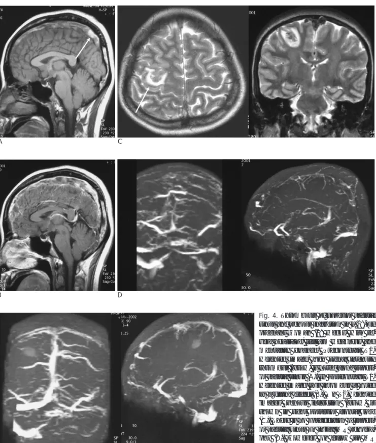
(RPLS))의 형태로 나타난다. 이 질환은 특징적으로 후두엽과 두정엽의 피질하백질과 인접한 회백질에 반점형의 T2 고신호 강도를 보이며 대부분 확산강조영상과 ADC map에서도 고신 호강도의 혈관성 부종(vasogenic edema)의 양상을 보인다 (Fig. 2). 비전형적인 경우 기저핵, 전두엽과 측두엽의 뒤쪽 부 분, 대뇌부챗살(corona radiate), 뇌간과 소뇌에도 신호강도 변 화가 올 수 있다. 이러한 영상 소견은 대개 임상증상과 증후 가 호전되면 정상으로 회복되나 드물게 영구적으로 남을 수 있다(4).
베르니케 뇌병증(Wernicke Encephalopathy)
베르니케 뇌병증은 수용성 비타민 B 복합체인 티아민 (thiamine or vitamine B1) 결핍에 의한 드문 질환으로 특징 적으로 고전적 세증후(classic triad)인 정신착란(mental confusion), 안구운동 마비(paralysis of ocular movement)와 조화운동불능(ataxia)을 보인다. 만성 알코올 중독증이 가장 흔 한 원인이며 장기간 음식을 거부하거나 먹지 못하는 환자에서 발생한다. 그 외에도 잦은 구토, 영양 섭취 부족 그리고 임신 으로 인한 대사 증가에 따른 임신 입덧(hyperemesis
A B C
Fig. 1. Cerebral hemorrhage in a 38-year-old pregnant (38 weeks) patient with preeclampsia complained headache. Her blood pres- sure was 220/170 mmHg. After delivery, she complained aphasia and right hemiparesis. Ovoid or enlongated high density is noted in left basal ganglia on pre-contrast enhanced CT (A), typical finding of acute hypertensive hemorrhage. On MR, this hematoma appears heterogenously high signal intensity on T2-weighted image (B) and dark signal intensity representing hemosiderin deposi- tion on gradient echo image (C).
A B C
Fig. 2. Posterior reversible encephalopathy syndrome (PRES) in a 32-years-old pregnant woman (34 weeks) with preeclampsia and symptoms of seizures and mental confusion. Multifocal high signal intensities (arrows) on T2-weighted image are demonstrated in cortical and subcortical region of both frontal and parietal lobes (A). On diffusion images, there is no abnormally increased signal intensity (B) and the high signal intensities on T2-weighted images appear to be increased value on ADC map (C), representing va- sogenic edema.
gravidarum)이 베르니케 뇌병증의 원인으로 알려져 있다.
발생기전은 정확히 알려져 있지 않지만 특징적 병리소견으 로 신경세포와 말이집구조(myelinated structures)의 괴사를 보인다.
영상소견은 특징적으로 내측 시상(medial thalamus), 중간 괴(massa intermedia), 시상하부 핵(hypothalamic nuclei), 수 관 주변 회색질(periaqueductal gray matter), 유두체 (mamillary bodies), 중간뇌(midbrain), 덮개(tectum)에 T2강 조 MR영상에서 대칭적인 고신호강도를 보인다(Fig. 3). 급성 기에는 제 3뇌실 주변, 유두체 그리고 수관 주변 회백질에 세 포 괴사에 의한 조영증강이 보일 수 있으며 만성기에는 유두 체가 위축되고 제 3뇌실이 확장된다. 확산강조영상에서는 대 부분 세포독성(cytotoxic) 부종에 의한 고신호강도를 보인다 (5).
뇌정맥굴혈전(Cerebral Venous Sinus Thrombosis)
임신 시에는 순환하는 혈액응고 인자(clotting factor)가 약 120-300%까지 증가하며 인자 II, VII과 X 이 증가하고 단백 질 S는 감소한다. 이러한 변화는 임신과 산욕기에 섬유소용해 기전(fibrinolytic system)을 방해하고 섬유소 합성(fibrin generation)을 증가시켜 과응고상태(hypercoagulable state)를 만들게 된다. 따라서 임신과 산욕기에 탈수가 되면 혈전 합병 증의 위험도가 증가한다. 뇌정맥굴혈전은 모성 나이(maternal age), 과다구토(hyperemesis), 제왕절개에 의한 분만, 감염과 모성 고혈압(maternal hypertension)과 관련이 있다. 대부분 의 뇌정맥굴혈전은 임신 중보다는 산욕기에 발생하며 두통을 흔히 호소한다(2). 그 외 초점 발작(focal seizure), 불완전마 비(paresis), 유두부종(papilledema), 의식변화(alteration of consciousness) 나 뇌내고혈압(isolated intracranial hypertension)이 나타나기도 한다(6). 진단은 CT에서 정맥굴
내에 고음영의 혈전을 확인할 수 있으며 특히 조영 전 CT에 서 위시상정맥동굴(superior sagittal sinus)에 그리스 문자 delta와 유사한 삼각형모양의 고음영 혈전을 볼 수 있어 이것 을‘delta sign’으로 불린다. 조영증강 후에는 위시상정맥동굴 내에 조영제에 둘러싸인 저음영의 혈전이 있어‘empty delta sign’으로 불린다. MR에서는 정맥굴내에 비정상 신호강도가 있고 자기공명정맥조영술(MR venography)에서 흐름이 없거 나 조영증강영상에서 조영증강이 없는 것으로 알 수 있다(Fig.
4).
쉬한증후군(Sheehan Syndrome)
산후 심한 출혈에 의해 발생한 저혈압 후에 생기는 허혈성 뇌하수체 괴사(ischemic pituitary necrosis)가 쉬한 증후군이 다. 뇌하수체는 혈관과다분포(highly vascularized)를 보이는 조직 중 하나로 임신 중 크기가 증가하여 안장가로막 (diaphragm sella)에 의해 위뇌하수체동맥(superior hypophyseal artery)이 눌려 경한 허혈(mild ischemia)을 일 으킬 수 있으며 분만시의 갑작스런 혈압의 변화에 의해서 동 맥연축(spasm)이 생겨 뇌하수체졸중(pituitary apoplexy)가 생 기게 된다.
전형적인 쉬한 증후군은 뇌하수체 전엽의 부분 또는 완전파 괴에 의해 수유실패(failure of lactation)나 무월경 (amenorrhea), 부신 기능부전(adrenal insufficiency), 갑상샘 저하증(hypothyroidism) 같은 뇌하수체 기능부전 증상이 나타 나게 된다. 급성기에는 갑작스런 종괴효과로 인한 두통이 생 기며 뇌하수체 기능부전에 의한 증상들이 나타나게 된다. 급 성기의 MR 소견은 뇌하수체가 커져 있으며 경색에 의한 중심 부 T1 저신호강도와 T2 고신호강도를 보인다. 조영증강 후 주 변부에 조영증강을 보이며 중심부분의 경색부위가 비균질한 조영증강을 보이게 된다(7). 이러한 비출혈성 중심부 허혈성
A B C
Fig. 3. Wernicke encephalopathy in a 31-year-old pregnant woman (16 weeks) with long history of vomiting for more than 2 months. She complained ophthalmoplegia and altered mentality. On FLAIR images, high signal intensities (arrows) are noted in both medial thalami, periaqueductal gray matter, and tectum.
A C
B D
Fig. 4. Thrombosis of superior sagittal sinus and venous infarction in a 28-old pregnant woman (28 weeks) with se- vere headache, left arm weakness, and mentality change. Precontrast T1- weighted image, high signal intensity thrombus (arrow) is noted along superi- or sagittal sinus (A). In postcontrast T1- weighted image, this thrombus is noted as a filling defect (B). On T2-weighted images, venous infarction (arrow) is shown in right posterior frontal lobe (C). There is no opacification of superi- or sagittal sinus on initial MR venogra- phy (D). However, on follow up MR venography after treatment, the superi- or sagittal sinus is well delineated after 15 days (E).
E
경색이 쉬한 증후군의 특징이며 수주 후에 뇌하수체의 부기가 빠지면서 정상 크기를 보이게 되며 수개월 후 뇌하수체가 위 축되고 빈안장증후군(empty sella)이 나타난다(Fig. 5).
폐양수색전증에 의한 이차성 저산소성허혈 (Hypoxic Encephalopathy Secondary to
Amniotic Fluid Embolism)
양수색전증은 갑작스런 저혈압, 저산소증, 소모성 응고병증 (consumptive coagulopathy)이 특징인 질환으로 치명률 (fatality)이 높아 모든 모성 사망의 약 10%를 차지하며 8,000- 12,000 분만 중 한 명의 빈도로 발생한다. 발생기전은 논란이 많으나 폐색전(pulmonary emboli)의 기계적인 폐색 (mechanical obstruction)에 의한 혈역학적 변화(hemody-
namic change)보다는 양수(amniotic fluid)에 대한 면역 면역 글로불린 E 매개 초과민반응(immune immunoglobulin E- mediated anaphylactoid reaction) 또는 비면역 초과민반응 (non-immune anaphylactoid reaction), 보체(complement)의 활성과 양수의 직접적인 유입이 중요한 원인으로 추정되고 있 다. 양수로부터 태아항원(fetal antigen)과 잠재적인 bioactive substances가 모체 순환으로 유입되면 갑작스런 심혈관부전과 호흡부전에 빠질 수 있으며 응고연쇄반응과 트롬빈 합성을 활 성화 시켜 파종혈관내응고(DIC)에 빠지게 된다. 정확한 위험 인자가 정립되어 있지 않지만 양수과다증(polyhydramnios), 태반조기박리(placental abrup-tion), 전치태반(placenta praevia), 유착태반(placenta accrete), 양막파열(rupture of the amniotic membranes), 산도파열(rupture of birth canal), 큰몸증(macrosomia), 임신유발고혈압(pregnancy-induced
A B
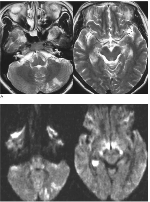
Fig. 6. Cerebral infarction in a 32-years-old woman who had a caesarean section at 37 week gestational period. After delivery, car- diovascular and respiratory failure was suddenly developed, and thereafter comatous mentality was developed. Diffuse high signal intensities are demonstrated in both frontal lobes on T2-weighted images (A) and FLAIR (B) images with diffuse atrophic changes involving both cerebral hemispheres. Additionally multiple small nodular ischemic foci are noted in both basal ganglia regions.
Fig. 5. Sheehan’s syndrome in a 57- years-old woman with a history of large amount postpartum hemorrhage and pituitary necrosis after twin deliv- ery, 27 years ago. On T1- weighted im- ages, CSF invagination is seen in the sella turcica with no remaining pitu- itary gland, representing empty sella (arrow). Typical obstetrical history of severe postpartum bleeding and emp- ty sella on MRI are suggested for the diagnosis of Sheehan’s syndrome.
hypertension), 다분만(multiparous), 알레르기 또는 아토피 (allergy or atopy), 제왕절개(caesarean section) 등이 관련이 있다는 보고가 있다(8).
확진은 특징적 임상 증상과 검사 소견, 그리고 조직학적으 로 혈관 내에 양수색전 성분을 확인하는 것이다(Fig. 6).
다발성 뇌경색(Multifocal Infarctions)
임신 제3기와 산욕기에 다양한 원인에 의해 허혈성 경색이 발생할 수 있으며 특히 출산 후 첫 1주에 높은 빈도를 보인다.
정확한 원인은 알려져 있지 않으나 임신 시에는 수분 정체 (water retention)로 혈중 나트륨 농도와 삼투압 감소하게 되 는데 출산 후 나트륨 농도와 삼투압이 증가하고 혈장량(plasma
volume)이 감소하면서 상대적인 혈관수축이 생겨 허혈성 경 색의 위험도가 증가할 것이라 추측하고 있다(9). 산욕기의 허 혈성 경색의 원인으로 심혈전과 전자간이나 자간이 높은 빈도 를 보였으나, 정확한 연관성은 알려져 있지 않다(Fig. 7).
교뇌 밖 수초용해증(Extrapontine Myelinolysis)
중심 교뇌(central pontine myelinolysis, CPM) 및 교뇌 밖 수초용해증(EPM)은 약 75% 이상에서 저나트륨혈증 (hyponatremia)의 급속한 교정이나 만성 알콜중독증과 관계 가 있으며 그 밖에 고나트륨혈증(hypernatremia), 영양실조 (malnutrition), 애디슨병(Addison’s disease), 신부전(renal failure), 그리고 이뇨제 사용(diuretic usage)과 관계가 있다.
A
B
Fig. 7. Multifocal infarctions in a 35- years-old woman who had a delivery one week ago at 38 weeks gestational period. At admission, she complained blurred vision, headache, dizziness, and left side tingling sensation.
Multifocal high signal intensities are noted in left cerebellar hemisphere, right hippocampus and parahip- pocampal gyrus on T2-weighted im- ages (A) and bright signal intensities on diffusion weighted images, repre- senting acute infarctions (B).
병 리 적 으 로 이 질 환 은 경 계 가 분 명 한 말 이 집 탈 락 (demyelination)이 특징적이라고 알려져 있으나 정확히 말하 면 축삭파괴에 의한 말이집 소실이며, 특히 중심부에는 괴사 와 동반된 축삭용해(axonal lysis)를 보인다. 이러한 병변은 교
뇌(pons)에 가장 흔히 생기며 교뇌 밖에 생길 때에는 특징적 으로 기저핵, 시상, 속섬유막(internal capsule), 겉섬유막 (external capsule), 대뇌 백질에 생기며 드물게 주변부 피질, 해마, 외측무릎체(lateral geniculate bodies)에 생길 수 있다.
A
B
C
Fig. 8. Extrapontine myelinolysis in a 29-years-old woman who had a cae- sarean section two days ago. At admis- sion, her blood sodium concentration was very low (115 mmEq). After very slow correction of her sodium concen- tration and osmolarity, semicomatous mentality was developed. Bilateral high signal intensities are noted in both periventricular deep white mat- ter, splenium of corpus callosum, and both posterior limbs of internal cap- sule on FLAIR images (A). On diffu- sion images, they appear to be very bright signal intensity (B), and de- creased values on ADC maps, repre- senting cytotoxic edema (C).
교뇌에서 이러한 병리적 특징은 등쪽 뇌바닥다리뇌(basis pontis)에서 정중 솔기(median raphe)부터 원심방향으로 삼각 형 모양(triangular) 또는 박쥐날개(bat-wing) 모양으로 나타 나며, 심한 경우를 제외하고 대부분 교뇌뒤부분(tegmentum), 겉질척수로와 겉질연수로(corticospinal and corticobulbar tract)는 침범하지 않는다.
특징적인 임상증상은 처음에는 말더듬증(dysarthria), 삼킴 곤란(dysphagia), 외안근쇠약(extraocular muscle weakness) 을 보이다가 발작(seizure)이나 졸음(drowsi-ness)에서 혼수 (coma)에 빠질 수 있다.
중심교뇌수초용해증(CPM)과 교뇌밖수초용해증(EPM)의 영 상소견은 특징적으로 대칭적으로 나타나며 CT에서 저음영을 보이고 MR T1 강조영상에 저신호강도, T2 강조영상에서 고 신호강도를 보인다(Fig. 8). 조영증강이 될 수도 있으나 출혈 은 동반되지 않는다. 급성기에는 확산강조영상에서 고신호강 도의 세포독성부종을 보일 수 있다(10).
뇌내출혈, 자간에 의한 가역적 후백색질뇌증, 베르니케 뇌병 증, 뇌정맥굴 혈전, 쉬한증후군, 폐양수색전증에 의한 이차성 저산소성허혈, 다발성 뇌경색과 교뇌 밖 수초용해증 등 임신 과 출산에 연관된 다양한 신경학적 질환의 원인 기전, 임상 증 상과 영상 소견을 알아보았다. 임신 및 출산에 관계된 이러한 질환의 영상의학적 소견을 정확히 숙지함으로써 조기진단과 정확한 감별진단 그리고 적절한 치료에 많은 도움을 줄 수 있 을 것으로 생각된다.
참 고 문 헌
1. Kittner SJ, Stern BJ, Feeser BR, Hebel R, Nagey DA, Buchholz DW, et al. Pregnancy and the risk of stroke. N Engl J Med 1996;335:768-774
2. Karnad DR, Guntupalli KK. Neurologic disorder in pregnancy. Crit Care Med 2005;33(10 Suppl):S362-S371
3. Schwartz RB, Feske SK, Polak JF, DeGirolami U, Iaia A, Beckner KM, et al. Preeclampsia-eclampsia: clinical and neuroradiographic correlates and insights into the pathogenesis of hypertensive en- cephalopathy. Radiology 2000;217:371-376
4. Finocchi V, Bozzao A, Bonamini M, Ferrante M, Romano A, Colonnese C, et al. Magnetic resonance imaging in Posterior Reversible Encephalopathy Syndrome?: report of three cases and review of literature. Arch Gynecol Obstet 2005;271:79-85
5. Callucci M, Bozzao A, Splendiani A, Masciocchi C, Passariello R.
Wernicke encephalopathy: MR findings in five patients. AJR Am J Roentgenol 1990;155:1309-1314
6. Brass SD, Copen WA. Neurological disorders in pregnancy from a neuroimaging perspective. Semin Neurol 2007;27:411-424 7. Kaplun J, Fratila C, Ferenczi A, Yang WC, Lantos G, Fleckman
AM, et al. Sequential pituitary MR imaging in Sheehan syndrome:
report of 2 cases. AJNR Am J Neuroradiol 2008;29:941-943 8. Davies S. Amniotic fluid embolus: a review of the literature. Can J
Anaesth 2001;48:88-98
9. Skidmore FM, Williams LS, Fradkin KD, Alonso RJ, Biller J.
Presention, etiology, and outcome of stroke in pregnancy and puerperium. J Stroke Cerebrovasc Dis 2001;10:1-10
10. Chua GC, Sitoh YY, Lim CC, Chua HC, Ng PY. MRI findings in osmotic myelinolysis. Clin Radiol 2002;57:800-806
J Korean Radiol Soc 2008;59:65-73
Address reprint requests to : Kook Jin Ahn, M.D., Department of Radiology, College of Medicine, The Catholic University of Korea 505, Banpo-dong, Seocho-gu, Seoul 137-701, Korea.
Tel. 82-2-2142-7492 Fax. 82-2-599-6771 E-mail: [email protected]
CT and MR Findings of Neurological Disorders Associated with Pregnancy and Childbirth
1Jee Young Kim, M.D., Kook Jin Ahn, M.D., Young Joo Kim, M.D., Bum-soo Kim, M.D., Seong Tae Hahn, M.D.
1Department of Radiology, College of Medicine, The Catholic University of Korea
The onset of pregnancy may predispose women to a variety of neurological diseases due to changes in their hemodynamics, hormonal effects, and complications associated with childbirth. The spectrum of neurological disorders associated with pregnancy and childbirth include hypertensive intracerebral hemorrhaging, posteri- or reversible encephalopathy syndrome (PRES) (secondary to eclampsia), Wernicke encephalopathy, cerebral venous sinus thrombosis, Sheehan’s syndrome, hypoxic ischemic encephalopathy (secondary to pulmonary amniotic fluid embolism), multifocal infarctions, and extra-potine myelinolysis. The recognition of the various imaging findings of these diseases, along with the clinical presentations, should aid in their early diagnosis and prompt treatment. The purpose of this pictorial assay is to describe the characteristic CT and MR findings of these diseases with a literature review to explain the mechanisms and clinical symptoms.
Index words :Brain
Pregnancy Complications Childbirth
Tomography, X-Ray computed Magnetic resonance (MR)