대한정형외과학회지:제 43 권 제 1 호 2008 J Korean Orthop Assoc 2008; 43: 131-134
131
통신저자:권 석 현
전북 익산시 신용동 344-2 원광대학교병원 정형외과
TEL: 063-859-1360ㆍFAX: 063-852-9329 E-mail: [email protected]
Address reprint requests to Seok Hyun Kweon, M.D.
Department of Orthopedic Surgery, Wonkwang University Hospital, 344-2, Sinyong-dong, Iksan 570-711, Korea
Tel: +82.63-859-1360, Fax: +82.63-852-9329 E-mail: [email protected]
*This paper was supported by Wonkwang University in 2008.
Synovial Chondromatosis of the Metacarpophalangeal Joint
- A Case Report -
Jeung Woo Kim, M.D., Seok Hyun Kweon, M.D., and Dong Chul Kim, M.D.*
Department of Orthopedic Surgery, Wonkwang University Hospital*, Hana Hospital†, Iksan, Korea
중수지관절에 발생한 활액막 연골종증 - 증례 보고-
김정우ㆍ권석현ㆍ김동철
* 원광대학교병원 정형외과, 하나 정형외과*Synovial chondromatosis is an uncommon lesion, which is characterized by cartilaginous and osseous metaplasia of the joint synovium. Synovial chondromatosis usually involves a large joint and rarely occurs in the hand. Intra-articular synovial chondromatosis during the hand should be considered in the differential diagnosis of swollen, stiff or painful joints. Other possible diagnoses include osteoarthritis, rheumatoid arthritis, gout, trauma and chronic infection. Moreover, if enchondral ossification of loose bodies is not seen a diagnosis of synovial chondromatosis can not be made preoperatively. Intra-articular synovial chondromatosis is a benign condition and surgical synovectomy remains the most effective treatment. The authors report a case of synovial chondromatosis of the fifth metacarpophalangeal joint.
Key Words: Fifth metacarpophalangeal joint, Synovial chondromatosis
Synovial chondromatosis is an uncommon disorder of joints, bursae, and tendon sheaths. The etiology of the disease is unclear, but it involves the metaplasia of normal synovial cells to chon- droblasts5). Synovial chondromatosis is charac- terized by the formation and growth of cartilagi- nous bodies that may then calcify or ossify within synovial tissue1). The usual treatment involves loose body removal and complete synovectomy.
Here, we describe a case of synovial chondro- matosis of the fifth metacarpophalangeal (MP) joint.
CASE REPORT
A 45-year-old, right-handed man presented with a 3-year history of pain, swelling and stiffness of the MP joint of the little finger of his right hand. He had no history of accident trauma or of a problem with other joints. Discomfort and progressive limitation of motion interfered with his daily activities. On examination, a non-tender, hard, and nodular mass about 2×2 cm in size was found and moderate tenderness along the joint line and a range of motion from 5o to 70o was evident, but sensation and perfusion in the little finger were
132 Jeung Woo Kim, Seok Hyun Kweon, Dong Chul Kim
Fig. 2. (A, B) T1-weighted MR images depicting an iso-intense intra-articular lesion (arrows). (C, D) T2-weighted MR images show- ing an intra-articular body of low signal intensity (arrowhead). The central area has the signal cha- racteristics of hyaline cartilage.
Fig. 1. Hand radiographs showing bony erosion of fifth meta- carpal head and several calcifications of 1 to 2 millimeters in size.
normal. A plain X-ray showed diffuse calcification in the area of the swelling (Fig. 1), and magnetic resonance imaging (MRI) scan findings were con-
sistent with synovial chondromatosis, which ap- peared to be localized to joint synovium and not to invade any surrounding structure (Fig. 2). We planned definitive surgery, i.e., loose body removal and complete synovectomy.
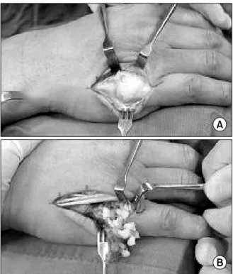
Under subscalenous regional anesthesia, with a tourniquet, surgical incision was performed through a longitudinal dorsal incision over the MP joint to expose the joint capsule. Division of the capsule revealed thickened inflammatory synovium, with numerous chondrous nodules in the capsule wall and lying free (Fig. 3A). These loose bodies were well contained within the dorsal pouch of synovium and capsule and between the joint surfaces. In edition the peripheral articular cartilage was eroded (Fig. 3B). Complete synovectomy was carried out and all loose bodies were removed.
Postoperatively, the patient was followed up for one year had a full active range of motion, with no MP joint instability.
Synovial Chondromatosis of the Metacarpophalangeal Joint 133
Fig. 3. Intraoperative clinical photographs. (A) Approximately a 3×2 cm sized mass was exposed from the proximal third of the 5th metacarpal bone to the metacarpophalangeal joint. (B) Numerous chondrous nodules in the capsule wall were flowed out.
DISCUSSION
Synovial chondromatosis is an uncommon condi- tion characterized by the formation of multiple cartilaginous nodules in synovial tissue. It gene- rally involves a large joint4), and its etiology has not been determined. The hand is rarely involved, but when it is, extra-articular structures, such as, the tenosynovium are usually affected8). Synovial chondromatosis tends to present between the ages of 20 and 50 and it is twice as common in men5). Its natural history is variable, and at presentation symptoms have usually been present from 3 months to 10 years. A correct diagnosis may not be made because of its rarity, especially if no calcification or ossification of cartilage is evident2). MRI is the best imaging modality as it shows the cartilaginous portion of the lesion in addition to any ossified areas3,7). Destruction of the joint space
is rare, but if present, is confirmed at surgery. Our case had a positive radiologic finding, which included ossified loose bodies and periarticular erosion. However, the joint space was well pre- served.
Treatment in most cases of intra-articular chondromatosis is by arthrotomy, loose body re- moval and synovectomy. This treatment is uni- formly successful; all reported patients have ex- perienced symptom resolution and an improved range of motion at follow-up.
Intra-articular synovial chondromatosis in the hand is a rare but benign condition, which should be considered in the differential diagnosis of patients presenting with a swollen, stiff or painful joint. The most effective treatment is surgical synovectomy.
REFERENCES
1. Constant E, Harebottle NH, Davis DG: Synovial chon- dromatosis of the hand. Plast Reconstr Surg, 54: 353-358, 1974.
2. Crotty JM, Monu JU, Pope TL Jr: Synovial osteochon- dromatosis. Radiol Clin North Am, 34: 327-342, 1996.
3. Hettiaratchy SP, Nanchahal J: Synovial chondromatosis of the metacarpophalang eal Joint. J Hand Surg Br, 27: 104-106, 2002.
4. Maurice H, Crone M, Watt I: Synovial chondromatosis. J Bone Joint Surg Br, 70: 807-811, 1988.
5. Milgram JW: Synovial osteochondromatosis: a histopathological study of thirty cases. J Bone Joint Surg Am, 59: 792-801, 1977.
6. Murphy FP, Dahlin DC, Sullivan CR: Articular synovial chondromatosis. J Bone Joint Surg, 44: 77-86, 1962.
7. Kramer J, Recht M, Deely DM, et al: MR appearance of idiopathic synovial osteochondromatosis. J Comput Assist Tomogrt, 17: 772-776, 1993.
8. Reed SC, Wright CS: Synovial chondromatosis of the metacarpophalangeal joint: case report and review of the literature. Can J Surg, 39: 407-409, 1996
134 Jeung Woo Kim, Seok Hyun Kweon, Dong Chul Kim
= 국문초록=
활액막 세포의 연골모세포로의 화생에 의해 발생하는 활액막 연골종증은 주로 슬관절, 골관절, 견관절, 주관절 등의 큰 관절을 침범하는 질환으로 수부에서는 매우 드문 것으로 알려져 있다. 수부 관절내 활액막 연골종증은 부종, 강직 및 동통의 증상을 나타냄으로, 관절염, 류마토이드 관절염, 통풍, 외상, 만성 감염 등이 질환과 감별 진단이 필요하다. 유리체의 연골내 석회화가 보이지 않는다면, 술전에 활액막 연골종증으로 진단하는 것이 어렵 다. 관절내 활액막 연골종증은 양성으로 수술적 활액막 절제술이 가장 효과적인 치료로 알려져 있다. 저자들은 제5 중수지관절에 발생한 활액막 연골종증을 1예 경험하여 이를 문헌 고찰과 함께 보고하고자 한다.