Received November 12, 2015, Revised April 8, 2016, Accepted for publication May 9, 2016
*These authors contributed equally to this study.
Corresponding author: Kyu Joong Ahn, Department of Dermatology, Konkuk University School of Medicine, 120 Neungdong-ro, Gwangjin-gu, Seoul 05029, Korea. Tel: 82-2-2030-5181, Fax: 82-2-2030-5179, E-mail: kjahn@
kuh.ac.kr
Soo-Yeon Kim, Korea Institute for Skin and Clinical Sciences, Konkuk University, 120 Neungdong-ro, Gwangjin-gu, Seoul 05029, Korea. Tel: 82- 2-450-4054, Fax: 82-2-3436-3778, E-mail: [email protected] This is an Open Access article distributed under the terms of the Creative Commons Attribution Non-Commercial License (http://creativecommons.
org/licenses/by-nc/4.0) which permits unrestricted non-commercial use, distribution, and reproduction in any medium, provided the original work is properly cited.
Copyright © The Korean Dermatological Association and The Korean Society for Investigative Dermatology
Ann Dermatol Vol. 29, No. 1, 2017 https://doi.org/10.5021/ad.2017.29.1.6
ORIGINAL ARTICLE
Kinetin Improves Barrier Function of the Skin
by Modulating Keratinocyte Differentiation Markers
Sungkwan An, Hwa Jun Cha, Jung-Min Ko, Hyunjoo Han, Su Young Kim, Kyung-Suk Kim, Song Jeong Lee, In-Sook An1, Sangwon Kim2, Hae Jeong Youn3, Kyu Joong Ahn3,*, Soo-Yeon Kim*
Korea Institute for Skin and Clinical Sciences, Konkuk University, Seoul, 1KISCS Incorporated, Cheongju, Korea, 2Orangewood Christian School, Maitland, FL, USA, 3Department of Dermatology, Konkuk University School of Medicine, Seoul, Korea
Background: Kinetin is a plant hormone that regulates growth and differentiation. Keratinocytes, the basic building blocks of the epidermis, function in maintaining the skin barrier. Objective: We examined whether kinetin induces skin barrier functions in vitro and in vivo. Methods: To eval- uate the efficacy of kinetin at the cellular level, expression of keratinocyte differentiation markers was assessed. Moreover, we examined the clinical efficacy of kinetin by evaluating skin moisture, transepidermal water loss (TEWL), and skin surface roughness in patients who used kinetin-containing cream. We performed quantitative real-time polymerase chain reaction to measure the expression of keratinocyte dif- ferentiation markers in HaCaT cells following treatment. A clinical trial was performed to assess skin moisture, TEWL, and evenness of skin texture in subjects who used ki- netin-containing cream for 4 weeks. Results: Kinetin in- creased involucrin, and keratin 1 mRNA in HaCaT cells.
Moreover, use of a kinetin-containing cream improved skin moisture and TEWL while decreasing roughness of skin
texture. Conclusion: Kinetin induced the expression of kera- tinocyte differentiation markers, suggesting that it may affect differentiation to improve skin moisture content, TEWL, and other signs of skin aging. Therefore, kinetin is a potential new component for use in cosmetics as an anti-aging agent that improves the barrier function of skin. (Ann Dermatol 29(1) 6∼
12, 2017)
-Keywords-
Cell culture techniques, Differentiation, Keratinocytes, Kine- tin, Skin barrier
INTRODUCTION
The effects of human aging are mostly visible in the skin in such forms as increased wrinkling and sagging, as well as decreased elasticity1. Aging is also associated with physical disorders of the skin because its barrier function is dis- rupted, leading to a dry appearance and enhanced risk of skin disorders2,3. Understanding the mechanisms of skin ag- ing is important for developing skin care products that de- lay aging and reduce damage4,5. Skin aging is induced by both intrinsic and extrinsic factors4,6 that lead to a reduction of structural integrity and loss of physiological function6. Kinetin, a cytokine isolated in 1955, is an essential plant growth hormone that regulates cell growth and differ- entiation7,8. Kinetin has also been reported to be present in human cell extracts9 and urine10, and has been identi- fied as a naturally occurring base modification agent of DNA11. Kinetin has been reported to have multiple func- tions, including anti-aging effects in cultured cells12,13 and fruit flies14, antioxidant properties15,16, antithrombotic ac- tivity17,18, and cell differentiation effects19-21. Therefore, in
this study, we demonstrate that kinetin may induce an- ti-aging effects in skin by improving its barrier function.
MATERIALS AND METHODS
Cell culture
HaCaT human keratinocytes (American Type Culture Collection, Boulecard Manassas, VA, USA) were cultured in Dulbecco’s modified Eagle medium (Gibco/Life Tech- nologies, Carlsbad, CA, USA) supplemented with 10% fetal bovine serum (Sigma-Aldrich, St. Louis, MO, USA) and 1%
penicillin/streptomycin (Gibco/Life Technologies) at 37oC in an atmosphere of 5% CO2. Kinetin was purchased from Sigma-Aldrich and dissolved in dimethyl sulfoxide.
Cell viability assay
HaCaT cells were seeded at a density of 3×10³ cells in 96-well plates and incubated for 24 h. The cells were then incubated with kinetin (0∼600 μM) for 24 h. HaCaT cell toxicity due to kinetin was evaluated using a water-soluble tetrazolium salt (WST)-1 assay (EZ-Cytox Cell Viability Assay Kit; Itsbio, Seoul, Korea). WST-1 solution was added to cultured cells at a volume equal to 10% that of the cul- ture medium, and then the cells were incubated at 37oC for 1 h. Cell viability was evaluated by measuring the ab- sorbance at 450 nm using an iMark microplate reader (Bio-Rad, Hercules, CA, USA).
Isolation of total RNA and quantitative real-time poly- merase chain reaction
Total RNA was isolated using TRIzol reagent (Invitrogen Life Technologies, Carlsbad, CA, USA) according to the manufacturer's protocol. The purity and concentration of the RNA were evaluated using a MaestroNanoⓇ, a micro- volume spectrophotometer (Maestrogen, Las Vegas, NV, USA). The recommended parameters of RNA quality for cDNA synthesis were OD 260/230 >1.8 and an OD 260/280 ratio within the range of 1.8∼2.0. cDNAs were synthesized using the miScript II RT Kit (Qiagen, Hilden, Germany) according to the manufacturer's protocol. To evaluate the expression of INV (forward primer: 5'- GGGTGGTTATTTATGTTTGGGTGG-3', reverse primer:
5'-GCCAGGTCCAAGACATTCAAC-3') and KRT1 (forward primer: 5'-ATTTCTGAGCTGAATCGTGTGATC-3', reverse primer: 5'-CTTGGCATCCTTGAGGGCATT-3'), quantitative real-time polymerase chain reaction was performed using EvaGreen dye (Solis BioDyne, Tartu, Estonia) with Line-Gene K software (Bioer Technology Co., Ltd., Hangzhou, China).
The CT value for each gene was normalized to β-actin (forward primer: 5'-GGATTCCTATGTGGGCGACGA-3', reverse primer: 5'-CGCTCGGTGAGGATCTTCATG-3'). Re-
lative expression levels of each gene were calculated us- ing the 2−ΔΔCt method22.
Subjects for clinical evaluation
The study protocols were approved by the Institutional Review Board of KISCS Incorporated (Cheongju, Korea) (IRB no. 1-70005239-A-N-01-2013-KISCS-ACA001-KSH).
All subjects were informed about the objective of the study and provided informed consent and agreed to use products for skin care during the study. Forty women greater than 40 years of age were enrolled in a random- ized, double-blind clinical trial (control group: 46.80±
4.83 years, experiment group: 46.70±0.83 years). The subjects were selected based on age, the absence of skin conditions other than age-related conditions, and were not pregnant or nursing. All subjects were informed about the objective of the study, provided signed informed consent, and agreed to use only products for skin care during the study. Reasons for dropping out were itching, erythema, or excessive drinking or smoking. Subjects were divided into control and experimental groups consisting of 20 sub- jects each. All conditions were identical, other than the exposure of the experimental group to the test material.
The study lasted four weeks, except no drop out. Clinical parameters were evaluated three times, namely, before ap- plication, and after 2 and 4 weeks of use. The investigator asked subjects about the condition of their skin and per- formed a visual examination of their skin condition, such as erythema, itching, scale, edema, tingling, and burning sensation, at every visit. The cream was prepared by in- corporating the ingredients in the three phases (A, B, C).
Ingredients in the A phase (distilled water, glycerin, 1,3-butylene glycol) were combined and heated until all the components were melted, and ingredients in the B phase (distilled water, dipotassium phosphate, sodium hy- droxylate, kinetin) were combined and heated to the same temperature, to ensure homogeneity. The A and B phases were combined and emulsified using a homo mixer (Tokushu Kika Kogyo Co., Ltd., Osaka, Japan) at 5,000 rpm for 10 min. The mixture was cooled to 60oC and blended with the homogenized phase C (emulium delta, sepipuls 400) at 5,000 rpm for 10 min. By then, the tem- perature of the mixture had dropped to 45oC. The mixture was combined and homogenized, while maintaining the pH at 6.2. The cream provided to the experimental group contained 2% (wt%) kinetin and the cream provided to the control group was prepared using the same volume of water in place of kinetin.
Experimental procedures
To investigate the improvement in skin barrier function,
Fig. 1. Cytotoxicity of kinetin in HaCaT keratinocytes. HaCaT keratinocytes were treated with kinetin at the indicated con- centrations for 24 h. The results are representative of three independent experiments (means±standard deviation are shown).
*p<0.05 and **p<0.001 as determined by the Student’s t-test.
subjects were instructed to apply 2 g of test material to the face every morning and night for 4 weeks. Subjects and investigators were blinded to test and control treatments.
Moisture and transepidermal water loss (TEWL) were measured on the right cheek and skin texture was eval- uated on the left side of the forehead. At every visit, before measurements were taken, all subjects washed with the cleanser provided and lay quietly in a room with constant temperature (22oC±1oC) and humidity (45%±5%) so that all subjects would be evaluated under the same conditions.
Evaluation of skin moisture
To evaluate improvement in skin moisture, the DermaLab USB moisture probe (Cortex Technology Inc., Hadsund, Denmark) was applied and data were analyzed using the associated application software version 1.09. All subjects were measured on the same region of the right cheek five consecutive times, and the mean, maximum, and mini- mum values were determined. Measurements were taken three times, namely, before application, and after 2 and 4 weeks of use. The device applies the conductance meas- urement principle to measure the water-binding capacity of the stratum corneum (SC). This value correlates with skin moisture and is expressed in microsiemens (μS).
TEWL measurements
To evaluate improvement in TEWL, the DermaLab USB TEWL probe (Cortex Technology Inc.) was applied and da- ta were analyzed using the associated application software version 1.09. Five consecutive measurements were taken on the subject's right cheek, and the mean, maximum, and minimum values were determined. Measurements were taken three times, namely, before application and at 2 and 4 weeks after the application.
Measurement of facial skin evenness
Facial skin evenness measured by PRIMOS Lite (field of view 45×30; GFMesstechnik GmbH, Teltow, Germany) was used to obtain clinical images. Captured images were analyzed using the associated imaging software, PRIMOS Lite version 5.6E. Three consecutive clinical images of the left side of the subject's forehead were captured. Facial skin roughness was assessed based on the Ra value, which is the average of all heights and depths relative to the ref- erence plane. Measurements were taken three times, namely, before application, and at 2 and 4 weeks after the application. The Ra value is the most widely used parame- ter of facial skin roughness and is the arithmetic mean of the maximum values of all measurements.
Statistical and mathematical analysis
In cellular efficacy tests, all results are presented as the mean percentage±standard deviation of three indepen- dent experiments. Differences with a p-value of less than 0.05 or 0.001, as determined by Student’s t-test, were con- sidered statistically significant. In clinical efficacy tests, statistical analyses were conducted using SPSS software (PASW Statistics ver. 17.0 for Windows; IBM Co., Armonk, NY, USA). Paired t-tests were performed in cases of re- peated measurements on the same subject. In clinical test, all statically analysis was compared between 0 week and 2 or 4 weeks using paired t-test which analyze statically significant by a comparison of experimental measured val- ue of each entity in 0 weeks and 2 or 4 weeks. To analyze subject questionnaires, the mean values, standard devia- tion, and percentage were used. The formula used to measure the percent change for each skin parameter was
Percent change= ×100,
where A is defined as the individual value of any parame- ter at the 2 and 4 week visits and B represents the zero hour of the assessed parameter.
RESULTS
Cytotoxicity of kinetin in human keratinocytes
To determine whether kinetin affects HaCaT cell viability, cells were exposed to kinetin at concentrations ranging from 0∼600 μM for 24 h. As shown in Fig. 1, kinetin re- duced cell viability by 8.88% at 200 μM, 18.99% at 400
Fig. 3. The effect of kinetin on keratin 1 mRNA expression in HaCaT keratinocytes. Relative expression level of keratin 1 mRNA in CaCl2-treated (A) and CaCl2 plus kinetin-treated HaCaT keratinocytes was determined by quantitative real-time polymerase chain reaction.
The results are representative of three independent experiments (means±standard deviation are shown).
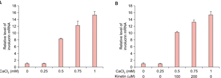
Fig. 2. The effect of kinetin on involucrin mRNA expression in HaCaT keratinocytes. Relative expression level of involucrin mRNA in CaCl2-treated (A) and CaCl2 plus kinetin-treated (B) HaCaT keratinocytes was determined by quantitative real-time polymerase chain reaction. The results are representative of three independent experiments (means±standard deviation are shown).
μM, and 29.78% at 600 μM. Kinetin-induced cytotox- icity increased significantly at concentrations greater than 200 μM. Thus, we used 200 μM as the maximum con- centration in subsequent experiments.
Effect of kinetin on expression of keratinocyte differen- tiation markers
Expression of involucrin, a marker of keratinocyte differ- entiation, was not altered by the treatment of HaCaT cells with 0.25 mM CaCl2. But when cells were treated with ki- netin, involucrin mRNA increased in a dose-dependent manner. The addition of kinetin following treatment with 0.25 mM CaCl2 increased the level of involucrin mRNA to that of cells treated with 2 mM CaCl2 alone (Fig. 2).
Simultaneously, we investigated the gene expression of
another keratinocyte differentiation maker, keratin 1. As seen for involucrin, keratin 1 expression was not altered by treatment of HaCaT cells with 0.25 mM CaCl2. However, when these cells were treated with kinetin, keratin 1 ex- pression increased markedly (Fig. 3).
Kinetin-containing cream improves skin hydration Keratinocyte moisture content is pivotal for maintaining moisture in the skin. Normal keratinocytes maintain 10%
∼30% moisture; however, when moisture content drops below 10%, keratinocytes are unable to maintain the skin's barrier function as skin becomes dry, acquires an uneven texture, and produces wrinkles, which accelerate senescence23. In this study, to evaluate the efficacy of ki- netin treatment on skin, we investigated the improvement
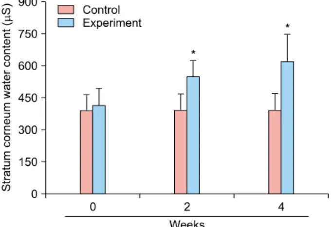
Fig. 4. Improvement in skin moisture over time. Measurements were taken three times, namely before application and after 2 and 4 weeks of application. To evaluate improvement in skin moisture, DermaLab USB moisture probe was applied and data were analyzed using the associated software application version 1.09. *p<0.001 as determined by the Student’s t-test.
Fig. 5. Percentage of improvement in transepidermal water loss (TEWL). Measurements were taken three times, namely, before application and after 2 and 4 weeks of use. *p<0.01 and **p< 0.001 as determined by the Student’s t-test.
in skin moisture after the treatment with a kinetin-contain- ing cream.
We analyzed skin moisture content using the DermaLab USB moisture probe. Our data demonstrate that the mois- ture content of the control group, which used the non-ki- netin-containing cream, was 388.10 μS before use, 389.96 μS after 2 weeks, and 390.72 μS after 4 weeks of appli- cation (Fig. 4). To compare the improvement at 2 and 4 weeks, we calculated and expressed the degree of im- provement (Fig. 4). Consequently, the moisture content of the control group increased by 0.48% and 0.68% after 2 and 4 weeks, respectively. These changes were not statisti- cally significant (p>0.05), indicating that the non-ki- netin-containing cream had no measurable effect on mois- ture content. In contrast, the moisture content in the ex- perimental group, which used kinetin-containing cream, was 413.34 μS before use, 551.39 μS after 2 weeks, and 619.98 μS after 4 weeks (Fig. 4). Interestingly, the use of the kinetin-containing cream significantly improved skin moisture by 33.40% and 49.99% after 2 and 4 weeks, re- spectively (p<0.001). These experiments demonstrate that the use of the kinetin-containing cream resulted in im- proved skin moisture content.
Improvement in TEWL following use of kinetin- containing cream
Next, to determine the efficacy of kinetin as a skin mois- turizer, we used the DermaLab USB TEWL probe to inves- tigate TEWL in the skin of subjects who had used control or kinetin-containing cream. The TEWL of control subjects was 9.59 g m−2 h−1 before use, 9.48 g m−2 h−1 after 2
weeks, and 9.38 g m−2 h−1 after 4 weeks (Fig. 5). To com- pare the improvement at 2 and 4 weeks, we calculated the improvement as a percentage based on the value before application. Consequently, the TEWL in the control group increased by 1.15% and 2.19% after 2 and 4 weeks of use, respectively. These changes were not statistically sig- nificant (p>0.05), indicating that the non-kinetin-contain- ing cream had no measurable effect on TEWL. In contrast, TEWL in the experimental group was 9.96 g m−2 h−1 be- fore use, 8.92 g m−2 h−1 after 2 weeks, and 7.08 g m−2 h−1 after 4 weeks (Fig. 5). To compare the improvement at 2 and 4 weeks, we calculated the improvement in TEWL as a percentage based on the value before application. Use of the kinetin-containing cream significantly improved TEWL by 10.40% and 28.88% after 2 and 4 weeks, re- spectively (p<0.001). Through these experiments, we identified the improvement in TEWL as an outcome of us- ing kinetin-containing cream.
Use of kinetin-containing cream improves evenness of skin texture
The thickness of the SC changes depending on its mois- ture content. Insufficient moisture in this layer roughens skin texture gradually24. Therefore, we investigated the ef- ficacy of kinetin-containing cream on skin texture. Facial skin evenness was measured using PRIMOS Lite. Evenness in the control group was 20.10 Ra before use, 19.95 Ra af- ter 2 weeks, and 19.79 Ra after 4 weeks (Fig. 6). To com- pare the improvement at 2 and 4 weeks, we calculated the improvement as a percentage based on the value before application. Consequently, skin texture in the control group improved by 0.77% and 1.54%, after 2 and 4 weeks of use, respectively. These data were not statistically sig-
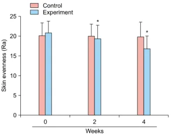
Fig. 6. Percentage of improvement in rates of skin roughness.
Evenness of the skin surface was measured by PRIMOS Lite.
Measurements were taken three times, namely, before applica- tion, and after 2 and 4 weeks of use. The captured images were analyzed using the associated imaging software PRIMOS Lite version 5.6E. *p<0.001 as determined by the Student’s t-test.
nificant (p>0.05), indicating that the non-kinetin-contain- ing cream had no measurable effect on improving skin texture. In contrast, skin evenness in the experimental group was 20.84 Ra before use, 19.30 Ra after 2 weeks, and 16.77 Ra after 4 weeks (Fig. 6). To compare the im- provement after 2 and 4 weeks, we calculated the im- provement as a percentage based on the value before application. Using the kinetin-containing cream signifi- cantly improved the evenness of skin texture by 7.39%
and 19.54% after 2 and 4 weeks (p<0.001), respectively.
Through these experiments, we revealed that use of ki- netin-containing cream can improve skin texture.
Analysis of adverse effects of kinetin-containing cream In this study, investigators asked the subjects individually about the condition of their skin and performed a visual evaluation of skin reactions, such as erythema, itching, scaling, tingling, tightness, prickling, and burning sensa- tion at every visit. No extraordinary reactions were reported based on either visual evaluation or the questionnaire.
DISCUSSION
Kinetin has been reported to confer antioxidant and an- ti-aging effects in cultured cells12,13,15,16. However, wheth- er kinetin plays a role in the barrier function of the skin re- mains to be investigated. Kinetin contributes to the delay in the skin aging process13,19. In the present study, we ex- amined whether kinetin reduces the aging process by im- proving skin barrier function. First, we evaluated the cyto-
toxicity of kinetin. HaCaT cell viability was affected at ki- netin concentrations greater than 200 μM. Thus, 200 μM was used as the maximum concentration in subsequent experiments (Fig. 1). Our data demonstrate that the ex- pression of keratinocyte differentiation markers, involucrin and keratin 1, was induced by kinetin in a dose-depend- ent manner (Fig. 2, 3). Furthermore, the expression of the major transcription factor p63 was upregulated by kinetin treatment (data not shown). Skin generates epidermal bar- rier by keratinocyte differentiation, which prevents water loss and maintain moisture25.
To evaluate the clinical efficacy of kinetin, we prepared a kinetin-containing cream and tested it on human skin.
Because TEWL is used for assessing the epidermal barrier function, we used it as one of the parameters for evaluat- ing the anti-aging effect of kinetin25 in a randomized, dou- ble-blind clinical trial. The control and experimental groups (n=20 each) used non-kinetin-containing and ki- netin-containing creams, respectively. The skin moisture content and TEWL improved following the use of the ki- netin-containing cream (Fig. 4, 5). Taken together, our re- sults indicate that kinetin induced the formation of the skin barrier by accelerating keratinocyte differentiation.
Human skin acts as a barrier between the internal and ex- ternal environment. Therefore, the skin protects the body from mechanical damage, noxious substances, and pene- tration by pathogens and radiation. The skin also plays a vital role in regulating body homeostasis by reducing TEWL to a minimum via the SC. Dysfunctional keratino- cyte differentiation, such as in aging skin, leads to the thin- ning of the epidermis and destruction of the skin barrier, which can be caused by a reduction in filaggrin, a natural moisturizing factor synthesized by differentiating keratino- cytes26. In addition, topical application of kinetin reduced spots, pores, and wrinkles, as well as improved the even- ness of the skin texture27. Evenness of skin texture in- creased with the use of the kinetin-containing cream in a time-dependent manner (Fig. 6). Compared to pre-applica- tion roughness, evenness of the skin texture increased by 7.39% after 2 weeks of application and by 19.53% after 4 weeks (Fig. 6). Moreover, kinetin-containing cream was ef- fective in treating wrinkles around the eye, such as crow's feet or under-eye wrinkles. In fact, using the kinetin-con- taining cream for 4 weeks reduced the number of un- der-eye wrinkles and the length and area of crow's feet (data not shown). These data demonstrate the clinical effi- cacy of kinetin on wrinkle elimination.
In conclusion, we identified a potential new cosmetic component derived from nature. We demonstrate the effi- cacy of kinetin for moisturizing and reducing the aging ef- fects in both human keratinocytes in vitro and in the
clinic. These findings indicate that kinetin reversed the ef- fects of skin aging by modulating the skin barrier func- tions, suggesting that kinetin is a potentially useful compo- nent for various cosmetic uses.
ACKNOWLEDGMENT
This work was supported by Konkuk University in 2014.
REFERENCES
1. Jenkins G. Molecular mechanisms of skin ageing. Mech Ageing Dev 2002;123:801-810.
2. Zouboulis CC, Makrantonaki E. Clinical aspects and mole- cular diagnostics of skin aging. Clin Dermatol 2011;29:
3-14.
3. Hashizume H. Skin aging and dry skin. J Dermatol 2004;
31:603-609.
4. Farage MA, Miller KW, Elsner P, Maibach HI. Intrinsic and extrinsic factors in skin ageing: a review. Int J Cosmet Sci 2008;30:87-95.
5. Elsner P, Fluhr JW, Gehring W, Kerscher MJ, Krutmann J, Lademann J, et al. Anti-aging data and support claims-- consensus statement. J Dtsch Dermatol Ges 2011;9 Suppl 3:S1-S32.
6. Landau M. Exogenous factors in skin aging. Curr Probl Dermatol 2007;35:1-13.
7. Miller CO, Skoog F, von Saltza MH, Strong FM. Kinetin, a cell division factor from deoxyribonucleic acid. J Am Chem Soc 1955;77:1392.
8. Amasino R. 1955: kinetin arrives: the 50th anniversary of a new plant hormone. Plant Physiol 2005;138:1177-1184.
9. Barciszewski J, Siboska GE, Pedersen BO, Clark BF, Rattan SI. Evidence for the presence of kinetin in DNA and cell extracts. FEBS Lett 1996;393:197-200.
10. Barciszewski J, Mielcarek M, Stobiecki M, Siboska G, Clark BF. Identification of 6-furfuryladenine (kinetin) in human urine. Biochem Biophys Res Commun 2000;279:69-73.
11. Barciszewski J, Siboska GE, Pedersen BO, Clark BF, Rattan SI. Furfural, a precursor of the cytokinin hormone kinetin, and base propenals are formed by hydroxyl radical damage of DNA. Biochem Biophys Res Commun 1997;238:317-319.
12. Rattan SI, Clark BF. Kinetin delays the onset of ageing characteristics in human fibroblasts. Biochem Biophys Res Commun 1994;201:665-672.
13. Sharma SP, Kaur P, Rattan SI. Plant growth hormone kinetin delays ageing, prolongs the lifespan and slows down development of the fruitfly Zaprionus paravittiger. Biochem Biophys Res Commun 1995;216:1067-1071.
14. Sharma SP, Kaur J, Rattan SI. Increased longevity of kinetin-fed Zaprionus fruitflies is accompanied by their reduced fecundity and enhanced catalase activity. Biochem Mol Biol Int 1997;41:869-875.
15. Olsen A, Siboska GE, Clark BF, Rattan SI. N(6)-Furfury- ladenine, kinetin, protects against Fenton reaction-mediated oxidative damage to DNA. Biochem Biophys Res Commun 1999;265:499-502.
16. Verbeke P, Siboska GE, Clark BF, Rattan SI. Kinetin inhibits protein oxidation and glycoxidation in vitro. Biochem Biophys Res Commun 2000;276:1265-1270.
17. Hsiao G, Shen MY, Lin KH, Chou CY, Tzu NH, Lin CH, et al. Inhibitory activity of kinetin on free radical formation of activated platelets in vitro and on thrombus formation in vivo. Eur J Pharmacol 2003;465:281-287.
18. Sheu JR, Hsiao G, Shen MY, Chou CY, Lin CH, Chen TF, et al. Inhibitory mechanisms of kinetin, a plant growth- promoting hormone, in platelet aggregation. Platelets 2003;
14:189-196.
19. Lee JH, Chung KY, Bang D, Lee KH. Searching for aging- related proteins in human dermal microvascular endothelial cells treated with anti-aging agents. Proteomics 2006;6:
1351-1361.
20. Ishii Y, Sakai S, Honma Y. Cytokinin-induced differentiation of human myeloid leukemia HL-60 cells is associated with the formation of nucleotides, but not with incorporation into DNA or RNA. Biochim Biophys Acta 2003;1643:11-24.
21. Berge U, Kristensen P, Rattan SI. Kinetin-induced differen- tiation of normal human keratinocytes undergoing aging in vitro. Ann N Y Acad Sci 2006;1067:332-336.
22. Livak KJ, Schmittgen TD. Analysis of relative gene expression data using real-time quantitative PCR and the 2(-Delta Delta C(T)) Method. Methods 2001;25:402-408.
23. Muendnich K, Spann W, Reichenbach M, JACOBI. The clinical and forensic significance of complications after puncture of the maxillary sinuses. HNO 1959;8:24-28.
24. Tagami H, Ohi M, Iwatsuki K, Kanamaru Y, Yamada M, Ichijo B. Evaluation of the skin surface hydration in vivo by electrical measurement. J Invest Dermatol 1980;75:500-507.
25. Wikramanayake TC, Stojadinovic O, Tomic-Canic M. Epi- dermal differentiation in barrier maintenance and wound healing. Adv Wound Care (New Rochelle) 2014;3:272-280.
26. Contet-Audonneau JL, Jeanmaire C, Pauly G. A histological study of human wrinkle structures: comparison between sun-exposed areas of the face, with or without wrinkles, and sun-protected areas. Br J Dermatol 1999;140:1038-1047.
27. Chiu PC, Chan CC, Lin HM, Chiu HC. The clinical anti- aging effects of topical kinetin and niacinamide in Asians: a randomized, double-blind, placebo-controlled, split-face comparative trial. J Cosmet Dermatol 2007;6:243-249.