Myocardial Assessment in School-Aged Children with Past Kawasaki Disease
Coronary artery involvement remains the most important complication with Kawasaki disease (KD). Additional myocardial injury can be caused by inflammatory response and ischemic event. However, the long-term outcome of myocardial function has not been fully known in KD. The purpose of this study is to evaluate myocardial function in school- aged children who had the past history of KD. Sixty-seven children in the second grade of elementary schools, who had the past history of KD, were included. Echocardiographic measurements of each coronary artery and myocardial function were obtained as the long- term follow-up data, and compared with the baseline data at the time of initial
presentation of KD. The mean age of the subjects was 8.6 ± 2.4 years, and 4.3 ± 3.4 years have passed since the diagnosis of KD. Among the echocardiographic data, interventricular septum thickness at end-diastole (IVSd), LV internal diameters at end-systole (LVIDs), maximal velocity of late diastolic filling across mitral valve (mitral A) flow, maximal velocity of early diastolic filling across mitral valve (mitral E)/A ratio, mitral inflow E wave to peak early diastolic tissue velocity (E/E’) ratio showed significant differences between the baseline and follow-up measurements. Coronary Z-score of left main artery (LMA), left anterior descending (LAD), and right coronary artery (RCA) showed no significant difference. The school-aged children with the past history of KD may have diastolic dysfunction.
Therefore, appropriate assessment of myocardial function would be recommended during the follow-up period in children with KD.
Keywords: Myocardial Assessment; School Aged Children; Kawasaki Disease Heeyoung Lee, Jaeeun Shin,
and Lucy Eun
Division of Pediatric Cardiology, Department of Pediatrics, Gangnam Severance Hospital, Seoul, Korea
Received: 28 April 2017 Accepted: 29 July 2017 Address for Correspondence:
Lucy Eun, MD, PhD
Division of Pediatric Cardiology, Department of Pediatrics, Gangnam Severance Hospital, 211 Eonju-ro, Gangnam-gu, Seoul 06273, Korea
E-mail: [email protected]
https://doi.org/10.3346/jkms.2017.32.11.1835 • J Korean Med Sci 2017; 32: 1835-1839
INTRODUCTION
Kawasaki disease (KD) is a systemic vasculitis first described by Kawasaki in 1967 (1). KD is the leading cause of acquired heart diseases in childhood. Coronary arteriopathy is the most im- portant complications of KD, ranging from no invasion to mul- tiple giant coronary aneurysms (2). The Japanese Ministry of Health criteria defines coronary arteriopathy as a maximum in- ternal diameter > 3 mm in children < 5 years of age and > 4 mm in children > 5 years, or a segment of > 1.5 times greater than the adjacent segments, or the presence of luminal irregu- larity. Coronary aneurysms occur in 15%–25% of untreated pa- tients; and 2%–3% of untreated patients die from coronary arte- riopathy (3). The American Heart Association proposed that patients with KD receive the therapeutic and follow-up man- agements according to the degree of coronary involvement (2,4).
The American Heart Association’s proposal classifies the thera- peutic managements into the five risk levels depending on the degree of coronary involvement: 1) risk level I, no coronary ab- normalities at any time of KD; 2) risk level II, transient coronary artery ectasia or dilatation; 3) risk level III, solitary small-to-me-
dium-sized (3–6 mm) coronary artery aneurysm in one or more coronary arteries; 4) risk level IV, one or more large (> 6 mm) or giant (> 8 mm) coronary artery aneurysm and/or multiple or complex coronary aneurysms without obstruction; and 5) risk level V, coronary artery obstruction and/or myocardial ischemia.
Moreover, other cardiac complications may occur in KD. Myo- carditis and valvulitis may occur, resulting in abnormal func- tion of myocardium and cardiac valves. Myocarditis may occur in up to 50% of patients with KD. Myocardial injury can be devi- ded into two types: inflammatory and ischemic lesions. Inter- stitial myocarditis and pericarditis are the inflammatory lesion with neutrophilic predominance. Coronary aneurysms and mi- crocirculatory disorders may cause ischemic myocardial dam- age, and the patients with ischemic myocardial damage may have cardiac wall motion abnormalities that can be confirmed by echocardiography (2).
There are previous studies on the systolic or diastolic dysfunc- tion in acute KD. However, the long-term outcome of myocar- dial function has not been fully known in KD. The purpose of this study is to evaluate myocardial function in school-aged chil- dren who had the past history of KD.
Pediatrics
2017-03-16 https://crossmark-cdn.crossref.org/widget/v2.0/logos/CROSSMARK_Color_square.svg
MATERIALS AND METHODS Patient characteristics
This is a retrospective study on the children who visited the pe- diatric cardiac outpatient clinic of Gangnam Severance Hospi- tal from January 2013 to December 2015, and had the past his- tory of KD. Sixty-seven patients in the second grade of elemen- tary schools were included in this study. Echocardiographic measurements of each coronary artery and myocardial func- tion were obtained as the long-term follow-up data, and com- pared with the baseline data at the time of initial presentation of KD.
Echocardiography and coronary artery measurement All patients were diagnosed with cardiac lesions based on find- ings from two-dimensional echocardiography with spectral Doppler and tissue Doppler examination. We used the diag- nostic criteria for cardiac lesions in KD defined by the Japanese Ministry of Health (5). The internal diameters of coronary arte- rial segments were measured from inner edge to inner edge.
The right coronary artery (RCA) and left anterior descending coronary artery were measured 3 to 5 mm distal to their origins in the parasternal short-axis view (6). Routinely examined car- diac structures, including valves, left ventricular (LV) internal diameters at end-diastole (LVIDd), LV internal diameters at end-systole (LVIDs), LV ejection fraction (LVEF), LV fraction shortening (LVFS), interventricular septum thickness (IVS) and LV posterior wall thickness (LVPW), were also measured ac- cording to the guidelines and standards for performance of pe- diatric echocardiogram by the Amedican Society of Echocar- diography (7).
Equation of coronary artery Z-score
Coronary arterial diameters were normalized for the body sur- face area (BSA) as Z-scores (standard deviations [SDs] from a predicted normal mean) based on non-linear regression equa- tions derived from a normal non-febrile population. The BSA was computed by the equation of Haycock et al. (8).
The coronary arterial Z-score was computed by the McCrin- dle’s equation and Dallaire Z-scoring Calculator (9,10). The Z- score (left main artery [LMA], left anterior descending [LAD], and RCA) was obtained by dividing the difference between the
actual measurement and the predicted measurement by the SD:
LMA = 0.31747 × (BSA0.36008) − 0.02887, SD
= 0.03040 + (0.01514 × BSA) LAD = 0.26108 × (BSA0.37893) − 0.02852, SD
= 0.01465 + (0.01996 × BSA) RCA = 0.26117 × (BSA0.39992) − 0.02756, SD
= 0.02407 + (0.01597 × BSA)
Statistical analysis
Statical analyses were performed using SAS version 9 (SAS In- stitute, Cary, NC, USA). The statistically significant level was set at P < 0.05. Data were expressed as mean ± SD. The Pearson correlation and paired t-test were used to compare the mean values of echocardiographic indices between the baseline and long-term measurements.
Ethics statement
The present study protocol was reviewed and approved by the Institutional Review Board of Yonsei University College of Med- icine (No. 2017-0108-001) with waive of informed consent.
RESULTS
A total of 67 KD patients were analysed. The mean age at the di- agnosis of KD was 4.74 ± 2.35 years and the mean age at the fol- low-up study was 8.6 ± 2.4 years. At the time of diagnosis, the average height was 117.0 ± 17.0 cm and the weight was 22.0 ± 8.0 kg. The average height and weight at the follow-up study was 135.0 ± 14.3 cm and 32.3 ± 10.5 kg (Table 1).
We performed blood tests two times during the acute phase of KD (before the diagnosis of KD and 3 days after immunoglobulin administration). The mean white blood cell count was 9,910.51 ± 4,667.57, hemoglobin was 11.94 ± 1.05, and the platelet count
Table 1. Patient characteristic and laboratory findings
Parameters Onset of KD At follow-up study
Age, yr 4.74 ± 2.35 8.60 ± 2.40
Sex (M:F) 34:33 34:33
Height, cm 117.0 ± 17.9 135.0 ± 14.3
Weight, kg 22.00 ± 8.02 32.30 ± 10.50
BSA, m2 1.00 ± 0.21 1.09 ± 0.23
Data are shown as mean ± standard deviation.
KD = Kawasaki disease, BSA = body surface area.
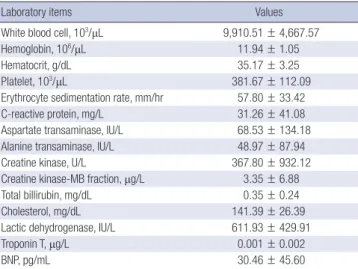
Table 2. The laboratory findings at diagnosis of KD
Laboratory items Values
White blood cell, 103/μL 9,910.51 ± 4,667.57
Hemoglobin, 106/μL 11.94 ± 1.05
Hematocrit, g/dL 35.17 ± 3.25
Platelet, 103/μL 381.67 ± 112.09
Erythrocyte sedimentation rate, mm/hr 57.80 ± 33.42
C-reactive protein, mg/L 31.26 ± 41.08
Aspartate transaminase, IU/L 68.53 ± 134.18
Alanine transaminase, IU/L 48.97 ± 87.94
Creatine kinase, U/L 367.80 ± 932.12
Creatine kinase-MB fraction, μg/L 3.35 ± 6.88
Total billirubin, mg/dL 0.35 ± 0.24
Cholesterol, mg/dL 141.39 ± 26.39
Lactic dehydrogenase, IU/L 611.93 ± 429.91
Troponin T, μg/L 0.001 ± 0.002
BNP, pg/mL 30.46 ± 45.60
Data are shown as mean ± standard deviation.
KD = Kawasaki disease, BNP = brain natriuretic peptide.
was 381,670 ± 112,090. The mean erythrocyte sedimentation rate was 57.8 ± 33.42 mm/hr and the mean C-reactive protein was 31.26 ± 41.08 mg/dL (Table 2).
Table 3 shows echocardiographic findings of KD patients. In- terventricular septum thickness at end-diastole (IVSd; 6.17 ± 1.08 mm at baseline vs. 6.55 ± 1.31 mm at follow-up, P = 0.005), LVIDs (23.4 ± 3.08 mm at baseline vs. 25.4 ± 3.20 mm at follow-up, P = 0.003), maximal velocity of late diastolic filling across mitral valve (mitral A) flow (0.53 ± 0.18 m/s at baseline vs. 0.47 ± 0.10 m/s at follow-up, P = 0.006), maximal velocity of early diastolic filling across mitral valve (mitral E)/A ratio (2.07 ± 0.51 at base line vs. 2.27 ± 0.53 at follow-up, P = 0.008), mitral E wave to peak early diastolic tissue wave (E/E’) ratio (7.14 ± 1.63 at base line vs. 6.93 ± 1.41 at follow-up, P = 0.013) showed significant differ- ences in the follow-up study. LVEF, LVFS, interventricular sep- tum thickness at end-systole (IVSs), LVIDd, LV posterior wall thickness at end-diastole (LVPWd), LV posterior wall thickness at end-systole (LVPWs), mitral E flow, mean value theorem for derivatives (MVTD), peak early diastolic tissue (E’), peak late diastolic tissue (A’), peak systolic tissue (S’) showed no signifi- cant differences. Table 4 shows coronary Z-scores of KD patients.
The LMA, LAD, and RCA showed no statistically significant dif- ferences. But delta value of LMA, LAD, and RCA showed signif- icant differences in the follow-up study.
DISCUSSION
The long-term cardiovascular outcome of KD is an important concern. Coronary artery aneurysm can be developed in 20%–
25% of untreated patients, and 5% of treated patients. Giant an- eurysms can cause coronary artery stenosis and increase the risk of myocardial infarction or death (11). Severe coronary ar- tery disease can develop into coronary stenosis, which can lead to acute myocardial infarction. Coronary aneurysms, greater than 6 mm, are highly likely to cause myocardial ischemia. Oth- er inflammatory heart diseases, such as myocarditis, endocar- ditis, valvulitis and pericarditis are also common during the acute phase of KD. The characteristics of myocarditis in KD are 1) mild decrease in LV systolic function, 2) transient pericardial effusion, 3) transient inflammatory changes in cardiac valves, and 4) mild or no symptoms. The severity of myocardial dysfunc- tion is associated with myocardial inflammation. More than Table 3. Echocardiographic indices in the KD patients with diastolic data
Indices Initial Follow-up data P value r
LVEF, % 66.54 ± 4.96 65.48 ± 5.11 0.390 0.15
LVFS, % 45.62 ± 52.65 35.55 ± 3.92 0.900 −0.02
IVSd, mm 6.17 ± 1.08 6.55 ± 1.31 0.005 0.51
IVSs, mm 8.09 ± 1.41 8.72 ± 1.48 0.010 0.45
LVIDd, mm 35.90 ± 4.34 39.00 ± 3.87 0.210 0.23
LVIDs, mm 23.41 ± 3.08 25.31 ± 3.20 0.003 0.52
LVPWd, mm 5.69 ± 1.38 5.74 ± 1.24 0.250 0.22
LVPWs, mm 8.60 ± 1.41 9.09 ± 1.70 0.270 0.21
Mitral E, m/s 1.02 ± 0.29 1.03 ± 0.15 0.760 0.053
Mitral A, m/s 0.53 ± 0.18 0.47 ± 0.10 0.006 0.46
Mitral E/A 2.07 ± 0.51 2.27 ± 0.53 0.008 0.44
DT, m/s 128.88 ± 29.20 146.10 ± 27.78 0.320 0.23
Doppler velocity of mitral annulus, m/s
E' 0.15 ± 0.02 0.15 ± 0.03 0.110 0.28
A' 0.09 ± 0.11 0.06 ± 0.01 0.790 −0.05
S' 0.11 ± 0.16 0.07 ± 0.02 0.060 0.35
E/E' 7.15 ± 1.63 6.93 ± 1.41 0.013 0.46
Data are shown as mean ± standard deviation.
KD = Kawasaki disease, LVEF = left ventricular ejection fraction, LVFS = left ventricular fraction shortening, IVSd = interventricular septum thickness at end-diastole, IVSs = inter- ventricular septum thickness at end-systole, LVIDd = left ventricular internal diameters at end-diastolic, LVIDs = left ventricular internal diameters at end-systolic, LVPWd = left ventricular posterior wall thickness at end-diastole, LVPWs = left ventricular posterior wall thickness at end-systole, mitral E = maximal velocity of early diastolic filling across mitral valve, mitral A = maximal velocity of late diastolic filling across mitral valve, mitral E/A = ratio of mitral E to A waves, DT = mitral valvular deceleration time, E' = peak early diastolic tissue, A' = peak late diastolic tissue, S' = peak systolic tissue, E/E' = ratio of mitral E to E' waves.
Table 4. Echocardiographic findings of coronary arteries
Findings Initial data Follow-up data P value r Delta value* SD P value
LMA (Z) 3.01 ± 0.20 0.65 ± 0.86 0.415 −0.174 −2.48 1.99 < 0.001
LAD (Z) 1.70 ± 1.07 0.55 ± 0.91 0.739 0.072 −0.92 1.33 0.003
RCA (Z) 0.82 ± 1.35 0.15 ± 0.77 0.688 −0.086 −0.91 1.50 0.007
LMA = left main artery, LAD = left anterior descending, RCA = right coronary artery, SD = standard deviation.
*Delta value: mean differences between initial and follow-up data.
50% of patients with KD have myocarditis showing symptoms, electrocardiogram changes, and echocardiographic changes within the first 3 weeks. Inflammatory myocardial changes can be detected with gallium-67 cardiac scan or Tc-99m labeled car- diac scan (12).
Deteriorated LV contractility can be improved after adminis- tration of intravenous immunoglobulin (13-15). Kurotobi et al.
(16) reported LV diastolic dysfunction in children with acute KD, which may be associated with increased brain natriuretic peptide (BNP) levels. Arnold et al. (17) examined asymptomatic children with persistent coronary artery lesions after KD, and revealed diastolic dysfunction in the segments supplied by ste- notic coronary arteries under conditions of exercise. Takeuchi et al. (18) reported decreased E’ and increased E/E’ ratio in the tissue Doppler study in the acute phase of KD, which were nor- malized in the convalescent phase. The BNP levels were signifi- cantly associated with velocity of circumferential fiber shorten- ing and S’ at the lateral mitral annulus in the tissue Doppler study, but not significantly associated with E′ lateral, E/E′, and E/A ra- tios. Selamet Tierney et al. (19) demonstrated decreased E’ and impaired diastolic function in patients with coronary artery an- eurysms. Decreased diastolic function may result in the increased BNP level. Most of previous studies on myocardial dysfunction in KD were performed in the acute phase of KD. The long-term follow-up study on myocardial function in KD had rarely been reported.
We performed a long-term follow-up study on the second- grade elementary school children who had been treated due to KD, including investigation of myocardial function. In the result of our study, the diastolic function was within reasonable rang- es both in the acute phase and long-term follow-up period. How- ever, there were significant differences in diastolic parameters, such as mitral A flow, mitral E/A, and E/E’ ratios. The decreased E/A ratio is an expected finding in the presence of impaired re- laxation, which slows diastolic tissue velocity (20). As a result, the blood flow from the atrium to the ventricle is delayed and the duration of diastole is prolonged. Defects in the diastolic phase in these patients may create small changes in the myo- cardial blood flow and reflect inflammatory changes in myo- cardium and coronary arteries (21).
The mean LMA Z-score in the acute phase was 3.1 and de- creased to 0.6 at the time of study, but it was not statistically sig- nificant. However, the delta values of individual coronary artery Z-score changes were statistically significant. Some of echocar- diographic measurements, such as IVSd, LVIDs, mitral A flow, mitral E/A, and E/E’ ratios, showed statistically significant dif- ferences in the follow-up period. Therefore, the follow-up echo- cardiography should be performed to ensure that the relaxation function be improved again. Currently, the follow-up guidelines of KD are mainly based on the presence of coronary artery an- eurysms. Children without coronary artery aneurysms have
been followed up for less than 2 years in many hospitals. How- ever, we think that even though the follow-up echocardiogra- phy is normal, significant alteration or reductions in myocardial function may be present. We intend to follow-up these patients after three and five years and see if functional abnormalities will be recovered. We think that the follow-up guidelines of KD need to be elaborated with respect to the diastolic function and the follow-up period.
DISCLOSURE
The authors have no potential conflicts of interest to disclose.
AUTHOR CONTRIBUTION
Conceptualization: Lee H, Eun L. Data curation: Lee H, Shin J, Eun L. Investigation: Lee H, Shin J, Eun L. Writing - original draft:
Lee H, Eun L. Writing - review & editing: Eun L.
ORCID
Heeyoung Lee https://orcid.org/0000-0002-8294-0914 Jaeeun Shin https://orcid.org/0000-0003-3089-9347 Lucy Eun https://orcid.org/0000-0002-4577-3168 REFERENCES
1. Kawasaki T. Acute febrile mucocutaneous syndrome with lymphoid in- volvement with specific desquamation of the fingers and toes in children.
Arerugi 1967; 16: 178-222.
2. Newburger JW, Takahashi M, Gerber MA, Gewitz MH, Tani LY, Burns JC, Shulman ST, Bolger AF, Ferrieri P, Baltimore RS, et al. Diagnosis, treatment, and long-term management of Kawasaki disease: a statement for health professionals from the Committee on Rheumatic Fever, Endocarditis, and Kawasaki Disease, Council on Cardiovascular Disease in the Young, American Heart Association. Pediatrics 2004; 114: 1708-33.
3. Eleftheriou D, Levin M, Shingadia D, Tulloh R, Klein NJ, Brogan PA. Man- agement of Kawasaki disease. Arch Dis Child 2014; 99: 74-83.
4. Alexoudi I, Kanakis M, Kapsimali V, Vaiopoulos G. Kawasaki disease: cur- rent aspects on aetiopathogenesis and therapeutic management. Auto- immun Rev 2011; 10: 544-7.
5. Akagi T, Rose V, Benson LN, Newman A, Freedom RM. Outcome of coro- nary artery aneurysms after Kawasaki disease. J Pediatr 1992; 121: 689- 94.
6. Weng KP, Hsieh KS, Huang SH, Ou SF, Ma CY, Ho TY, Lai CR, Ger LP. Clini- cal relevance of the risk factors for coronary artery lesions in Kawasaki disease. Kaohsiung J Med Sci 2012; 28: 23-9.
7. Lai WW, Geva T, Shirali GS, Frommelt PC, Humes RA, Brook MM, Pig- natelli RH, Rychik J; Task Force of the Pediatric Council of the American Society of Echocardiography; Pediatric Council of the American Society of Echocardiography. Guidelines and standards for performance of a pe- diatric echocardiogram: a report from the Task Force of the Pediatric Coun-
cil of the American Society of Echocardiography. J Am Soc Echocardiogr 2006; 19: 1413-30.
8. Haycock GB, Schwartz GJ, Wisotsky DH. Geometric method for measur- ing body surface area: a height-weight formula validated in infants, chil- dren, and adults. J Pediatr 1978; 93: 62-6.
9. Dallaire F, Dahdah N. New equations and a critical appraisal of coronary artery Z scores in healthy children. J Am Soc Echocardiogr 2011; 24: 60- 74.
10. McCrindle BW, Li JS, Minich LL, Colan SD, Atz AM, Takahashi M, Vetter VL, Gersony WM, Mitchell PD, Newburger JW, et al. Coronary artery in- volvement in children with Kawasaki disease: risk factors from analysis of serial normalized measurements. Circulation 2007; 116: 174-9.
11. Brogan PA, Bose A, Burgner D, Shingadia D, Tulloh R, Michie C, Klein N, Booy R, Levin M, Dillon MJ. Kawasaki disease: an evidence based approach to diagnosis, treatment, and proposals for future research. Arch Dis Child 2002; 86: 286-90.
12. Matsuura H, Ishikita T, Yamamoto S, Umezawa T, Ito R, Hashiguchi R, Saji T, Matsuo N, Takano M. Gallium-67 myocardial imaging for the detection of myocarditis in the acute phase of Kawasaki disease (mucocutaneous lymph node syndrome): the usefulness of single photon emission com- puted tomography. Br Heart J 1987; 58: 385-92.
13. Newburger JW, Sanders SP, Burns JC, Parness IA, Beiser AS, Colan SD.
Left ventricular contractility and function in Kawasaki syndrome. Effect of intravenous gamma-globulin. Circulation 1989; 79: 1237-46.
14. Kao CH, Hsieh KS, Wang YL, Chen CW, Liao SQ, Wang SJ, Yeh SH. Tc-99m HMPAO WBC imaging to detect carditis and to evaluate the results of high- dose gamma globulin treatment in Kawasaki disease. Clin Nucl Med 1992;
17: 623-6.
15. Moran AM, Newburger JW, Sanders SP, Parness IA, Spevak PJ, Burns JC, Colan SD. Abnormal myocardial mechanics in Kawasaki disease: rapid response to gamma-globulin. Am Heart J 2000; 139: 217-23.
16. Kurotobi S, Kawakami N, Shimizu K, Aoki H, Nasuno S, Takahashi K, Koga- ki S, Ozono K. Brain natriuretic peptide as a hormonal marker of ventric- ular diastolic dysfunction in children with Kawasaki disease. Pediatr Car- diol 2005; 26: 425-30.
17. Arnold R, Goebel B, Ulmer HE, Gorenflo M, Poerner TC. An exercise tis- sue Doppler and strain rate imaging study of diastolic myocardial dysfunc- tion after Kawasaki syndrome in childhood. Cardiol Young 2007; 17: 478- 86.
18. Takeuchi D, Saji T, Takatsuki S, Fujiwara M. Abnormal tissue doppler im- ages are associated with elevated plasma brain natriuretic peptide and increased oxidative stress in acute Kawasaki disease. Circ J 2007; 71: 357- 62.
19. Selamet Tierney ES, Newburger JW, Graham D, Baker A, Fulton DR, Col- an SD. Diastolic function in children with Kawasaki disease. Int J Cardiol 2011; 148: 309-12.
20. Lester SJ, Tajik AJ, Nishimura RA, Oh JK, Khandheria BK, Seward JB. Un- locking the mysteries of diastolic function: deciphering the Rosetta Stone 10 years later. J Am Coll Cardiol 2008; 51: 679-89.
21. Yoshida S, Takeuchi K, del Nido PJ, Ho C. Diastolic dysfunction coincides with early mild transplant rejection: in situ measurements in a heteroto- pic rat heart transplant model. J Heart Lung Transplant 1998; 17: 1049- 56.