Coronary Heart Disease in Moyamoya Disease: Are They Concomitant or Coincidence?
The purpose of this study was to determine the prevalence and characteristics of
symptomatic coronary heart disease (CHD) in patients with moyamoya disease (MMD). This retrospective study evaluated 456 patients who received examination for MMD between 1995 and 2012. We reviewed the patients’ medical history and coronary imaging, including conventional coronary angiography and coronary computed tomography angiogram (CTA). Among 456 patients with MMD, 21 (4.6%) patients were found to have symptomatic CHD. Ten patients were treated with coronary artery bypass graft or percutaneous coronary intervention for unstable angina or myocardial infarction. Eleven were treated with medication for stable angina (n = 6) and variant angina with mild degree of stenosis (n = 5).The median age of these patients was 44 yr (range, 27-59). The median Framingham score at diagnosing MMD was < 1% (range, < 1%-16%). The old age was associated with CHD in uni- and multivariate analyses (P = 0.021, OR, 1.053; 95% CI, 1.008-1.110). Considering low age of onset and low stroke risk factor, CHD might be a systemic manifestation that is clinically relevant to MMD.
Keywords: Moyamoya Disease; Coronary Disease; Prevalence; Characteristics Taek Min Nam, Kyung Il Jo,
Je Young Yeon, Seung Chyul Hong, and Jong Soo Kim
Department of Neurosurgery, Samsung Medical Center, Sungkyunkwan University School of Medicine, Seoul, Korea
Received: 8 July 2014 Accepted: 3 December 2014 Address for Correspondence:
Jong Soo Kim, MD
Department of Neurosurgery, Samsung Medical Center, Sungkyunkwan University School of Medicine, 81 Irwon-ro, Gangnam-gu, Seoul 135-710, Korea
Tel: +82.2-3410-3499, Fax: +82.2-3410-0048 E-mail: jsns.kim@samsung.com
http://dx.doi.org/10.3346/jkms.2015.30.4.470 • J Korean Med Sci 2015; 30: 470-474
INTRODUCTION
Moyamoya disease (MMD) is known as a progressive steno-oc- clusive intracranial angiopathy with small, fragile, and multiple collateral formation (1). The prevalence of MMD was found to be 16.1 per 100,000 in the Republic of Korea, when calculated with the 2011 population (2). The MMD is usually known to in- volve intracranial arteries, but systemic involvement of endo- thelial hyperplasia was raised in an autopsy study (3). Also, the renal artery is often involved with this disease in around 8% of the cases (4). It has been reported that the systemic arterial in- volvement in MMD can be a manifestation of certain genetic mutation such as ACTA 2 (5). Up to date, coronary artery dis- ease in MMD has been described sporadically in several case reports, and the association between MMD and coronary heart disease (CHD) has been constantly suggested in the literature (6-14). However, to the best of our knowledge, there have been no reports determining the prevalence of symptomatic CHD in MMD. In this study, we retrospectively reviewed the adult pa- tients in MMD registry to determine the prevalence of CHD.
Also, we explored the potential risk factors and analyzed the characteristics of CHD in MMD.
MATERIALS AND METHODS Patients
From January 1995 to December 2012, 456 consecutive adult
patients (≥ 20 yr of age) with MMD visited our hospital and were registered as adult MMD patients. The diagnosis of MMD was made based on idiopathic steno-occlusion at the terminal por- tion of internal carotid artery with concomitant abnormal vas- cular networks in the vicinity of the steno-occlusive lesions, which was confirmed by conventional angiography and/or magnetic resonance imaging.
All clinical data including coronary evaluation and medical history from our adult MMD registry were reviewed by two neu- rosurgeons. To obtain possible missing data from manual anal- yses, data collection of CHD was also performed using comput- erized queries, retrieving all coronary angiographic details and discharge summaries in the registry. The CHD patients were identified by searching with the keywords “coronary artery”,
“angina”, or “myocardiac infarction”. CHD was defined as typical chest pain with corresponding coronary artery lesion on imag- ing study.
The following explanatory variables were assessed: age, gen- der, type of MMD, and clinical risk factors that were known for causes of atherosclerosis and CHD, such as hypertension, dia- betes mellitus (DM), smoking and dyslipidemia (15). Framing- ham Score at MMD diagnosis was also assessed in the patients with CHD. The different types of MMD were categorized accord- ing to clinical symptoms at the initial diagnosis with MMD as follows: ischemic, hemorrhagic, asymptomatic, and atypical symptoms, such as headache and dizziness.
Statistical analysis
To explore the potential risk factors of CHD in MMD, statistical analyses were performed. The frequency comparisons were performed with Fisher’s exact test. For continuous variables, Mann-whitney U-test was performed, and multiple logistic re- gression test was performed for multivariate analysis. Statistical analysis was performed using SPSS 20.0 software (SPSS, Chica- go, IL, USA).
Ethics statement
This study was approved by the institutional review board of the Samsung Medical Center (IRB No. 2014-07-077-001). Informed
consent was waived by the board.
RESULTS
The baseline characteristics of the patients are described in Ta- ble 1. Twenty one (4.6%) out of 456 patients were diagnosed as symptomatic CHD. The detailed information on these patients with CHD are described in Table 2. The median follow-up peri- od was 32 months (range, 1-192). During the follow-up period, one patient was admitted to emergency room and dead upon arrival. The sudden death could have been caused by coronary heart disease, but there was no definite evidence associated with it as a cause of death, because medical examination could not be performed. Thus, we excluded the patient from CHD Table 1. Baseline characteristics of the patients
Characteristics No. of
without CHD (n = 435)
Patients with CHD
(n = 21)
P value by univariate analysis Age, median years (range)* 39 (20-76) 46 (20-63) 0.003
Gender (Male/Female)† 126/309 7/14 0.631
Diabetes† 35 (8.8%) 5 (23.8%) 0.031
Hypertension† 130 (30.7%) 10 (47.6%) 0.145
Dyslipidemia† 52 (12.6%) 3 (14.2%) 0.743
Smoking† 43 (10.3%) 4 (19.0%) 0.177
Presenting Symptom† Hemorrhagic Ischemic Atypical Asymptomatic
93 (20.4%) 272 (59.6%) 77 (16.9%) 14 (3.7%)
3 (14.2%) 13 (61.9%) 4 (19.0%) 2 (9.5%)
0.876
*Estimated by Mann-whitney U-test; †Estimated by Fisher’s exact test. CHD, coronary heart disease.
Table 2. Detailed characteristics of the moyamoya disease patients with coronary heart disease Patient
No. Sex Age*
(yr) Type of CAD DM HTN Dyslipidemia Smoking Framingham
score*
Time from MMD to CAD (yr)
Premature CAD
Treatment for CAD
1 F 39 Unstable angina Yes Yes Yes No < 1% 8 Yes CABG
2 M 45 Unstable angina Yes Yes Yes Yes 10% 8 Yes PCI
3 F 41 Unstable angina No Yes No Yes 3% 1 Yes PCI
4 F 53 Stable angina No No No No < 1% 2 Yes Medication
5 M 49 Stable angina No No No No 3% 5 Yes Medication
6 F 45 Unstable angina Yes Yes Yes No < 1% 1 Yes PCI
7 F 52 Unstable angina No Yes No No 1% 1 Yes CABG
8 M 36 Myocardiac infarction No Yes No No < 1% 5 Yes PCI
9 F 32 Stable angina No No No No < 1% 3 Yes Medication
10 M 57 Stable angina No Yes No Yes 16% 2 Yes Medication
11 M 38 Stable angina No No No No < 1% 3 Yes Medication
12 F 52 Stable angina No No No No 2 Yes Medication
13 F 44 Unstable angina No Yes No No 3% -12 Yes CABG
14 F 38 Myocardiac infarction Yes No No No < 1% -1 Yes CABG
15 F 41 Unstable angina No No No No < 1% 2 Yes PCI
16 M 41 Unstable angina No No No Yes 4% 1 Yes PCI
17 F 41 Variant angina No Yes No No < 1% 1 Yes Medication
18 F 20 Variant angina No No No No NA 7 Yes Medication
19 F 45 Variant angina No Yes No No < 1% 5 Yes Medication
20 M 58 Variant angina No No No No 6% 1 Yes Medication
21 F 55 Variant angina No No No No 1% 0 Yes Medication
*At the time of MMD Diagnosis. CAD, coronary artery disease; DM, diabetes mellitus; HTN, hypertension; CABG, coronary artery bypass surgery; PCI, percutaneous coronary intervention.
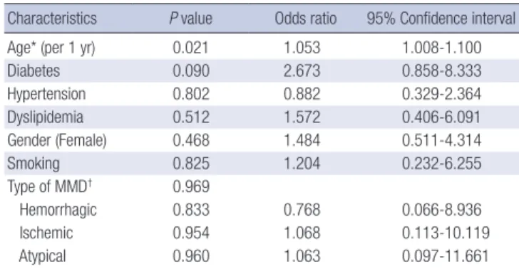
Table 3. Results of multivariate analysis using multiple logistic regression test Characteristics P value Odds ratio 95% Confidence interval
Age* (per 1 yr) 0.021 1.053 1.008-1.100
Diabetes 0.090 2.673 0.858-8.333
Hypertension 0.802 0.882 0.329-2.364
Dyslipidemia 0.512 1.572 0.406-6.091
Gender (Female) 0.468 1.484 0.511-4.314
Smoking 0.825 1.204 0.232-6.255
Type of MMD† 0.969
Hemorrhagic 0.833 0.768 0.066-8.936
Ischemic 0.954 1.068 0.113-10.119
Atypical 0.960 1.063 0.097-11.661
*At the time of MMD Diagnosis; †Type of MMD was categorized according to initial presenting symptom.
group. The median age at CHD diagnosis was 44 yr (range, 27- 59). Three (Patient No. 7, 13, and 14) of them were unilateral disease and 4 (Patient No. 2, 3, 15, and 16) were hemorrhagic subtype of MMD.
The treatment for CHD were as follows: 11 medical treatments (5 variant angina with mild degree of stenosis, 6 stable angina), 4 coronary artery bypass graft (CABG), and 6 percutaneous coro- nary intervention (PCI). One patient (Patient No. 7) who under- went CABG surgery had a postoperative ischemic stroke.
The patients with CHD were older in age (median, 46; range, 20-63) when diagnosed with MMD than the patients who were diagnosed as MMD without CHD (median age, 39; range, 20- 76; P = 0.003, Mann-Whitney U-test). The patients with MMD and CHD had higher percentage of DM than the MMD patients without the CHD (23.8% vs. 8.2%, P = 0.031 Fisher’s exact test).
Multivariate analyses using multiple logistic regression test showed that only the age factor was associated in those with both CHD and MMD (P = 0.021, OR, 1.053; 95% CI, 1.008-1.110).
A B
C D
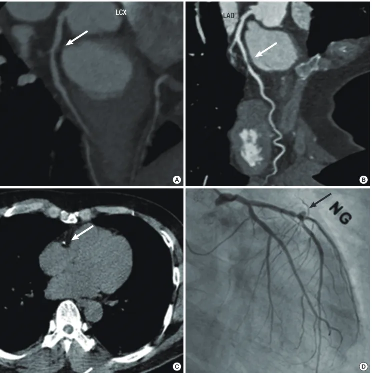
Fig. 1. Angiographic findings of coronary heart disease in moyamoya disease. (A) and (B) show coronary stenosis (arrows) without calcification. (C) shows calcified plaque (arrow) on a non-contrast coronary computed tomography, and (D) shows aneurysmal dilatation and focal stenosis (arrow). Nitroglycerin (NG) was administered to the patient.
LCX LAD
Other variables, such as gender, hyperlipidemia, hypertension, smoking, and subtype of MMD (hemorrhagic vs. ischemic vs.
atypical vs. asymptomatic) were not statistically different be- tween those with and without CHD (Table 3).
Coronary computed tomography angiography (CTA) was available for 7 patients, and 4 patients showed no calcification of affected coronary artery (Fig. 1), and one patient showed an- eurysmal dilatation.
DISCUSSION
Several of the previous reported articles demonstrated CHD in MMD, and most of them were reported from East Asian coun- tries (7-14, 16). Ikeda demonstrated that MMD is involved with the extra-cranial vessels as well as the intracranial vessels, and there are systemic etiologic factors, which cause intimal thick- ening in the systemic vessels (3). Histopathologic studies of the involved internal carotid arteries in MMD showed fibrocellular thickening of the intima and proliferated smooth muscle cells (SMC) as the cause of the arterial occlusion (17, 18). Also, the SMC proliferation is part of the mechanism of atherosclerosis in CHD, which is similar to the histopathology of MMD (19, 20).
Several mutant genes which can cause stenosis in both MMD and CHD have already been reported (5, 17).
Both MMD and CHD can cause life-threatening events. If these two diseases had significant association, screening test should be performed to reduce the morbidity and mortality. In our adult MMD registry, 4.6% showed CHD. However, this per- centage could be underestimated because only the symptom- atic patients who underwent diagnostic study at our hospital were retrospectively included. The prevalence of CHD in the Asian population is known to be less than 1% person per year in the population of 65 or less years of age (21), which could be significant.
In this study, all of the CHD were developed at relatively young age (< 65 yr), and Framingham scores were low at the initial di- agnosis of MMD. Also, half of the patients showed no calcifica- tion on CTA, which reflects the atherosclerotic burden of the coronary artery. These findings support the idea that MMD is a causative factor of CHD rather than pure atherosclerosis (16, 22).
Previous case reports about CHD in MMD described the minimal atherosclerotic burden on the affected coronary artery (10, 16), and half of our data supported this except the coronary artery lesions were heterogeneous in this study. The prevalence was not high enough to designate MMD as the main culprit.
Also, in this study the classic risk factors for atherosclerosis, such as old age and diabetes, were associated with CHD in MMD.
Therefore, we hypothesized that the underlying endothelial hy- perplasia in the coronary artery is not so significant in the most cases of MMD, but smaller atherosclerotic burden seemed to be causing the symptomatic CHD in MMD patients at younger age
than normal population. In our study, 5 (23.8%) of 21 CHD pa- tients were diagnosed as variant angina, and this proportion was high compared with general CHD patients. In 1998, Ikeda et al., reported variant angina associated with MMD, and 2 (22.2%) out of 9 MMD patient were diagnosed as variant angina (23).
There seems to be an association between MMD and variant angina. However, there has been no evidence that supports this idea, considering the mechanism of variant angina and MMD.
Several limitations must be noted in this study. First, this was an uncontrolled, retrospective study. Therefore, the data cannot provide the exact prevalence of CHD in MMD. Also, not all of the patients underwent coronary CTA, which could have pro- vided more information about atherosclerotic components, such as calcium score or plaque content. The MMD is a rare disease and there is no adequate evidence regarding the association between CHD and MMD, which makes it difficult to perform a prospective, controlled study. This study also reflects mainly the patients who were treated in our hospital. Since we have con- sidered those with CHD treated in other hospitals, there are some loss of data from outside medical records. Therefore, the preva- lence suggested in this study might be underestimated.
In conclusion, MMD does not seem to be the main culprit of CHD, but it can lead to CHD with small atherosclerosis proba- bly due to the underlying endothelial proliferation. The screen- ing test for CHD in the MMD patients with older age is in need, and DM might have a role for this matter. The future study with a larger sample size is warranted to reveal the general charac- teristics of CHD in MMD and to determine the usefulness of coronary screening test in MMD.
DISCLOSURE
Relevant conflicts of interest/financial disclosure : Nothing to report.
AUTHOR CONTRIBUTION
Study design: Nam TM, Jo KI, Kim JS. Data collection and anal- ysis: Nam TM, Jo KI. Writing manuscript: Nam TM, Jo KI. Kim JS. Discussion and manuscript revision: Nam TM, Jo KI. Yeon JY, Hong SC, Kim JS. Manuscript arrpoval: Kim JS.
ORCID
Nam Taek Min http://orcid.org/0000-0002-9596-4659 Jo Kyung Il http://orcid.org/0000-0001-7585-1916 Kim Jong Soo http://orcid.org/0000-0001-5144-3336 REFERENCES
1. Suzuki J, Takaku A. Cerebrovascular “moyamoya” disease. Disease show-
ing abnormal net-like vessels in base of brain. Arch Neurol 1969; 20: 288- 99.
2. Ahn IM, Park DH, Hann HJ, Kim KH, Kim HJ, Ahn HS. Incidence, preva- lence, and survival of moyamoya disease in Korea: a nationwide, popu- lation-based study. Stroke 2014; 45: 1090-5.
3. Ikeda E. Systemic vascular changes in spontaneous occlusion of the circle of Willis. Stroke 1991; 22: 1358-62.
4. Yamada I, Himeno Y, Matsushima Y, Shibuya H. Renal artery lesions in patients with moyamoya disease: angiographic findings. Stroke 2000; 31:
733-7.
5. Guo DC, Papke CL, Tran-Fadulu V, Regalado ES, Avidan N, Johnson RJ, Kim DH, Pannu H, Willing MC, Sparks E, et al. Mutations in smooth muscle alpha-actin (ACTA2) cause coronary artery disease, stroke, and Moyamoya disease, along with thoracic aortic disease. Am J Hum Genet 2009; 84: 617-27.
6. von Bary C, Liebig T, Gaa J, von Beckerath N. Ischaemic stroke and myo- cardial infarction in a Caucasian patient with Moya-Moya disease. Eur Heart J 2008; 29: 842.
7. St Goar FG, Gominak SC, Potkin BN. Bilateral aortoostial coronary ar- tery disease: moyamoya of the heart? Am J Cardiol 1999; 83: 1296-9, a10.
8. Kim DK, Yoo KJ. Off-pump coronary artery bypass grafting in moyamo- ya disease. Yonsei Med J 2007; 48: 876-8.
9. Komiyama M, Ishikawa T, Takanashi S, Shimizu Y. Minimal invasive di- rect coronary artery bypass in moyamoya disease. Interact Cardiovasc Thorac Surg 2003; 2: 65-7.
10. Komiyama M, Nishikawa M, Yasui T, Otsuka M, Haze K. Moyamoya dis- ease and coronary artery disease--case report. Neurol Med Chir (Tokyo) 2001; 41: 37-41.
11. Murakami T, Ueno M, Takeda A, Yakuwa S, Kuroda S. Image in cardio- vascular medicine. Multiple coronary stenosis in infantile Moyamoya disease. Circulation 2009; 119: 1689.
12. Wang N, Kuluz J, Barron M, Perryman R. Cardiopulmonary bypass in a patient with moyamoya disease. Anesth Analg 1997; 84: 1160-3.
13. Akasaki T, Kagiyama S, Omae T, Ohya Y, Ibayashi S, Abe I, Fujishima M.
Asymptomatic moyamoya disease associated with coronary and renal artery stenoses--a case report. Jpn Circ J 1998; 62: 136-8.
14. Ahn YK, Jeong MH, Bom HS, Park JC, Kim JK, Chung DJ, Chung MY, Cho JG, Kang JC. Myocardial infarction with Moyamoya disease and pi- tuitary gigantism in a young female patient. Jpn Circ J 1999; 63: 644-8.
15. Wilson PW, D’Agostino RB, Levy D, Belanger AM, Silbershatz H, Kan- nel WB. Prediction of coronary heart disease using risk factor categories.
Circulation 1998; 97: 1837-47.
16. Lee JH, Youn TJ, Yoon YE, Park JJ, Hong SJ, Chun EJ, Choi SI, Cho YS, Cho GY, Chae IH, et al. Coronary artery stenosis in moyamoya disease: tissue characterization by 256-slice multi-detector CT and virtual histology. Cir- culation 2013; 127: 2063-5.
17. Roder C, Peters V, Kasuya H, Nishizawa T, Takehara Y, Berg D, Schulte C, Khan N, Tatagiba M, Krischek B. Common genetic polymorphisms in moyamoya and atherosclerotic disease in Europeans. Childs Nerv Syst 2011; 27: 245-52.
18. Achrol AS, Guzman R, Lee M, Steinberg GK. Pathophysiology and genet- ic factors in moyamoya disease. Neurosurg Focus 2009; 26: E4.
19. Sata M, Saiura A, Kunisato A, Tojo A, Okada S, Tokuhisa T, Hirai H, Makuuchi M, Hirata Y, Nagai R. Hematopoietic stem cells differentiate into vascular cells that participate in the pathogenesis of atherosclerosis.
Nat Med 2002; 8: 403-9.
20. Davies MJ, Woolf N, Rowles PM, Pepper J. Morphology of the endotheli- um over atherosclerotic plaques in human coronary arteries. Br Heart J 1988; 60: 459-64.
21. Asia Pacific Cohort Studies Collaboration. The impact of cardiovascular risk factors on the age-related excess risk of coronary heart disease. Int J Epidemiol 2006; 35: 1025-33.
22. Greenland P, LaBree L, Azen SP, Doherty TM, Detrano RC. Coronary ar- tery calcium score combined with Framingham score for risk prediction in asymptomatic individuals. JAMA 2004; 291: 210-5.
23. Ikeda U, Fujikawa H, Shimada K. Variant angina pectoris associated with moyamoya disease. Lancet 1998; 351: 183-4.