INTRODUCTION
In addition to hematopoietic stem cells (HSCs), the bone marrow contains a second type of stem cells that are capable of giving rise to multiple mesenchymal cell lineages, namely, mesenchymal stem cells (MSCs) (1, 2). Bone marrow stromal cells have been shown to include MSCs, which are thought to support hematopoiesis by creating an optimal microenvi- ronment (the marrow matrix), and by providing cytokines and other regulatory factors that stimulate and enhance pro- liferation of the hematopoietic elements (3). Moreover, mul- tipotent human MSCs can be isolated from bone marrow and expanded more than 1-billion-fold in cell culture without loss of stem cell capacity (4). These cells are capable of differ- entiating into osteoblasts, adipocytes, chondrocytes, myocytes, neural elements, and stromal fibroblasts under defined in vitro or in vivo conditions, and show an identical phenotype when
grown from a single cell clone (5-7). In the area of hemato- poiesis, MSCs are being used clinically to support hematopoi- etic recovery by replacing stroma. Moreover, a growing body of clinical evidence suggests the existence of a link between the efficiency of hematopoietic recovery and the degree of stromal damage that occurs as a result of myeloablative can- cer therapy (8). As a result, therapy-ablated marrow may not be able to sustain hematopoietic stem cell maintenance or their differentiation into specific lineages, such as, megakary- ocytes and platelets. In addition, a prolonged stromal defect in growth factor production, like early-stage cytokines, such as, interleukin-6, leukemia inhibitory factor, stem cell factor and granulocyte-colony stimulating factor, after autologous bone marrow transplantation has been observed for as long as a year in patients with hematologic disorders (9).
Independent laboratories have shown that recipients of un- manipulated allogeneic bone marrow transplants have only
Seong-Kyu Park, Jong-Ho Won, Hyun-Jung Kim, Sang-Byung Bae, Chan-Kyu Kim, Kyu-Taeg Lee, Nam-Su Lee, You Kyoung Lee*, Dae-Chul Jeong�, Nak-Gyun Chung�, Hyun-Soo Kim�, Dae-Sik Hong, Hee-Sook Park
Departments of Internal Medicine and Laboratory Medicine*, Soon Chun Hyang University College of Medicine, Seoul; Department of Pediatrics�, The Catholic University of Korea College of Medicine, Seoul;
Research Institute, Pharmicell Inc.�, Seoul, Korea
Address for correspondence Jong-Ho Won, M.D.
Division of Hematology-Oncology, Soon Chun Hyang University Hospital, 657-58 Hannam-dong, Yongsan-gu, Seoul 140-743, Korea Tel : +82.2-709-9182, Fax : +82.2-709-9200 E-mail : jhwon@hosp.sch.ac.kr
*This study was supported by a grant from the Korean Health 21 R&D Project (No. 02-PJ1-PG10-20699-0001), Ministry of Health and Welfare, Republic of Korea.
*The authors declare that they have no vested interest that could be construed to have inappropriately influenced this study.
412
Co-transplantation of Human Mesenchymal Stem Cells Promotes Human CD34+ Cells Engraftment in a Dose-dependent Fashion in NOD/SCID Mice
Mesenchymal stem cells (MSCs) have recently been identified and characterized in humans. Moreover, MSC secrete cytokines that can support hematopoietic pro- genitor growth. In the present study, we evaluated whether the efficacy of hemato- poietic stem cell transplantation is improved by their co-transplantation with MSC, and whether this is positively correlated with the dose of infused MSCs. Accordingly, irradiated NOD/SCID mice were transplanted with 1××105human CD34+ cells in the presence or absence of culture expanded MSCs (1××106or 5××106). We eval- uated human hematopoietic cell engraftment by flow cytometry and assessed MSC tissue distributions by fluorescence in situ hybridization. We found that CD45+ and CD34+ cell levels were significantly elevated in a dose-dependent manner in co- transplanted mice 4 weeks after transplantation. The engraftments of CD33+ and CD19+ cells also increased dose-dependently. However, the engraftment of CD3+
cells did not increase after co-transplantation with MSCs. Human Y chromosome+
cells were observed in multiple tissues and were more frequently observed in mice co-transplanted with 5××106rather than 1××106MSCs. These results suggest that MSCs are capable of enhancing hematopoietic cell engraftment and distribution in multiple organs in a dose-dependent fashion.
Key Words : Mesenchymal Stem Cells; Hematopoietic Stem Cells; Transplantation
Received : 11 August 2006 Accepted : 21 November 2006
host-type marrow stromal cells and MSCs in bone marrow (10, 11). These results are attributed to the inability of the conditioning regimen to ablate host marrow stroma and/or the inability of stromal progenitors of donor origin to engraft.
In addition, the number of MSCs has been estimated to be low in an average bone marrow graft (400-1,000 MSCs/kg).
On the other hand, a recent report noted that allogeneic osteo- blasts could be detected in recipients with osteogenesis imper- fecta (OI) after sibling-matched allogeneic bone marrow trans- plantation (12). These findings suggest that osteoblasts may be transplanted successfully under certain conditions. Stro- mal chimerism has been achieved using infusions of culture- expanded stromal progenitors in murine models, and xeno- transplantation models have been developed to determine the homing of human MSCs in animals. Human MSCs were detected predominantly in bone marrow, and less frequently in other organs of immunodeficient NOD/SCID mice 8 weeks after infusion. Moreover, recent data indicate that MSC co- infusion enhances human hematopoiesis in NOD/SCID mice (13, 14). Based on encouraging data obtained in both rodent and large animal models, clinical studies of MSC transplan- tation are underway in both autologous and allogeneic set- tings. Koc et al. (15) found that the co-infusion of autologous MSCs together with autologous HSCs is free of toxicity and that it is associated with rapid hematopoietic recovery in 28 women receiving high dose chemotherapy for advanced breast carcinoma. In this report, we demonstrate that co-transplan- tation with human MSCs can enhance the engraftment of human hematopoietic stem cells in NOD/SCID mice, and that this engraftment-enhancing effect increases according to the number of infused MSCs. We also show that an increased dose of MSCs enhances the tissue distribution of human MSCs in multiple tissues in NOD/SCID mice. Our results show that MSCs are capable of enhancing hematopoietic engraft- ment and distribution to multiple organs in a dose-depen- dent fashion.
MATERIALS AND METHODS
Collection and isolation of CD34++cells from umbilical cord blood (UCB)
UCB was obtained at the time of full-term deliveries, after receiving informed consent for the study protocol by our hos- pital’s ethics committee. UCB was collected after clamping and cutting of cords by draining blood into sterile collection tubes containing the anticoagulant citrate-phosphate dextrose.
Mononuclear cells (MNCs) were isolated from UCB using Ficoll-Hypaque (1.077 g/cm3, Sigma, St Louis, MO) densi- ty centrifugation (400 g for 25 min). CD34+ cells were iso- lated using a magnetic cell sorting system, Mini-MACS (Mil- tenyi Biotec, Auburn, CA), according to the manufacturer’s instructions. Purity was determined by flow cytometry using
a FITC-conjugated anti-CD34 (anti-HPCA-2, Becton-Dick- inson, Mountain View, CA), and was found to be 90 to 95%.
Human MSCs isolation and ex vivo culture
Ten to twenty mililiters of bone marrow aspirate was ob- tained under sterile conditions by posterior iliac crest punc- ture from bone marrow transplantation donors with given informed consent. Human MSCs were isolated and culture- expanded according to the method described by Pittenger et al. (1). MNCs were isolated from bone marrow, and cells were cultured in human MSC medium composed of Dulbec- co’s modified Eagle’s low glucose medium (DMEM-LG; Gib- coBRL, Grand Island, NY) containing 10% fetal bovine serum (FBS; GibcoBRL) and 1% antibiotic-antimycotic solution (GibcoBRL). Human MSCs were confirmed to be negative for hematopoietic markers by flow cytometry and to be capable of differentiating into osteocytes, chondrocytes, and adipocytes in vitro (Fig. 1, 2).
Differentiation of MSCs
MSCs were induced to differentiate into adipocytes by treat- ing confluent second passage monolayer cultures with 1 M dexamethasone (Sigma, St. Louis, U.S.A.), 0.5 mM methyl- isobutylxanthine (Sigma), 10 g/mL insulin (Sigma), and 100 mM indomethacin (Sigma) in DMEM containing 10%
FBS. Adipogenic differentiation was demonstrated by the accumulation of lipid vesicles stained red by Oil red O (Sig- ma). The osteogenic differentiation of MSCs was induced by culturing confluent monolayers in DMEM containing 10%
FBS, 0.1 M dexamethasone, 50 M ascorbate (Sigma), and 10 M -glycerol- phosphate (Sigma). MSC osteogenic dif- ferentiation over 21 days was demonstrated by increases in alkaline phosphatase and calcium accumulation. Alkaline phosphatase was detected histologically, and calcium was stained by the Kossa’s method. The chondrogenic differen- tiation of MSCs was in- duced by pellet culture in 500 L of DMEM containing 10% FBS, 0.1 M dexamethasone, 50 M ascorbic acid 2-phosphate (Sigma), and 1 g/mL trans- forming growth factor- ( TGF- R&D System, Minnea- polis, MN). Under low speed centrifugation (1,000 g for 5 min), a dense mass of cells formed at the bottom of conical centrifuge tubes. Chondrogenic differentiation was demon- strated by toluidine blue staining after 3 weeks of culture.
Mice
Female NOD/SCID mice (5-6 weeks old), purchased from Charles-River Laboratory (Tokyo), were housed in micro-iso- lator cages on laminar flow racks at the animal facilities of the Catholic University of Korea. Mice were provided a ster- ile diet and autoclaved acidified water.
Xenotransplantation of human cord blood CD34++cells and MSCs into mice
NOD/SCID mice were sublethally irradiated (3.5 Gy) 4 to 6 hr before transplantation. Human female cord blood
CD34+ cells were injected at a final volume of 100 L Iscove’s Modified Dulbecco’s Media (IMDM; GibcoBRL) per mouse and human male MSCs were injected with IMDM at a vol- ume of 100 L/106MSCs per mouse. Both CD34+ cells and MSCs were injected via a tail vein. Two mice were assigned to each experimental group. In three separate experiments, mice were assigned to receive 1×10⁵CD34+ cells, CD34+
cells plus 1×10⁶MSCs, or CD34+ cells plus 5×10⁶MSCs.
One mouse treated with 5×10⁶MSCs in the first experiment died after MSCs infusion due to myocardial infarction (patho- logically confirmed, data not shown).
Detection of human hematopoietic cells in NOD/SCID mice by flow cytometry
Four weeks after transplantation, mice were sacrificed by carbon dioxide inhalation, and bone marrow (BM) cells were collected by flushing both femurs with RPMI medium (Gibco BRL). Cell suspensions were stained with mouse anti-human monoclonal antibodies, fluorescein isothiocyanate (FITC)- conjugated CD45 antibody and phycoerythrin (PE)-conju- gated CD34, CD13, CD33, CD3, and CD19 antibodies (Bec- ton-Dickinson, Mountain View, CA). Flow cytometry to detect human CD45+ and CD34+, CD13+, CD33+, CD3+, or CD19+ cells was performed on a FACScan flow cytometer (Becton-Dickinson). FITC- and PE-conjugated mouse iso- type control antibodies were used for each culture, and 5,000-
Osteoblasts
Mesenchymal stem cells
Chondrocytes Adipocytes Fig. 2.Culture-expanded human mesenchymal stem cells exhib- ited a fibroblastic morphology (top panel). Under specific differ- entiation-inducing conditions, mesenchymal stem cells differenti- ated into osteoblasts, chondrocytes, and adipocytes.
Events
11
0
FITC-control
99.17%
Events
53
0
CD105-FITC
Events
4
0
PE-control
Events
59
0
CD166-PE
Events
11
0
FITC-control
Events
10
0
CD14-FITC
Events
11
0
CD34-FITC
Events
9
0
CD45-FITC Fig. 1.Immunophenotype of cultured human mesenchymal stem cells. Mesenchymal stem cells expressed CD105 and CD166, but were negative for CD34, CD45, and CD14.
93.15%
M1: 93.15%
99.20%
90.95%
0.60%
0.77%
0.93%
99.17%
M1: 90.95%
M1: 0.93% M1: 0.77% M1: 0.60%
10,000 events were counted for each analysis. Differences in engraftment percentages were calculated using the Student’s t-test. p values of <0.05 were considered statistically signifi- cant.
Detection of human MSCs in NOD/SCID mice by fluorescence in situ hybridization
Fluorescence in situ hybridization (FISH) for the human Y chromosome was used to detect transplanted MSCs in BM, spleen, liver, lung, kidney, heart, skeletal muscle, intestine, and skin of NOD/SCID mice at four weeks after transplan- tation in the second and third experiments. Total numbers of Y chromosome positive cells in ten×high-power fields were counted.
RESULTS Differential potential of MSCs
Culture of bone marrow MNCs produced a monomorphic confluent adherent layer of elongated fibroblast-like cells that survived multiple passages under mesenchymal culture condi- tions. At the end of the second passage, bone marrow derived MSCs had successfully differentiated along osteogenic, chon- drogenic, and adipogenic lineages, using methods described above (Fig. 2).
Effect of MSCs on human hematopoietic cell engraftment in NOD/SCID mice
Transplantation of UCB CD34+ cells (1×10⁵/mouse) in the presence of MSCs (1×10⁶and 5×10⁶/mouse) resulted in significantly higher engraftment levels in the BM of NOD/
SCID mice than was observed after transplanting UCB CD34+
cells alone. The mean percentage of CD45+ cells in the BM of NOD/SCID mice according to the MSCs number, i.e., 1×
106or 5×10⁶, increased to 41.05±5.62 and 55.58±8.26, respectively, compared with 29.18±1.00 for transplantation with CD34+ cells without MSCs (p<0.05). The difference between the group infused with 1×10⁶MSCs and the group infused with 5×10⁶MSCs was statistically significant (p<
0.05) (Fig. 3). The mean percentage of CD45+-CD34+cells increased to 13.78±1.50 and 17.43±2.05 at these MSC dosages, compared with 7.76±0.37 for transplantation with CD34+ cells without MSCs (p<0.05). In addition, the groups infused with 1×10⁶or 5×10⁶were significantly different (p<0.05) (Fig. 3).
Effects of MSCs on human myeloid cell engraftment in NOD/SCID mice
Co-transplantation of MSCs significantly increased the level of human myeloid cell engraftment in NOD/SCID mouse BM. The mean percentage of CD45+-CD33+cells in NOD/
SCID mouse BM at MSC doses of 1×10⁶and 5×10⁶were 19.62±1.18 and 24.38±3.05, respectively, compared with 6.54±0.17 for transplantation with CD34+ cells alone (p<
0.05). Moreover, the level of myeloid cell engraftment in the group infused with a high dose of MSCs was significant higher than in the group infused with a lower dose (p<0.05) (Fig. 3).
Mean percentages of CD45+-CD13+cells increased to 21.64± 2.04 and 24.45±3.41 for high and low MSC doses, compared with 10.34±0.57 for transplantation with CD34+ cells alone (Fig. 3). Although there was a distinct difference bet- ween the group co-transplanted with MSCs and the group transplanted with HSC alone (p<0.05), we did not notice a difference between levels of HSC engraftment according to the number of MSCs co-transplanted.
Effects of MSCs on human lymphoid cell engraftment in NOD/SCID mice
The mean percentages of CD45+-CD19+cells in NOD/
SCID mouse BM according to the MSC dose were signifi- cantly increased to 9.93±4.77 and 24.39±11.22, respec- tively, compared with 7.08±1.46 of CD34+ cells alone (p<
0.05). Co-transplantation of MSCs at 5×10⁶ significantly increased the level of human B lymphoid cell engraftment in NOD/SCID mouse BM (p<0.05). Mean percentages of CD45+-CD3+cells engrafted by low and high MSC doses were 2.89±1.90 and 4.43±1.64, respectively, compared
CD34+
Percentage of positive cells
80 70 60 50 40 30 20 10 0
CD45
+
CD45
+34+ CD45
+33+ CD45
+13+ CD45
+19+ CD45
+3+
Fig. 3.Effects of MSCs on human UCB 34+ cell engraftment in NOD/SCID mice. Co-transplantation with UCB CD34+ cells and MSCs resulted in higher engraftment levels in NOD/SCID mouse bone marrow 4 weeks after transplantation than after transplan- tation with UCB CD34+ cells alone. Moreover, this engraftment- promoting effect was related to the MSC dosage and increased myeloid and B lymphoid cell numbers but not those of T-lymphoid cells. *A significant difference (p<0.05) between the cotransplant- ed group and the group transplanted with CD34+ alone. �Signifi- cant (p<0.05) between the group infused with 1×10⁶MSCs and the group infused with 5×10⁶MSCs.
*,�
*
*,�
*
*,�
*
*,�
*
*
*
CD34++MSC (1×10⁶) CD34++MSC (5×10⁶)
with 3.02±1.17 for CD34+ cells alone. Co-transplantation of MSCs did not increase the level of human T lymphoid cell engraftment in NOD/SCID mouse BM (Fig. 3).
Engraftment of human MSCs in NOD/SCID mice Human Y chromosome positive cells were found in BM,
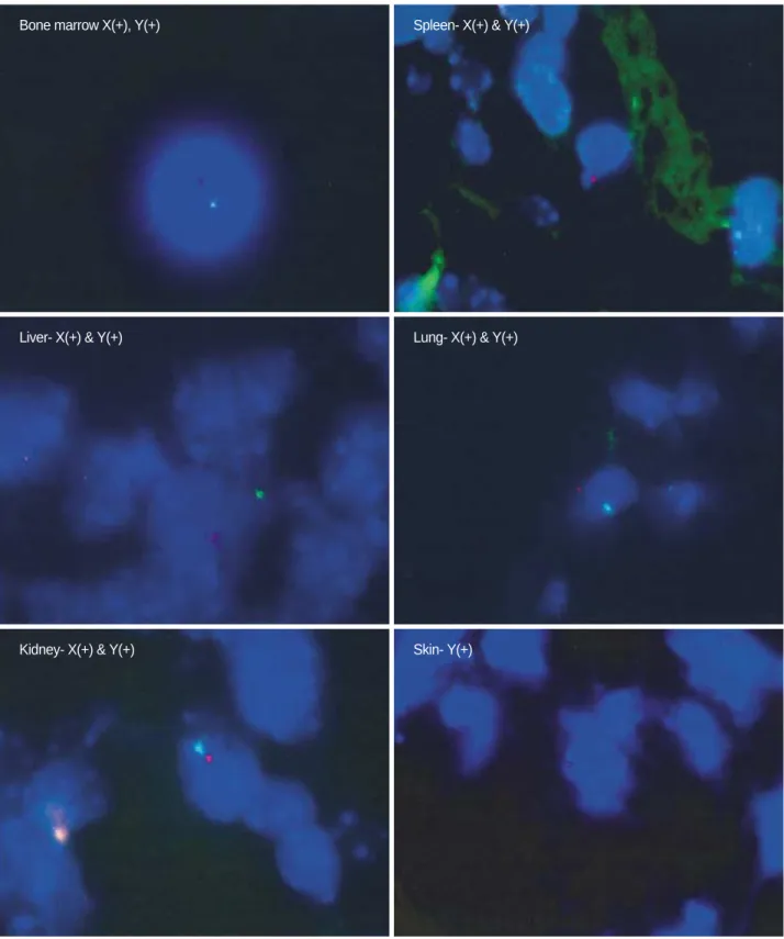
Fig. 4.Human X chromosome (green) and Y chromosome (red) expression by fluorescence in situ hybridization (FISH) analysis. Human X and Y chromosomes were detected in bone marrow, spleen, liver, lung, and kidney. Y chromosomes were detected in the heart, intes- tine, and skin (results of heart and intestine are not shown).
Bone marrow X(+), Y(+) Spleen- X(+) & Y(+)
Liver- X(+) & Y(+) Lung- X(+) & Y(+)
Kidney- X(+) & Y(+) Skin- Y(+)
spleen, liver, lung, kidney, heart, intestine, and skin of NOD/
SCID mice at four weeks after transplantation but not in skeletal muscle. The distribution of human Y chromosome positive cells differed by tissue; Y chromosome positive cells were found more frequently in the liver, lung, and kidney than in other tissues (Fig. 4, Table 1). In addition, numbers of these cells were higher in mice that received a higher dose of MSCs in same tissues.
DISCUSSION
We infused relatively low doses of CD34+ cells (1×10⁵ cells) into NOD/SCID mice in this study. We tried to evalu- ate the dose-dependent effect of MSCs on HSC engraftment, and thus, we used relatively high doses of MSCs (5×10⁶/ani- mal) as compared with the doses (1×10⁶/animal) usually applied. We observed that the high-dose MSC group showed significantly higher levels of HSC engraftment than the low- dose (1×10⁶/animal) MSC group, thus confirming that MSCs have a dose-dependent effect on HSC engraftment.
Noort et al. (13) demonstrated in NOD/SCID mice that engraftment levels were higher in the co-transplantation of MSCs (1×10⁶) and UCB CD34+ cells than in transplanta- tion with UCB CD34+ cells alone. They also demonstrated that the effects of MSC co-transplantation on BM engraft- ment were more pronounced after transplantation with rela- tively low doses of CD34+ cells (0.03-0.1×10⁶). The eng- raftment level increased three- to four-fold with MSCs co- transplantation with low doses of UCB CD34+ cells. How- ever, no further additive effect was observed at higher doses of UCB CD34+ cells. Angelopoulos et al. (14) reported that MSC co-transplantation enhanced human cell engraftment when CD34+ cell infusion was limited, and that the effica- cy of engraftment was blunted when the CD34+ cell dose was increased to 1.5×10⁶. These data suggested that MSC
co-transplantation can enhance the efficacy of engraftment if only a low dose of CD34+ cells is available. We observed similar effects in the present study.
Few reports have evaluated whether the engraftment-en- hancing effect of MSCs is related to the MSC dose, which is why we decided to use a higher dose in this study. However, problems occur when animal models are administered higher doses. Initially, we wondered whether higher doses increase the risk of lethal vascular complications, such as, pulmonary embolism. In fact, the frequency of sudden death within 24 hr after MSC infusion did tend to increase slightly, but this was not significant (not shown data). Additionally, the use of a high cell dose could be criticized for not being clinically relevant. However, the objective of this study was to deter- mine the effect that MSCs have on HSC engraftment, and therefore we chose a somewhat extreme model.
The effect of MSCs on HSC engraftment is not lineage-res- tricted, with grafts being comprised B lymphoid cells as well as myeloid cells. However, T lymphoid cell levels were not increased by co-transplanting HSCs with MSCs. This sup- portive activity may have been derived from the specific char- acteristics of MSCs, which have a suppressive effect on the proliferation of T lymphocytes and a modulating effect on the immune system (16, 17). However, no mechanism exp- laining the immunoregulatory effects of MSCs has been elu- cidated (16-20).
Several studies in animal models have demonstrated that stromal cells not only seed the bone marrow but also enhance hematopoietic recovery (13, 21, 22). However, the transplan- tation ability of bone marrow stromal elements still remains an open issue in humans (23). Several independent clinical studies have shown that recipients transplanted with unma- nipulated allogeneic bone marrow have only host-type mar- row stromal cells and MSCs in bone marrow (11, 24). This finding suggests that neither stromal progenitors nor MSCs are able to engraft into bone marrow because a ‘‘normal’’ bone marrow graft contains too few MSCs to support engraftment.
However, others have reported contrary results (25, 26). This discordance may originate from either methodological differ- ences concerning the detection of donor-derived mesenchy- mal cells, or from differences between doses of reinfused stro- mal cells. To detect engrafted donor derived cells in various tissue types, green fluorescence protein (GFP) is most widely used as a reporter. However, even though the GFP method has a higher sensitivity than the monoclonal antibody method for membrane antigen, it is difficult to determine whether GFP expressing cells are quiescent. Therefore, we decided to use FISH for the human X and Y chromosomes to detect engrafted donor derived cells in the organs of NOD/SCID mice. The data obtained provide evidence that human HSCs can be engrafted into various organs in NOD/SCID mice, and that the number of engrafted cells increases in propor- tion to the number of MSCs infused.
Our results reveal that MSCs have an accelerating effect
FISH, fluorescence in situ hybridization; MSCs, mesenchymal stem cells;
NF, not found.
CD34+ cells (1×10⁵)+
MSCs (1×10⁶)
1 2 3 4
CD34+ cells (1×10⁵)+
MSCs (5×10⁶)
1 2 3 4
Heart NF 1 NF 2 NF 1 NF NF
Lung NF NF >5 NF 1 >5 2 4
Kidney NF NF 4 2 >5 NF >5 2
Liver NF NF >5 NF > 5 >5 >5 >5
Spleen NF NF >5 NF 1 1 1 NF
Intestine NF NF NF NF 1 >5 NF 4
Muscle NF NF NF NF NF NF NF NF
Skin NF NF NF NF 1 NF NF NF
Table 1.Results obtained by FISH for human Y chromosome four weeks after cotransplanting human CD34+ cells and MSCs in various organs of NOD/SCID mice (number of Y chromosome positive cells)
on HSC engraftment and that this is dose-dependent. It is also suggested that a graft engineered to increase the num- ber of MSCs may pave the way for further applications in the field of transplantation with respect to hematopoietic support and efficient engraftment.
REFERENCES
1. Pitterger MK, Mackay AM, Beck SC, Jaiswal RK, Douglas R, Mo- sca JD, Moorman MA, Simonetti DW, Craig S, Marshak DR. Mul- tilineage potential of adult human mesenchymal stem cells. Science 1999; 284: 143-7.
2. Jiang Y, Jahagirdar BN, Reinhardt RL, Schwartz RE, Keene CD, Ortiz-Gonzalez XR, Reyes M, Lenvik T, Lund T, Blackstad M, Du J, Aldrich S, Lisberg A, Low WC, Largaespada DA, Verfaillie CM.
Pluripotency of mesenchymal stem cells derived from adult marrow.
Nature 2002; 418: 41-9.
3. Deans RJ, Moseley AB. Mesenchymal stem cells; biology and poten- tial clinical uses. Exp Hematol 2000; 28: 875-84.
4. Koc ON, Lazarus HM. Mesenchymal stem cells: heading into clin- ic. Bone Marrow Transplant 2001; 27: 235-9.
5. Majumdar MK, Thiede MA, Mosca JD, Moorman M, Gerson SL.
Phenotype and functional comparison of cultures of marrow-derived mesenchymal stem cells (MSCs) and stromal cells. J Cell Physiol 1998; 176: 57-66.
6. Prockop DJ. Marrow stromal cells as stem cells for nonhematopoi- etic tissues. Science 1997; 276: 71-4.
7. Liechty KW, MacKenzie TC, Shaaban AF, Radu A, Moseley AB, Deans R, Marshak DR, Flake AW. Human mesenchymal stem cells engraft and demonstrate site-specific differentiation after in utero transplantation in sheep. Nat Med 2000; 6: 1282-6.
8. Galotto M, Berisso G, Delfino L, Podesta M, Ottaggio L, Dallorso S, Dudour C, Ferrara GB, Abbondandolo A, Dini G, Bacigalupo A, Cancedda R, Quarto R. Stromal damage as consequence of high-dose chemo/radiotherapy in bone marrow transplant recipients. Exp Hema- tol 1999; 27: 1460-6.
9. Roingeard F, Binet C, Lecron JC, Truglio D, Colombat P, Domenech J. Cytokines released in vitro by stromal cells from autologous bone marrow transplant patients with lymphoid malignancy. Eur J Haema- tol 1998; 61: 100-8.
10. Simmons PJ, Przepiorka D, Thomas ED, Torok-Storb B. Host origin of marrow stromal cells following allogeneic bone marrow transplan- tation. Nature 1987; 328: 429-32.
11. Koc ON, Peters C, Aubourg P, Raghavan S, Dyhouse S, DeGasperi R, Kolodny EH, Yoseph YB, Gerson SL, Lazarus HM, Caplan AI, Watkins PA, Krivit W. Bone marrow-derived mesenchymal stem cells remain host-derived despite successful hematopoietic engraft- ment after allogeneic transplantation in patients with lysosomal and peroxisomal storage diseases. Exp Hematol 1999; 27: 1675-81.
12. Horwitz EM, Prockop DJ, Fitzpatrick LA, Koo WW, Gordon PL, Neel M, Sussman M, Orchard P, Marx JC, Pyeritz RE, Brenner MK.
Transplantability and therapeutic effects of bone marrow derived mesenchymal cells in children with osteogenesis imperfecta. Nat Med
1999; 5: 309-13.
13. Noort WA, Kruisselbrink AB, in ’t Anker PS, Kruger M, van Be- zooijen RL, de Paus RA, Heemskerk MH, Lowik CW, Falkenburg JH, Willemze R, Fibbe WE. Mesenchymal stem cells promote en- graftment of human umbilical cord blood-derived CD34+ cells in NOD/ SCID mice. Exp Hematol 2002; 30: 870-8.
14. Angelopoulou M, Novelli E, Grove JE, Rinder HM, Civin C, Cheng L, Krause DS. Co-transplantation of human mesenchymal stem cells enhances human myelopoiesis and megakaryocytosis in NOD/SCID mice. Exp Hematol 2003; 31: 413-20.
15. Koc ON, Gerson SL, Cooper BW, Dyhouse SM, Haynesworth SE, Caplan AI, Lazarus HM. Rapid hematopoietic recovery after coinfu- sion of autologous-blood stem cells and culture-expanded marrow mesenchymal stem cells in advanced breast cancer patients receiv- ing high-dose chemotherapy. J Clin Oncol 2000; 18: 307-16.
16. Bartholomew A, Sturgeon C, Siatskas M, Ferrer K, McIntosh K, Patil S, Hardy W, Devine S, Ucker D, Deans R, Moseley A, Hoffman R.
Mesenchymal stem cells suppress lymphocyte proliferation in vitro and prolong skin graft survival in vivo. Exp Hematol 2002; 30: 42-8.
17. Di Nicola M, Carlo-Stella C, Magni M, Milanesi M, Longoni PD, Matteucci P, Grisanti S, Gianni AM. Human bone marrow stromal cells suppress T-lymphocyte proliferation induced by cellular or non- specific mitogenic stimuli. Blood 2002; 99: 3838-43.
18. Le Blanc K, Tammik L, Sundberg B, Haynesworth SE, Ringden O.
Mesenchymal stem cells inhibit and stimulate mixed lymphocyte cul- tures and mitogenic responses independently of the major histocom- patibility complex. Scand J Immunol 2003; 57: 11-20.
19. McIntosh K, Bartholomew A. Stromal cell modulation of the immune system: a potential role for mesenchymal stem cells. Graft 2000; 3:
324-8.
20. Krampera M, Glennie S, Dyson J, Scott D, Laylor R, Simpson E, Dazzi F. Bone marrow mesenchymal stem cells inhibit the response of naive and memory antigen-specific T cells to their cognate pep- tide. Blood 2003; 101: 3722-9.
21. in ’t Anker PS, Noort WA, Kruisselbrink AB, Scherjon SA, Bee- khuizen W, Willemze R, Kanhai HH, Fibbe WE. Nonexpanded pri- mary lung and bone marrow-derived mesenchymal cells promote the engraftment of umbilical cord blood-derived CD34(+) cells in NOD/SCID mice. Exp Hematol 2003; 31: 881-9.
22. Thalmeier K, Huss R. Highly efficient retroviral gene transfer into immortalized CD34(-) cells and organ distribution after transplan- tation into NOD/SCID mice. Cytotherapy 2001; 3: 245-51.
23. Bianco P, Robey PG. Marrow stromal stem cells. J Clin Invest 2000;
105: 1663-8.
24. Cilloni D, Carlo-Stella C, Falzetti F, Sammarelli G, Regazzi E, Colla S, Rizzoli V, Aversa F, Martelli MF, Tabilio A. Limited engraftment capacity of bone marrow-derived mesenchymal cells following T-cell- depleted hematopoietic stem cell transplantation. Blood 2000; 96:
3637-43.
25. Fouillard L, Bensidhoum M, Bories D, Bonte H, Lopez M, Moseley AM, Smith A, Lesage S, Beaujean F, Thierry D, Gourmelon P, Naj- man A, Gorin NC. Engraftment of allogeneic mesenchymal stem cells in the bone marrow of a patient with severe idiopathic aplastic ane- mia improves stroma. Leukemia 2003; 17: 474-6.
. .
26. Devine SM, Bartholomew AM, Mahmud N, Nelson M, Patil S, Hardy W, Sturgeon C, Hewett T, Chung T, Stock W, Sher D, Weissman S, Ferrer K, Mosca J, Deans R, Moseley A, Hoffman R. Mesenchymal
stem cells are capable of homing to the bone marrow of non-human primates following systemic infusion. Exp Hematol 2001; 29: 244-55.