J. Exp. Biomed. Sci. 2010, 16(4): 341~347
Inhibitory Effects of Extracts of Houttuynia cordata Thumb (Saururaceae) on Cytokine Release of Human Monocytic THP-1 Cells
and Human Eosinophilic EoL-1 Cells
Ji-Sook Lee†
Department of Clinical Laboratory Science, Wonkwang Health Science University, Iksan, Jeonbuk 570-750, Korea
In the present study, we investigated whether Houttuynia cordata Thumb (Saururaceae; HC) extracts have an anti-inflammatory effect in human monocytic THP-1 cells and human eosinophilic EoL-1 cells. The dried and powdered whole plants of HC were extracted with 80% EtOH. The combined extract (HC-1) was concentrated under reduced pressure. The residue was diluted with water, and then successively partitioned with n-hexane, EtOAc, and BuOH to produce the n-hexane (HC-2), EtOAc (HC-3), BuOH (HC-4), and the water-soluble fractions (HC-5), respectively. HC extracts have no cytotoxicity on THP-1 cells and EoL-1 cells at a high concentration of 10 μg/ml for 24 h, except HC-2 extract (10 μg/ml). Interleukin-6, Interleukin-8 and Monocyte chemoattractant protein-1 in THP-1 cells were increased after the treatment with the extract from house dust mite or LPS. The increase of cytokine production was strongly suppressed by HC-3 extract, in comparision with other extracts. HC-3 also had inhibitory effect on Interleukin-6 production increased by mite extract and LPS in EoL-1 cells. However, HC-3 extract increased Interleukin-8 production induced by mite extract and LPS in EoL-1 cells. These results suggest that HC extracts may be used as useful agents for treating allergic disorders such as asthma and atopic dermatitis.
Key Words: Houttuynia cordata, Cytokine, Anti-inflammatory effect
서 론
Houttuynia cordata Thumb (Saururaceae)는 한국에서는 어성초로 알려져 있으며, 동남아시아에서 광범위하게 분 포하는 식물이다. 어성초의 바이러스, 종양, 알러지에 대 한 억제효과는 여러 논문을 통해서 보고되었다 (Chang et al., 2001; Chiang et al., 2003; Li et al., 2005; Lu et al., 2006).
어성초는 비만세포를 매개로 하는 염증반응에 억제효과 가 있다는 내용이 최근에 발표되었지만, 단핵구와 호산 구의 사이토카인 분비에 대한 효과에 대해서는 아직까지 연구된 바가 없다 (Kim et al., 2007).
집먼지진드기 항원은 아토피피부염, 알러지천식과 같 은 알러지질환의 발병에 매우 중요한 역할을 한다. 특히
세로무늬진드기 (Dermatophagoides pteronissinus)는 집먼 지진드기 중에서 가장 중요한 병인원으로 알려져 있다 (Arian and Platts-Mils, 2001). IgE는 아토피피부염과 천식 환자의 혈청에서 높게 나오며, 이 환자들은 세로무늬진 드기 첩포시험 (patch test) 양성으로 나타난다 (Friedmann, 1999, Ring and Darsow, 2001). 세로무늬진드기는 IgE에 의 한 매개 면역반응을 유도하고, 직접적인 염증반응을 일 으키는 것으로 생각되고 있다. 일부 연구들은 세로무늬 진드기로부터 나오는 단백질분해효소들이 호산구와 기 관지 상피세포를 활성화시키고, 비만세포로부터 염증매 개물질 분비를 촉진시킨다고 보고하였다 (Machado et al., 1996; Wong et al., 2006).
사이토카인 조절작용은 면역관련 질환의 발병과정에 중요한 경로이다. Interleukin-6 (IL-6)는 B세포와 상피세포 를 포함한 피부관련 세포의 성장과 분화를 촉진시킨다 (Hirano, 1998). IL-6는 염증반응의 급성기에서 만성기로의 전환을 매개한다 (Gabay, 2006; Bellanti, 1998). monocyte chemoattractant protein-1 (MCP-1)과 IL-8는 단핵구와 중성 구에 대한 화학주성인자로서 작용하며, 알러지 염증질환
*접수일: 2010년 10월 19일 / 수정일: 2010년 12월 2일 채택일: 2010년 12월 10일
†Corresponding author: Ji-Sook Lee, Department of Clinical Laboratory Science Wonkwang Health Science University, 344-2 Sinyong-dong, Iksan, Jeonbuk 570-750, Korea.
Tel: +82-63-840-1216, Fax: +82-63-840-1219 e-mail: [email protected]
과 연관성이 있다 (Rossi and Zlotnik, 2000; Shakoory et al., 2004). 집먼지진드기 항원은 집먼지진드기에 감작된 천 식환자의 호염기구가 IL-4와 IL-13의 생산하도록 자극 시켜며, 상피세포에서 IL-8의 분비를 촉진시킨다 (Shimizu et al, 1998; Wong et al., 2006; Sohn et al., 2007). IL-8, tumor necrosis factor (TNF)-α, GM-CSF와 같은 사이토카인과 집 먼지진드기 항원은 호산구를 활성화시켜서, 사이토카인 생산을 일으킨다 (Coward et al., 2004; Wong et al., 2006). 본 연구에서는 단핵구와 호산구의 사이토카인 분비에 대한 어성초의 효과를 분석하고, 이를 통하여 알러지질환과 같은 염증질환에 대한 치료제로서의 어성초의 활용 가능 성을 규명하고자 했다.
재료 및 방법
세포 배양
사람의 단핵구 세포인 human acute monocytic leukemia cell line-1 (THP-1) 세포 (American Type Culture Collection, Manassas, VA)과 호산구 세포주인 eosinophilic leukemia- 1 (EoL-1) 세포 (Riken cell bank, Tsukuba, Japan)는 10%
소태아혈청 (fetal bovine serum, FBS), 100 U/ml의 페니실 린, 100 μg/ml의 스트렙토마이신 (Life technologies, Inc., Gaithersburg, MD)이 포함된 RPMI 1640 배지 (Life techno- logies, Inc.)에서 배양하였다. 세포는 37℃, 5% CO2 조건 으로 배양하였다.
어성초 추출물의 제조
어성초는 2004년 10월에 전남 여수에서 수집하였고, 표준 추출물 (voucher number 81)은 한국한의학연구원과 대전대학교 생명과학대학에 보관되어 있다. 어성초 30 g 을 80% 에탄올로 이틀 동안 실온에서 추출하였다. 얻 어진 추출물은 감압상태에서 농축하였다. 이 추출물을 HC-1이라고 명명하였다. 나머지 부분은 증류수로 희석 한 후에 n-헥산, 에틸아세테이트, 부탄올을 가지고 연속 적으로 분획하였다. 각각의 분획물을 HC-2, HC-3, HC-4 라고 명명하였으며, 나머지 친수성 분획은 HC-5라고 명 명하였다.
어성초 추출물들의 세포독성
THP-1과 EoL-1 세포에서 어성초 추출물 (HC-1 ~ HC-5) 이 세포독성 및 증식에 영향을 주는지 확인하기 위해 MTT (3-(4,5-Dimethylthiazol-2-yl)-2,5-diphenyltetrazolium bro-
mide) 측정법 (Roche, Penzberg, Germany)을 이용하였다.
세포는 5 × 10⁴ cells/100 μl로 배양액에 부유하여 96 well plate에 분주하였다. 어성초 추출물을 0.1 μg/ml, 1 μg/ml, 10 μg/ml의 농도로 24시간 동안 각 웰에 처리하였다.
MTT 용액을 10 μl 첨가하여 37℃, 5% CO2 배양기에서 4 시간을 배양한 다음 100 μl의 MTT 용해액을 추가로 첨 가하였다. 37℃, 5% CO2 배양기에서 하루 동안 둔 후에 550 nm에서 흡광도를 측정하였다.
효소면역측정법
어성초 추출물을 30분 동안 전 처리한 후에 THP-1 세 포와 EoL-1 세포에 세로무늬진드기 추출물 (연세대학 교 용태순 제공) 또는 LPS (Sigma, Seoul, Korea)로 처리 하였다. 세포 상층액에 있는 IL-6, IL-8, MCP-1의 농도는 매뉴얼에 따라서 OptEIA Set (BD Biosciences, San Diego, CA)를 이용하여 측정하였다.
통계처리
모든 실험 결과는 통계적인 유의성을 측정하기 위해 대조군과 양성대조군 (세로무늬진드기 처리군 또는 LPS 처리군), 양성대조군과 어성초 추출물 처리군의 결과를 Student's t-test로 분석하였고, 각 통계 처리의 P값이 0.05 미만인 경우 통계적으로 유의한 차이가 있다고 해석하 였다.
결 과
어성초 추출물의 THP-1과 EoL-1 세포에 대한 세포독성 효과
THP-1과 EoL-1 세포에 어성초 추출물 (HC-1 ~ HC-5) 를 농도별로 24시간 동안 처리하여 MTT 측정법을 통해 세포 생존율을 확인하였다. HC-1, HC-3, HC-4, HC-5는 10 μg/ml의 고농도에서도 세포독성을 일으키지 않았다 (Fig.
1). HC-2는 0.1 μg/ml과 1 μg/ml 에서는 세포 생존에 영향 을 주지 않았으며, 10 ug/ml 농도에서는 대조군에 비하여 20~30%의 세포 수가 감소하였다.
THP-1 세포의 사이토카인 분비에 대한 어성초 추출물 HC-3의 억제효과
THP-1 세포에서 분비되는 염증성 사이토카인에 대 한 어성초 추출물의 효과분석을 통하여 어성초 추출물 의 항염증효과를 조사하였다. 세로무늬진드기 추출물
은 THP-1 세포에서 IL-6, IL-8, MCP-1의 분비를 크게 촉 진시켰다 (Fig. 2). 어성초 추출물의 전 처리를 통하여 사 이토카인의 농도가 감소하였는데, 특히 HC-3는 IL-6와 MCP-1의 분비를 다른 추출물에 비하여 크게 감소시켰 으며, IL-8의 분비도 감소시켰다 (Fig. 2). 일반적으로 어
성초 추출물들이 사이토카인 감소를 일으켰으나, HC-4와 HC-5과 같은 추출물은 IL-6 분비를 진드기 추출물 처리 한 군보다 더 증가시켰다. HC-3의 억제효과를 확인하기 위하여 농도별로 전 처리하였고, HC-3 농도가 증가함에 따라서 사이토카인 억제효과를 더 크게 보여주었다 (Fig.
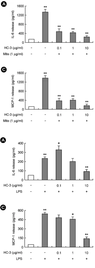
3). 염증성인자로 잘 알려져 있는 LPS 처리에 의하여 증 가하는 사이토카인 분비에 대하여 HC-3의 억제효과가 있는지를 조사하였다. Fig. 4에서 보여주는 것처럼, LPS에 서 의해서 증가한 IL-6와 MCP-1을 HC-3가 농도별로 크 게 감소시켰다. HC-3는 IL-8의 분비에도 억제효과를 보 여주었다. 이 결과를 통하여, 어성초 추출물 중에서 HC-3 가 THP-1 세포에서 분비되는 사이토카인의 양을 크게 감소키는 것으로 나타났으며, 그 효과는 추출물에 따라 서 다르게 나타났다.
]EoL-1 세포의 사이토카인 분비에 대한 어성초 추출물 HC-3의 억제효과
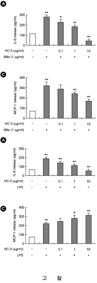
HC-3가 THP-1 세포에서 분비되는 사이토카인을 억제 시켰기 때문에 다른 염증성 백혈구인 EoL-1 세포에서 분 비되는 IL-6, IL-8, MCP-1에 대한 어성초 추출물의 효과 를 분석하였다. 세로무늬진드기 추출물과 LPS는 THP-1 세포에서와 마찬가지로 EoL-1 세포에서 IL-6, IL-8, MCP-1 의 분비를 증가시켰다 (Fig. 5 and 6). HC-3는 세로무늬진 드기 추출물에 의해 증가한 IL-6와 MCP-1의 분비를 농 도별로 유의하게 감소시켰다 (Fig. 5). 하지만, IL-8의 분
A B
C
Fig. 2. HC-3 inhibits the expression of IL-6, IL-8 and MCP-1
increased by house dust mite extract in THP-1 cells. THP-1 cells
were pre-incubated with HC-1, HC-2, HC-3, HC-4 and HC-5 at concentrations of 10 μg /ml for 30 min. THP-1 cells were stimulated with 1 μg/ml house dust mite extract (mite) for 24 h. The supernatant was collected and the expression of IL-6 (A), IL-8 (B) and MCP-1 (C) was analyzed by ELISA as described in the Methods section.**P<0.01 and *P<0.05 were considered a significant difference between the untreated group and mite-treated group or between the mite-treated group and HC-treated group. The data are expressed as the means ± S.D. of three independent experiments.
Fig. 1. The cytotoxic effects of Hc extracts on THP-1 cells and EoL-1 cells. THP-1 cells (A) and EoL-1 cells (B) were incubated
with HC-1, HC-2, HC-3, HC-4 and HC-5 at concentrations of 0.1 μg/ml, 1 μg/ml and 10 μg/ml for 24 h. The survival rates were then measured by a MTT assay. The data are expressed as the relative ratio to the absorbance of the untreated cells, which was set at 100%. The data are expressed as the means ± S.D. of three independent experiments.A
B
비는 오히려 증가시켰다. LPS를 처리에 의한 사이토카인 측정에서는 HC-3가 IL-6분비만 감소시켰으며, MCP-1과 IL-8을 증가시켰다 (Fig. 6). 이 결과를 통하여, HC-3는
EoL-1 세포에서 사이토카인 분비를 감소시켰으나, 세로 무늬진드기 추출물과 LPS에 의한 IL-8분비는 오히려 증 가시키는 것으로 나타났다.
A
C
B
Fig. 3. HC-3 inhibits the expression of Interleukin-6, Interleukin-8 and Monocyte chemoattractant protein-1 due to house dust mite extract in a dose-dependent manner. THP-1
cells were pre-incubated with HC-3 at concentrations of 0.1 μg/ ml, 1 μg/ml, or 10 μg/ml for 30 min. THP-1 cells were stimulated with 1 μg/ml house dust mite extract (mite) for 24 h. The supernatant was collected and the expression of IL-6 (A), IL-8 (B) and MCP-1 (C) was analyzed by ELISA as described in the Methods section.**P<0.01 was considered a significant difference between the untreated group and mitetreated group or between the mite-treated group and HC-treated group. The data are expressed as the means ± S.D. of three independent experiments.
A B
C
Fig. 4. HC-3 inhibits the expression of Interleukin-6, Interleukin-8 and Monocyte chemoattractant protein-1 increased by LPS in THP-1 cells. THP-1 cells were pre-incubated with HC-3
at concentrations of 0.1 μg/ml, 1 μg/ml, or 10 μg/ml for 30 min.THP-1 cells were stimulated with 1 μg/ml LPS for 24 h. The supernatant was collected and the expression of IL-6 (A), IL-8 (B) and MCP-1 (C) was analyzed by ELISA as described in the Methods section. **P<0.01 and *P<0.05 were considered a significant difference between the untreated group and mite-treated group or between the mite-treated group and HC-treated group. The data are expressed as the means ± S.D. of three independent experiments.
고 찰
본 연구는 한의학과 민속약학에서 알러지에 대한 억제
효과로 알려져 있는 어성초에 대한 항염증효과를 분석하 였다. 항염증효과는 염증세포인 단핵구와 호산구를 이용 하였고, 자극물질로 진드기항원 추출물과 LPS를 사용하 여 증가한 사이토카인에 대한 어성초 추출물의 억제효 A
C
B
Fig. 6. HC-3 alters the expression of Interleukin-6, Interleukin- 8 and Monocyte chemoattractant protein-1 increased by LPS in EoL-1 cells. EoL-1 cells were pre-incubated with HC-3 at concen-
trations of 0.1 μg/ml, 1 μg/ml, or 10 μg/ml for 30 min. EoL-1 cells were stimulated with 1 μg/ml LPS for 24 h. The supernatant was collected and expression of Interleukin-6 (A), Interleukin-8 (B) and Monocyte chemoattractant protein-1 (C) was analyzed by ELISA as described in the Methods section. **P<0.01 and *P<0.05 were considered a significant difference between the untreated group and mite-treated group or between the mite-treated group and HC-treated group. The data are expressed as the means ± S.D. of three inde- pendent experiments.A B
C
Fig. 5. HC-3 alters the expression of Interleukin-6, Interleukin- 8 and Monocyte chemoattractant protein-1 due to house dust mite extract in EoL-1 cells. EoL-1 cells were pre-incubated with
HC-3 at concentrations of 0.1 μg/ml, 1 μg/ml, or 10 μg/ml for 30 min. EoL-1 cells were stimulated with 1 μg/ml house dust mite extract (mite) for 24 h. The supernatant was collected and the expression of IL-6 (A), IL-8 (B) and MCP-1 (C) was analyzed by ELISA as described in the Methods section. **P<0.01 and *P<0.05 were considered a significant difference between the untreated group and mite-treated group or between the mite-treated group and HC-treated group. The data are expressed as the means ± S.D.
of three independent experiments.
과를 조사하였다. 이 과정은 이미 본 실험실에서 논문으 로 발표하여 항염증효과 측정방법으로의 가능성을 인정 받았다 (Lee et al., 2008; Lee et al., 2009).
어성초의 염증효과를 조사하기 전에 우리는 어성초를 유기용매에 의하여 분획을 실시하였다. 식물추출물은 부 위에 따라서, 화학적인 추출방법에 따라서 효능이 많이 다르게 나타난다 (Chattopadhyay D et al., 2002; Kim et al., 2007). 추출방법에 따라서 어성초의 효과가 차이가 있을 수 있기 때문에 5가지로 분획한 어성초 추출물을 각각 전 처리하여 사이토카인 억제효과를 살펴보았다. HC-3 가 다른 어성초 추출물에 비하여 진드기항원에 의해서 증가한 THP-1 세포의 IL-6, IL-8, MCP-1의 분비를 유의하 게 억제시켰고, 농도별 실험에서 유사한 결과를 보여주 었다 (Fig. 2 and 3). 이러한 억제효과는 정도의 차이는 있 었으나, LPS에 의해 증가한 사이토카인에 대해서도 나 타났다 (Fig. 4). 다음 실험으로 HC-3의 사이토카인 억제 효과를 호산구세포에서 분석하였다. 진드기항원과 LPS에 의해 증가한 Interleukin- 6는 HC-3에 의해서 억제되었다 (Fig. 5 and 6). 본 실험에서 HC-3의 사이토카인 억제효과 는 명확하게 확인하였지만, 앞으로 HC-3가 어떠한 세포 신호전달기전을 통해서 사이토카인 억제를 일으키는지 분석이 필요하다.
염증작용은 초기에 작용하는 선천면역의 주요 반응으 로서 알려져 있으나, 과도한 면역작용으로 인한 염증반응 은 알러지질환을 일으킨다. 알러지질환의 병인원으로 알 려져 있는 진드기 추출물은 염증을 일으키는 물질로 자 주 사용된다 (Lee et al., 2008; Lee et al., 2009). 항염증효과 는 염증을 억제시키는 효과로 정의하나, 복잡한 작용기전 이 연결되어 있어서 하나의 작용으로 정의 내리기는 매 우 어렵다. 본 실험에서는 진드기 추출물과 LPS를 동시 에 사용하여 사이토카인의 억제효과를 관찰하였다. 사이 토카인은 염증유발에 중요한 인자임에도 불구하고, 생산 되는 면역세포에 따라서 자극시키는 물질에 따라서 다 르게 반응할 수가 있다. HC-3는 단핵구에서는 조사되어 진 사이토카인에 대해서는 모두 억제시켰으나, 호산구 에서는 오히려 IL-8의 분비를 증가시켰다 (Fig. 5 and 6).
이러한 양면적인 결과는 향후 다른 ROS (reactive oxygen species) 생성, 케모카인 생성 등과 염증관련 억제효과의 분석과 알러지동물 모델과 같은 전임상모델을 이용한 효 과분석이 향후 필요할 것으로 판단된다.
본 연구의 결과는 HC-3가 단핵구와 호산구의 사이토 카인 분비를 억제시킴으로서 항염증 기능을 가지고 있으
며, 향후 HC-3를 포함한 다른 분획물의 항염증효과를 좀 더 조사한다면 항염증 치료제로서의 활용이 가능할 것으 로 판단된다.
감사의 글
이 논문은 2010년도 원광보건대학 교내연구비 지원에 의해서 수행됨.
REFERENCES
Bellanti JA. Cytokines and allergic diseases: clinical aspects.
Allergy Asthma Proc. 1998. 19: 337-341.
Chang JS, Chiang LC, Chen CC, Liu LT, Wang KC, Lin CC.
Antileukemic activity of Bidens pilosa L. var. minor (Blume) Sherff and Houttuynia cordata Thunb. Am J Chin Med. 2001.
29: 303 -312.
Chattopadhyay D, Arunachalam G, Mandal AB, Sur TK, Mandal SC, Bhattacharya SK. Antimicrobial and anti- inflammatory activity of folklore: Mallotus peltatus leaf extract.
J Ethnopharmacol. 2002. 82: 229-237.
Chiang LC, Chang JS, Chen CC, Ng LT, Lin CC. Anti-Herpes simplex virus activity of Bidens pilosa and Houttuynia cordata.
Am J Chin Med. 2003. 31: 355-362.
Coward WR, Sagara H, WInterleukinson SJ, Holgate ST, Church MK. Allergen activates peripheral blood eosinophil in nuclear factor-kappaB to generate granulocyte macrophage- colony stimulating factor, tumour necrosis factor-alpha and interleukin-8. Clin Exp Allergy 2004. 34: 1071-1078.
Friedmann PS. The role of dust mite antigen sensitization and atopic dermatitis. Clin Exp Allergy 1999. 29: 869-872.
Hirano T. Interleukin 6 and its receptor: ten years later. Int Rev Immunol. 1998. 16: 249-284.
Gabay C. Interleukin-6 and chronic inflammation. Arthritis Res Ther. 2006. 8: S3.
Kim IS, Kim JH, Kim JS, Yun CY, Kim DH, Lee JS. The inhibitory effect of Houttuynia cordata extract on stem cell factor-induced HMC-1 cell migration. J Ethnopharmacol.
2007. 112: 90-95.
Lee JS, Kim IS, Ryu JS, Yun CY. House dust mite, Dermatophagoides pteronissinus increases expression of Monocyte chemoattractant protein-1, Interleukin-6 and Interleukin-8 in human monocytic THP-1 cells. Cytokine 2008. 42: 365-371.
Lee JS, Kim IS, Yun CY. Secretion of Monocyte chemoattractant
protein-1, Interleukin-8 and Interleukin-6 increased by house dust mite, Dermatophagoides pteronissinus in human eosino- philic EoL-1 cells. Animal Cells Systems 2009. 13: 391-397.
Li GZ, Chai OH, Lee MS, Han EH, Kim HT, Song CH. Inhibitory effects of Houttuynia cordata water extracts on anaphylactic reaction and mast cell activation. Biol Pharm Bull. 2005. 28:
1864-1868.
Lu HM, Liang YZ, Yi LZ, Wu XJ. Anti-inflammatory effect of Houttuynia cordata injection. J Ethnopharmacol. 2006. 104:
245-249.
Machado DC, Horton D, Harrop R, Peachell PT, Helm BA.
Potential allergens stimulate the release of mediators of allergic response from cells of mast cell lineage in the absence of sensitization with antigen-specific IgE. Eur J Immunol.
1996. 26: 2972-2980.
Ring J, Darsow U. Role of aeroallergens in atopic eczema: proof of concept with the atopy patch test. J Am Acad Dermatol.
2001. 45: S49-S52.
Rossi D, Zlotnik A. The biology of chemokines and their receptors.
Annu Rev Immunol. 2000. 18: 217-242.
Shakoory B, Fitzgerald SM, Lee SA, Chi DS, Krishnaswamy G.
The role of human mast cell-derived cytokines in eosinophil biology. J Interferon Cytokine Res. 2004. 24: 271-281.
Shimizu Y, Shichijo M, Hiramatsu K, Takeuchi M, Nagai H, Takagi K. Mite antigen-induced Interleukin-4 and Interleukin- 13 production by basophils derived from atopic asthma patients. Clin Exp Allergy 1998. 28: 497-503.
Sohn MH, Lee KE, Kim KW, Kim ES, Park JY, Kim KE.
Calcium-calmodulin mediates house dust mite-induced ERK activation and Interleukin-8 production in human respiratory epithelial cells. Respiration 2007. 74: 447-453.
Wong CK, Li ML, Wang CB, Ip WK, Tian YP, Lam CW. House dust mite allergen Der p 1 elevates the release of inflam- matory cytokines and expression of adhesion molecules in co-culture of human eosinophils and bronchial epithelial cells. Int Immunol. 2006. 18: 1327-1335.