J. Exp. Biomed. Sci. 2013, 19(1): 48~54 pISSN : 1738-3226
Anti-inflammatory Effects of Extracts of Duchesnea chrysantha in Human Monocytic THP-1 Cells and Human Eosinophilic EoL-1 Cells
Ji-Sook Lee†
Department of Clinical Laboratory Science, Wonkwang Health Science University, Iksan, Jeonbuk 570-750, Korea
Atopic dermatitis is a recurrent or chronic eczematous skin disease with severe pruritus and has annually increased in Korea. In this study, we investigated whether Duchesnea chrysantha (Dc) extracts have an anti-inflammatory effect in human monocytic THP-1 cells and human eosinophilic EoL-1 cells. The dried and powdered whole plants of Dc were extracted with 80% EtOH (Dc-1). The residue was diluted with water, and then successively partitioned with n-hexane, EtOAc, and BuOH to produce the n-hexane (Dc-2), EtOAc (Dc-3), BuOH (Dc-4), and the water-soluble fractions (Dc-5), respectively. The mite extract and LPS increased the production of IL-6, IL-8 and MCP-1 in THP-1 cells and the increase was strongly suppressed by Dc-3 extract, as compare with other extracts. Dc-3 also inhibited the release of IL-6 increased by mite extract and LPS in EoL-1 cells. However, Dc-3 extract increased IL-8 production induced by the mite extract and LPS in EoL-1 cells. These results suggest that Dc extract may be used as anti-inflammatory agents in treating allergic disorders such as asthma and atopic dermatitis.
Key Words: Duchesnea chrysantha, Anti-inflammatory effect, THP-1 cells, EoL-1 cells
서 론
뱀딸기 (Duchesnea chrysantha)는 사매 (蛇苺) 또는 지 매 (地苺)라고도 하며, 장미과 (薔薇科 Rosaceae)에 속하 는 덩굴성 다년생초로서, 한국, 중국 등 동아시아에서 속 하는 나라에서 전통적으로 천식, 치통, 암, 태열과 같은 다양한 질환에 대한 예방 및 치료제로 사용되고 있다 (Lee and Yang, 1994; Kim et al., 2002; Kim et al., 2007).
아토피성 알레르기 질환은 치료가 어려운 난치성 면역 질환으로서 천식과 아토피 피부염이 포함된다 (Gavino et al., 2008). 알레르겐이 체내에 노출되면 Th2 세포의 증식 이 일어나며, Th2 세포에서 분비되는 IL-4에 의해 B 세포 에서 IgE 전환이 이루어진다. IgE는 IgE 수용체 (FcεR1) 를 통하여 비만세포의 활성화를 일으켜서 알레르기 질환
의 초기를 나타낸다. 후기와 만성시기에는 호산구 등의 백혈구가 조직으로 침윤되고, Th2 세포의 사이토카인으 로 알려져 있는 IL-4, IL-5, IL-13, IL-23과 eotaxin, TARC, MDC와 같은 케모카인이 증가하여 질환에 관련된다. 이 분자들은 IgE 전환, 호산구의 활성화와 생존력, 호흡과민 반응을 증가시킨다.
아토피성 알레르기 질환의 원인은 아직까지 명확하게 밝혀지지 않았으나, 흡입 항원에 대한 노출, 실내 외 공 기오염의 증가와 같은 환경적인 변화와 유전적인 소인으 로 생각된다. 천식 및 아토피 피부염 환자의 50% 이상이 집먼지진드기 항원에 대한 알레르기 피부반응 검사에서 양성이 나타나고, 소아 호흡기 알레르기 환자의 70% 이 상이 같은 양상을 나타냄을 통하여 집먼지진드기 항원이 주요한 원인으로 대두되고 있다 (Reinhold et al., 1990; Jujo et al., 1992; Robinson et al., 1992).
본 연구는 집먼지진드기 항원 추출물과 세균성 염증유 발물질인 지질다당류 lipopolysaccharide (LPS)를 사람의 단핵구와 호산구에 처리하여 염증성 사이토카인 (IL-6, IL-8과 MCP-1)을 증가시켰다. 증가된 사이토카인에 대하 여 뱀딸기 추출물들이 어떠한 효과를 나타내는지 분석하 였다 .
*Received: September 6, 2012 / Revised: September 28, 2012 Accepted: September 28, 2012
†Corresponding author: Ji-Sook Lee. Department of Clinical Laboratory Science Wonkwang Health Science University, 344-2 Sinyong-dong, Iksan, Jeonbuk, 570-750, Korea.
Tel: +82-63-840-1216, Fax: +82-63-840-1219 e-mail: [email protected]
○CThe Korean Society for Biomedical Laboratory Sciences. All rights reserved.
Original Article
재료 및 방법
뱀딸기 추출물의 제조
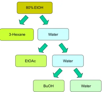
뱀딸기는 2004년 10월에 전남 여수에서 수집하였고, 표 준 추출물 (voucher number 81)은 한국한의학연구원과 대 전대학교 생명과학대학에 보관되어 있다. 뱀딸기 30 g을 80% 에탄올로 이틀 동안 실온에서 추출하였다. 얻어진 추출물은 감압상태에서 농축하였다. 이 추출물을 Dc-1이 라고 명명하였다. 나머지 부분은 증류수로 희석한 후에 n-헥산, 에틸아세테이트, 부탄올을 가지고 연속적으로 분 획하였다. 각각의 분획물을 Dc-2, Dc-3, Dc-4라고 명명 하였으며, 나머지 친수성 분획은 Dc-5라고 명명하였다 (Fig. 1).
세포 배양
사람의 단핵구 세포인 THP-1 (American Type Culture Collection, Manassas, VA)과 호산구 세포주인 EoL-1 세포 (Riken cell bank, Tsukuba, Japan)는 10% 소태아혈청 (fetal bovine serum, FBS), 100 U/ml의 페니실린, 100 μg/ml의 스 트렙토마이신 (Life technologies, Inc., Gaithersburg, MD)이
이 세포 독성 및 증식에 영향을 주는지 확인하기 위해 MTT 측정법 (Roche, Penzberg, Germany)을 이용하였다.
세포는 5 × 10
4cells/100 μl로 배양액에 부유하여 96 well plate에 분주하였다. 뱀딸기 추출물을 0.1 μg/ml, 1 μg/ml, 10 μg/ml의 농도로 24시간 동안 각 웰에 처리하였 다. MTT(3-(4,5-Dimethylthiazol-2-yl)-2,5-diphenyltetrazolium bromide) 용액을 10 μl 첨가하여 37℃, 5% CO
2배양기에 서 4시간을 배양한 다음 100 μl의 MTT 용해액을 추가 로 첨가하였다. 37℃, 5% CO
2배양기에서 하루 동안 둔 후에 550 nm에서 흡광도를 측정하였다.
효소면역측정법
뱀딸기 추출물을 30분 동안 전 처리한 후에 THP-1 세 포와 EoL-1 세포를 용태순 교수 (연세의대)로부터 제공받 은 세로무늬진드기 추출물 또는 LPS (Sigma Korea, Seoul, Korea)로 처리하였다. 세포 상층액에 있는 IL-6, IL-8, MCP-1의 농도는 회사의 매뉴얼에 따라서 OptEIA Set (BD Biosciences, San Diego, CA)를 이용하여 측정하였다.
통계 처리
모든 실험 결과는 통계적인 유의성을 측정하기 위해 대조군과 양성대조군 (세로무늬진드기 처리군 또는 LPS 처리군 ), 양성대조군과 뱀딸기 추출물 처리군의 결과를 Student's t-test로 분석하였고, 각 통계 처리의 P 값이 0.05 미만인 경우 통계적으로 유의한 차이가 있다고 해석하 였다.
결 과
딸기 추출물의 THP-1과 EoL-1 세포에 대한 세포 독성 효과
THP-1과 EoL-1 세포에 뱀딸기 추출물 (Dc-1~Dc-5)를 농도별로 24시간 동안 처리하여 MTT 측정법을 통해 세 포 생존율을 확인한 결과, 모든 분획물에서 10 μg/ml의 고농도에서도 세포 독성을 일으키지 않았다 (Fig. 2).
EtOAc Water
BuOH Water
Fig. 1. Preparation of Dc extract. The dried and powdered whole plants of Dc (100 g) were extracted with 80% ethanol (3 × 0.5 L) for 2 days at room temperature. The combined liquid extracts were concentrated under reduced pressure. The residue was diluted with water, and then successively partitioned with n-hexane, EtOAc, and BuOH to produce the n-hexane (Dc-2), EtOAc (Dc-3), BuOH (Dc-4), and the water-soluble fractions (Dc-5), respectively.
A B
Fig. 2. The cytotoxic effects of Dc extracts on THP-1 cells and EoL-1 cells. THP-1 cells (A) and EoL-1 cells (B) were incubated with Dc-1, Dc-2, Dc-3, Dc-4 and Dc-5 at concentrations of 0.1 μg/ml, 1 μg/ml and 10 μg/ml for 24 h. The survival rates were then measured by a MTT assay. The data are expressed as the relative ratio to the absorbance of the untreated cells, which was set at 100%. The data are expressed as the means ± S.D. of three independent experiments.
Fig. 3. Dc-3 inhibits the expression of IL-6, IL-8 and MCP-1 increased by LPS in THP-1 cells. THP-1 cells were pre-incubated with Dc-1, Dc-2, Dc-3, Dc-4 and Dc-5 at concentrations of 10 μg/ml for 30 min (A). THP-1 cells were pre-incubated with Dc-3 at concentrations of 0.1 μg/ml, 1 μg/ml, or 10 μg/ml for 30 min (B). THP-1 cells were stimulated with 1 μg/ml LPS for 24 h. The supernatant was collected and the expression of IL-6, IL-8 and MCP-1 was analyzed by ELISA as described in the Methods section. **P < 0.01 and *P < 0.05 were considered a significant difference between the untreated group and LPS-treated group or between the LPS-treated group and Dc-treated group. The data are expressed as the means ± S.D. of three independent experiments.
A B
Dc-3 (μg/ml)
Dc-3 (μg/ml)
Dc-3 (μg/ml)
LPS 의한 THP-1 세포의 사이토카인 분비에 대한 뱀딸기 추출물 Dc-3의 억제 효과
THP-1 세포에서 분비되는 염증성 사이토카인에 대한 뱀딸기 추출물의 효과분석을 통하여 뱀딸기 추출물의 항 염증 효과를 조사하였다. 염증성 인자로 잘 알려져 있는 LPS는 THP-1 세포에서 IL-6, IL-8, MCP-1의 분비를 크게 촉진시켰다 . 뱀딸기 추출물의 전 처리를 통하여 사이토카 인의 농도가 감소하였는데, 특히 Dc-3는 IL-6와 MCP-1의 분비를 다른 추출물에 비하여 크게 감소시켰으며, IL-8의 분비도 감소시켰다 (Fig. 3A). Dc-3를 농도별로 처리한 후, 농도가 증가함에 따라 염증성 사이토카인의 생산이 억제되었다 (Fig. 3B). 이 결과를 통하여, 뱀딸기 추출물 중에서 DC-3가 THP-1 세포에서 분비되는 사이토카인의 양을 크게 감소키는 것으로 나타났으며, 그 효과는 추출 물에 따라서 다르게 나타났다.
집먼지진드기 추출물에 의한 THP-1 세포의 사이토카인 분비에 대한 뱀딸기 추출물 Dc-3의 억제 효과
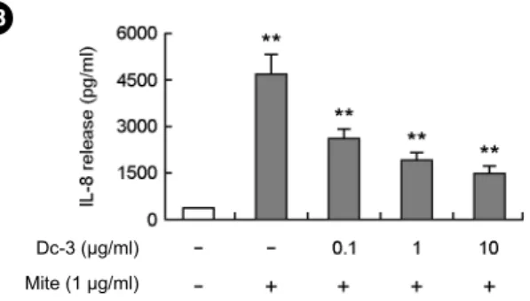
Dc-3는 LPS에 의해 일어나는 THP-1 세포의 염증성 사 이토카인 분비를 억제시켰다. 이번 실험에서는 아토피 피 부염의 주된 항원인 집먼지진드기에 의하여 IL-6, IL-8, MCP-1이 Dc-3에 의해서 억제가 되는지 분석하였다. Dc-3
sms 0.1 μg/ml, 1 μg/ml, 10 μg/ml의 농도에 따라서 염증성 사이토카인을 모두 억제하는 경향을 나타냈다. Dc-3는 LPS에 증가한 염증성 사이토카인의 억제에서 작용한 농 도보다 더 저농도에서도 집먼지진드기에 의해 유도된 염 증성 사이토카인을 억제시켰다 (Fig. 4).
LPS에 의한 EoL-1 세포의 사이토카인 분비에 대한 뱀딸 기 추출물 Dc-3의 억제 효과
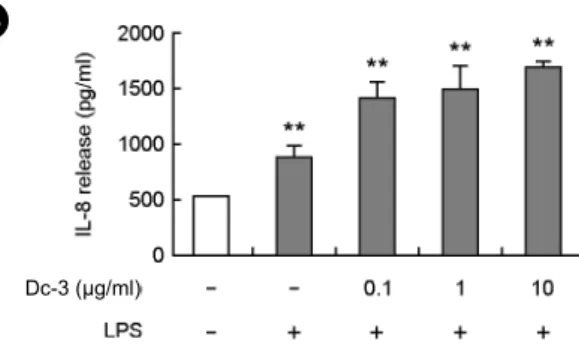
알레르기성 질환에서 증가하는 호산구의 실험을 위해 서 호산구성 백혈구인 EoL-1 세포를 이용하였고, LPS를 처리한 후 분비되는 IL-6, IL-8, MCP-1에 대한 뱀딸기 추 출물의 효과를 분석하였다. LPS는 THP-1 세포에서와 마 찬가지로 EoL-1 세포에서 IL-6, IL-8, MCP-1의 분비를 증 가시켰다 (Fig. 5). Dc-3는 Fig. 5A에서 보는 바와 같이, IL-6에 대해 농도별로 유의하게 감소시켰다. 그러나, IL-8 과 MCP-1의 분비는 오히려 증가시켰다 (Fig. 5B and C).
집먼지진드기 추출물에 의한 EoL-1 세포의 사이토카인 분비에 대한 뱀딸기 추출물 Dc-3의 억제 효과
집먼지진드기에 의해 증가하는 EoL-1 세포의 염증성 사이토카인 (IL-6, IL-8, MCP-1)에 대한 뱀딸기 추출물인 Dc-3의 효과를 분석하였다. Dc-3는 집먼지진드기에 의해 생성된 IL-6와 MCP-1에 대해 농도별로 유의하게 감소
CDc-3 (μg/ml) Mite (1 μg/ml)
Dc-3 (μg/ml) Mite (1 μg/ml)
Dc-3 (μg/ml) Mite (1 μg/ml)
Fig. 4. Dc-3 inhibits the expression of IL-6, IL-8 and MCP-1 due to house dust mite extract in a dose-dependent manner. THP-1 cells were pre-incubated with Dc-3 at concen- trations of 0.1 μg/ml, 1 μg/ml, or 10 μg/ml for 30 min. THP-1 cells were stimulated with 1 μg/ml house dust mite extract (mite) for 24 h. The supernatant was collected and the expression of IL-6 (A), IL-8 (B) and MCP-1 (C) was analyzed by ELISA as described in the Methods section. **P < 0.01 was considered a significant difference between the untreated group and mite- treated group or between the mite-treated group and Dc-treated group. The data are expressed as the means ± S.D. of three independent experiments.
시켰다 . 그러나, IL-8의 분비는 LPS에 의해 증가한 IL-8 에 대한 Dc-3의 효과와 마찬가지로 오히려 증가시켰다
(Fig. 6).
A B
C
Dc-3 (μg/ml) Mite (1 μg/ml)
Dc-3 (μg/ml) Mite (1 μg/ml)
Dc-3 (μg/ml) Mite (1 μg/ml)
Fig. 6. Dc-3 alters the expression of IL-6, IL-8 and MCP-1 due to house dust mite extract in EoL-1 cells. EoL-1 cells were pre-incubated with Dc-3 at concentrations of 0.1 μg/ml, 1 μg/ml, or 10 μg/ml for 30 min. EoL-1 cells were stimulated with 1 μg/ml house dust mite extract (mite) for 24 h. The supernatant was collected and the expression of IL-6 (A), IL-8 (B) and MCP-1 (C) was analyzed by ELISA as described in the Methods section. **P < 0.01 was considered a significant difference between the untreated group and mite-treated group or between the mite-treated group and Dc-treated group. The data are expressed as the means ± S.D. of three independent experiments.
A B
C
Dc-3 (μg/ml) Dc-3 (μg/ml)
Dc-3 (μg/ml)
Fig. 5. DC-3 inhibits the expression of IL-6, IL-8 and MCP-1 increased by LPS in EoL-1 cells. EoL-1 cells were pre-incubated with Dc-3 at concentrations of 0.1 μg/ml, 1 μg/
ml, or 10 μg/ml for 30 min. EoL-1 cells were stimulated with 1 μg/ml LPS for 24 h. The supernatant was collected and the expression of IL-6 (A), IL-8 (B) and MCP-1 (C) was analyzed by ELISA as described in the Methods section. **P < 0.01 and
*P < 0.05 were considered a significant difference between the untreated group and mite-treated group or between the mite- treated group and Dc-treated group. The data are expressed as the means ± S.D. of three independent experiments.
되며 한국, 중국, 및 아시아의 일부 지역에 야생하는 뱀 딸기 (Duchesnea chrysantha)에 대한 항염증 효과를 분석 하였다. 항염증 효과를 보기 위해 생체 내 염증 및 알레 르기성 질환에 작용하는 세포인 단핵구와 호산구를 이용 하였고, 자극물질로 진드기 항원 추출물과 LPS를 처리한 후 증가된 염증성 사이토카인에 대한 뱀딸기 추출물의 억제 효과를 조사하였다. 이 과정은 이미 본 실험실에서 논문으로 발표하여 항염증 효과 측정방법으로의 가능성 을 인정받았다 (Lee et al., 2008; Lee et al., 2009).
뱀딸기의 염증 효과를 조사하기 전에 우리는 뱀딸기를 유기용매에 의하여 분획을 실시하였다. 화학적인 추출 방법에 따라서 식물 추출물의 효능은 많이 다르게 나타 난다 (Chattopadhyay et al., 2002; Kim et al., 2007). 기존 연 구 결과를 통해 뱀딸기 에테르 추출물에서 미생물에 대 한 억제활성이 관찰되었으며, 인간종양세포주인 PC14와 MKN45에 의해 80% 아세톤 추출물에서 분리된 페놀성 화합물이 암세포 증식 억제활성을 나타낸다고 보고한 바 있다 (Lee and Kim, 1986; Lee and Yang, 1994). 또한, 아토 피 피부염의 병인기전은 명확하지 않으나, 피부의 염증작 용은 초기에 작용하는 선천면역의 주요 반응으로서 알려 져 있으나, 과도한 면역작용으로 인한 염증반응은 알러지 질환을 일으킨다. 특히, 지속적인 알러젠 또는 박테리아 성 수퍼항원 노출에 의해 CC chemokine ligand 18이 다량 생산되며 이것은 만성염증과 아토피 피부염의 만성화를 초래한다 (Pivarcsi et al., 2004). 본 실험에서는 아토피 피 부염의 주요 항원인 진드기 추출물 및 아토피 피부염의 만성염증을 초래하는 LPS를 처리하여 유발된 염증성 사 이토카인에 대한 뱀딸기의 효과를 살펴보았다. 뱀딸기 추출물은 추출방법을 다르게 하여 5가지로 분획하였다.
Dc-3가 다른 뱀딸기 추출물에 비하여 LPS 및 집먼지진드 기 항원에 의해서 증가한 THP-1 세포의 IL-6, IL-8, MCP-1 의 분비를 유의하게 억제시켰고, 농도별 실험에서 유사 한 결과를 보여주었다 (Fig. 3 and 4). 또한, 단핵구 세포 인 THP-1 세포에서는 진드기에 의해 유발된 IL-6, IL-8, MCP-1의 분비를 유의하게 Dc-3가 모두 억제시켰으나, 호 산구 세포인 EoL-1 세포에서 LPS에 유도된 염증성 사이 토카인 IL-6의 생산만 억제시켰다. 집먼지진드기에서 유
서 , 단핵구에서 작용하는 IL-8은 Dc-3의 억제 효과와 호 산구에 작용하는 기전이 다름을 알 수 있다. 또한, Dc-3가 LPS에 비해 집먼지진드기에 의해 유도된 염증성 사이토 카인에 대해 낮은 농도에서도 억제효과가 나타나는 것을 볼 때 집먼지진드기에 유도된 염증성 사이토카인에 대해 특이성 (specificity)을 보임을 알 수 있었다. 이러한 결과 는 본 실험에서 Dc-3의 염증성 사이토카인에 대한 억제 효과를 명확하게 확인하였지만, 앞으로 Dc-3가 집먼지진 드기에 대한 특이성 및 뱀딸기 추출물의 주요 생리활성 물질로 이미 알려진 리놀레산 (linolic acid), 베타-시스토 스테롤 (beta-sistosterol), 쿼시트린 (quercitrin) 등 이 외에 항염증 및 항아토피성 효과에 대한 유효성분의 분석이 필요하다.
본 연구의 결과는 Dc-3가 단핵구와 호산구의 사이토카 인 분비를 억제시킴으로서 항염증 기능을 가지고 있으며, 향후 Dc-3를 포함한 다른 분획물의 항염증 효과를 향후 더 연구한다면 항염증 치료제로서의 활용이 가능할 것으 로 판단된다.
감사의 글
이 논문은 2012년도 원광보건대학교 교내연구비 지원 에 의해서 수행됨.
REFERENCES
Chattopadhyay D, Arunachalam G, Mandal AB, Sur TK, Mandal SC, Bhattacharya SK. Antimicrobial and anti-inflammatory activity of folklore: Mallotus peltatus leaf extract. J Ethnopharmacol. 2002. 82: 229-237.
Gavino AC, Needham GR, High WA. Atopic dermatitis, patch testing, and house dust mites: a brief review. Dermatitis 2008.
19: 121-128.
Jujo K, Renz H, Abe J, Gelfand EW, Leung DY. Decreased interferon-γ and increased interleukin-4 production in atopic dermatitis promotes IgE synthesis. J Allergy Clin Immunol.
1992. 90(3 Pt1): 323-331.
Kim IG, Jung IL, Oh TJ, Kim KC, Shim HW. Polysaccharide- enriched fraction isolated from Duchesnea chrysantha protects
against oxidative damage. Biotech Lett. 2002. 24: 1299-1305.
Kim KC, Kim JS, Son JK, Kim IG. Enhanced induction of mitochondrial damage and apoptosis in human leukemia HL-60 cells by the Ganoderma lucidum and Duchesnea chrysantha extracts. Cancer Lett. 2007. 246: 210-217.
Kim SH, Cui CB, Kang IJ, Kim SY, Ham SS. Cytotoxic effect of buckwheat (Fagopyrum esculentum Moench) hull against cancer cells. J Med Food. 2007. 10: 232-238.
Lee IR, Kim YH. Studies on the antitumor activity of Duchesnea indicae Herba. Arch Pharm Res. 1986. 9: 1-4.
Lee IR, Yang MY. Phenolic compounds from Duchesnea chrysantha and their cytotoxic activities in human cancer cell.
Arch Pharm Res. 1994. 17: 476-479.
Lee JS, Kim IS, Ryu JS, Yun CY. House dust mite, Dermatophagoides pteronissinus increases expression of MCP-1, IL-6, and IL-8 in human monocytic THP-1 cell.
Cytokine 2008. 42: 365-371.
Lee JS, Kim IS, Ryu JS, Yun CY. House dust mite, Dermatophagoides pteronissinus increases expression of MCP-1, IL-6, and IL-8 in human eosinophilic leukemia EoL-1 cells. Animal Cells and Systems 2009. 13: 391-397.
Pivarcsi A, Gombert M, Dieu-Nosjean MC, Lauerma A, Kubitza R, Meller S, Rieker J, Muller A, Da Cunha L, Haahtela A, Sonkoly E, Fridman WH, Alenius H, Kemeny L, Ruzicka T, Zlotnik A, Homey B. CC chemokine ligand 18, an atopic dermatitis-associated and dendritic cell-derived chemokine, is regulated by staphylococcal products and allergen exposure.
J Immunol. 2004. 173: 5810-5817.
Reinhold U, Wehrmann W, Kukel S, Kreysel HW. Evidence that defective interferon-γ production in atopic dermatitis patients is due to intrinsic abnormalities. Clin Exp Immunol. 1990.
79: 374-379.
Robinson DS, Hamid Q, Ying S, Tsicopoulos A, Barkans J, Bentley AM, Corrigan C, Durham SR, Kay AB. Predominant TH2-like bronchoalveolar T-lymphocyte population in atopic asthma. N Engl J Med. 1992. 326: 298.
Rossi D, Zlotnik A. The biology of chemokines and their receptors.
Annu Rev Immunol. 2000. 18: 217-242.
Shakoory B, Fitzgerald SM, Lee SA, Chi DS, Krishnaswamy G.
The role of human mast cell-derived cytokines in eosinophil biology. J Interferon Cytokine Res. 2004. 24: 271-281.