155 This is an Open-Access article distributed under the terms of the Creative Commons Attribution Non-Commercial License (http://
creativecommons.org/licenses/by-nc/3.0) which permits unrestricted non-commercial use, distribution, and reproduction in any medium, provided the original work is properly cited.
J. Mushrooms 2016 December, 14(4):155-161 http://dx.doi.org/10.14480/JM.2016.14.4.155 Print ISSN 1738-0294, Online ISSN 2288-8853
© The Korean Society of Mushroom Science
*Corresponding author E-mail : [email protected]
Tel : +82-43-871-5585, Fax : +82-43-871-5589 Received November 29, 2016
Revised December 7, 2016 Accepted December 16, 2016
Inhibitory effect of Panax ginseng and Pleurotus osteratus
complex on expression of cytokine genes induced by extract of Dermatophagoides pteronissinus in human monocytic THP-1 and EoL-1 cells
Kyeong Hun Park
1, Eun Suk Lee
1, Yong Ik Jin
2, Kyung Sun Myung
1, Hong Woo Park
1, Chun Geon Park
1, Won Sik Kong
3, and Young Ock Kim
1,*
1Department of Herbal Crop Research, National Institute of Horticultural and Herbal Science, RDA, Eumseong, 27709, Korea.
2Highland Agriculture Research Institute, National Institute of Crop Science, Rural Development Administration, Pyeongchang 25342, Korea
3Mushroom Research Division, National Institute of Horticultural and Herbal Science, RDA, Eumseong, 27709, Korea.
ABSTRACT: A recent study reported that Pleurotus ostreatus has the potential to be used as a β-glucan-based cream for supportive complementary therapy of atopic dermatitis. KH054 is a new herbal prescription consisting of P. ostreatus and Panax ginseng. The effects of atopic dermatitis-induced materials on the expression of cytokine genes in human monocytes (THP-1, EoL- 1) have been examined. Some reports demonstrated that P. ginseng augments the activity of natural killer cells, which plays an important role in innate immunity against infection and tumor development. Monocyte chemotactic protein 1 (MCP-1), interleukin (IL)-6, and IL-8 have important roles in mediating the infiltration of various cells into the skin of atopic dermatitis and psoriasis. The present study investigated whether KH054 on induced IL-6, IL-8, and MCP-1 secretion by house dust mite (Dermatophagoides pteronissinus) in THP-1 (human acute monocytic leukemia) and EoL-1(Human eosinophilic leukemia) cell. D.
pteronissinus functions in the pathogenesis of allergic diseases, including atopic dermatitis and asthma. The inhibitory effect of KH054 on the induction of IL-6, IL-8, and MCP-1 secretion by D. pteronissinus extract in THP-1 and EoL-1 cells was examined.
KH054 potently suppressed the elevated production of IL-6 and IL-8 induced by D. pteronissinus treatment in THP-1 and EoL-1 cells. Based on the present results, KH054 may be useful for developing functional foods to treat atopic dermatitis.
KEYWORDS: Monocyte chemoattractant protein-1, Interleukin-6, Interleukin-8, Pleurotus ostreatus, Panax ginseng, Dermatophagoides pteronissinus
Introduction
Dermatophagoides pteronissinus (D. pteronissinus) has been most prevalent (Heymann et al., 1989). Mites of the family Pyroglyphidae (Dermatophagoides spp.) are the major source of allergens in house dust (Voorhorst et
al., 1967). Exposure to extract of D. pteronissinus is known to play a important role in the onset and aggravation of allergic diseases. In particular, allergen related to D. pteronissinus has been considered as one of important pathogens of atopic dermatitis(AD) and asthma (Sun et al., 1991). D. pteronissinus induces the production of immunoglobulin E (IgE), which is very important in the pathogenesis of AD (Bieber, 2008). The epidermis is largely consisted of keratinocytes, and they not only play an important structural part in forming a physical barrier to foreign antigens and microorganisms, but also are important functionally in mediating cutaneous immune reactions. Their nature and importance in Th2 responses is under active investigation and their role in skin diseases unknown( Brandt et al., 2011). T helper 2 (Th2) lymphocytes are the main cells associated with the pathogenesis of AD (Romagnani, 1994). The cells over secrete Th2 cytokines such as
interleukin IL-4, IL-5 and IL-13, which disrupt the Th1/
Th2 balance and increase the production of IgE. IL-6 is a pleiotropic cytokine (Le et al., 1989). The biologic effects of IL-6 are B-cell terminal differentiation, T-cell proliferation, and induction of cytotoxicity (Gabay, 2006). IL-6 has been shown to be a cofactor that potentiates IgE production from switched B cells by enhancing the effects of IL-4,10 suggesting a role for IL-6 in atopy and Th2-type responses(Lee et al., 2008).
MCP-1 and IL-8 act as chemotactic factors inducing monocytes and neutrophils (Shakoory et al., 2004).
However, few studies have reported details on the inflammatory regulation triggered by D. pteronissinus in monocytes (Liu et al., 2005). In the present study, we investigated whether D. pteronissinus extract (DpE) increases cytokine release in human monocytic THP- 1and EoL-1 cells. P. ginseng has been showed diverse biological activities including antiaging, antidiabetic, anticarcinogenic, analgesic, antipyretic, antistress, antifatigue, and tranquilizing effects (Kim et al., 1990).
There are a number of reports concerning the immunological effects of P. ginseng. Some reports have shown that P. ginseng augments the activity of natural killer (NK) cells, which play an important role in innate immunity against infection and tumor development (Smyth et al., 2002). β-Glucan-based cream (containing pleuran isolated from pleurotus ostreatus) in supportive treatment of mild-to-moderate atopic dermatitis (Jesenak et al., 2016). In the present study, the mixture of P.
ginseng and P. ostreatus is designed as formula KH054.
The effect of KH054 on increases cytokine release in human monocytic THP-1 and EoL-1 cells, with the aim to explore KH054 the thrapeutically for the treatment of allergies, including atopic dermatitis.
Materials and methods
D. pteronyssinus preparation
D. pteronyssinus was obtained from Jung-ang Exp.
Lab (Seoul Korea). Enzyme-linked immunosorbent assay (ELISA) kits for IgE was obtained from BD Biosciences (San Diego, CA, USA) and R&D Systems (Minneapolis, MN, USA). All chemicals and solvents were of the highest grade commercially available.
Preparation of KH054
The dried roots of P. ginseng and P. ostreatus were extracted separately with 100% water for 6 h in a reflux
apparatus. After refluxing, the extracts were filtered, the filtrates were evaporated in a rotary evaporator, and the samples were lyophilized in a freeze dryer (Operon, Gyeonggi, Republic of Korea). The extract yields of P.
ginseng, and P. ostreatus were 23%, 28%. Each herb of KH054 was mixed in the following ratio: P. ginseng: P.
ostreatus = 8:13. The KH053 was dissolved in distilled water and sequentially passed through 0.22-µm filters for sterilization.
Cell culture
The human monocytic cell line THP-1 was obtained from the American Type Culture Collection (Manassas, VA). The human eosinophilic leukemia cell line EoL-1 was obtained from the Riken Cell Bank (Tsukuba, Japan). Both cells were grown in RPMI 1640 supplemented with 10% heat-inactivated fetal bovine serum (FBS), penicillin (100 U/ml), and streptomycin (100 μg/ml).
Viability assay
Viability was assayed based on the conversion of (3- (4,5-dimethylthiazol-2-yl)-2,5-diphenyltetrazolium bromide (MTT) using a cell proliferation kit (Roche, Penzberg, Germany). Both THP-1 cells and EoL-1 cells in 100 μl of culture medium were seeded into wells of a 96-well plate. KH054 was added to each well as a concentration of 10 μg/ml, 30 μg/ml, or 100 μg/ml.
After incubation for 24 h at 37oC, 10 μl of MTT solution was added and incubated for 4 h. 100 uL of solubilization solution was added to each well. After 24 h incubation, the absorbance was measured at 550 nm using a BIO-TEK ELx808 enzyme-linked immunosorbent assay (ELISA) reader (Bio-Tek Instruments,Winooski, VT).
Enzyme-linked immunosorbent assay (ELISA) DpE was obtained from Jungang Lab(Korea). Mite powder was triturated in liquid nitrogen and allergens were extracted in 5 mM borate-buffered saline. After centrifugation (20,000g for 45 min at 4oC), the supernatant was dialyzed, filtered through a 0.22-μm membrane and protein concentration was determined.
Allergen extract aliquots were stored at −20oC until being used in serological and proliferation assays.
Cytokine release of THP-1 and EoL-1 cells was measured (Lee et al.,2008). After pre-treatment with KH054 for 30 min, THP-1 and EoL-1 cells were treated
with the extract of house dust mite, The concentrations of MCP-1, IL-6, and IL-8 in the supernatant were measured by a sandwich. ELISA using OptEIA Set (BD Biosciences, San Diego, CA), according to the manufacturer's instructions. All assays were performed in triplicate. The cytokine concentration was calculated using a linear-regression equation obtained from the standard absorbance values.
Quantitative real –time PCR
The reverse transcription test for cDNA was conducted with specific primers for each cytokine in 40 µl volume (including 33 µl of the studied sample). The reverse transcription test was carried out for 30 min at 40°C. Inactivation of the reverse transcriptase enzyme was performed for 5 minutes at 95 after the reaction.
Then, the sample of cDNA was diluted 5-fold in TE buffer. Real time RT-PCR was performed using the “DT- 96” instrument (DNK-Technologya, Russia) in 35 µl volume using the following amplification program:1 cycle-80°C for 30 seconds, then 94°C for 1.5 min; 50
cycles-94°C for 20 seconds, then 62°C for 20 seconds.
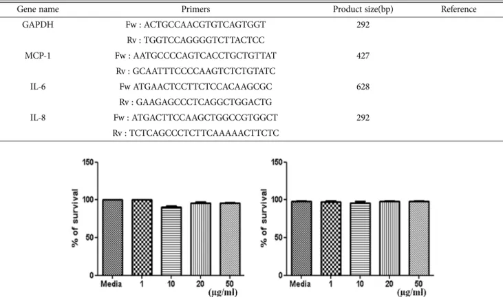
The primers and probes listed in Table.1. were designed using Primer3 software.
Statistical analysis
Data were expressed as the means ± SD. Data were analyzed by one-way analysis of variance (anova) followed by using the Mann–Whitney U-test. P<0.05 was considered to indicate a statistically significant difference.
Results and Discussion
KH054 inhibits the secretion of MCP1, IL6 and IL8 in THP1 and EoL1 cells. KH054 inhibits the secretion of MCP1, IL6 and IL8 in THP1 and EoL1 cells. A MTTbased assay was used to determine the effect of KH054 on the viability of THP1, and EoL1 cells. As shown in Fig. 1, the survival rate of EoL1 cells was not affected by KH054 concentration ranging between 1 and 50 μg/ml. The viability of THP1 cell Table 1. Primers of RT-PCR for various cytokines
Gene name Primers Product size(bp) Reference
GAPDH Fw : ACTGCCAACGTGTCAGTGGT 292
Rv : TGGTCCAGGGGTCTTACTCC
MCP-1 Fw : AATGCCCCAGTCACCTGCTGTTAT 427
Rv : GCAATTTCCCCAAGTCTCTGTATC
IL-6 Fw ATGAACTCCTTCTCCACAAGCGC 628
Rv : GAAGAGCCCTCAGGCTGGACTG
IL-8 Fw : ATGACTTCCAAGCTGGCCGTGGCT 292
Rv : TCTCAGCCCTCTTCAAAAACTTCTC
Fig. 1. Effect of KH054 on survival of THP1 and EoL1 cells. THP1, EoL1, cells were incubated in medium in the absence (medium alone) or presence of KH054 at the indicated concentrations for 24 h. The survival rate was measured by performing the MTTbased viability assay. Data are expressed as the relative ratio to the absorbance of the untreated cells, which was set at 100% and as the mean ± SD of three independent experiments. MTT, 3(4,5dimethylthiazol2yl)2,5Diphenyltetra zolium bromide.
The media is RPMI 1640 supplemented with 10% FBS, penicillin (100 U/ml), and streptomycin (100 μg/ml).
was weakly inhibited by KH054 concentration ranging 10 μg/ml. These results may be caused by a variety of factors, including cell culture conditions and variations in the skills of the different investigators (Kim et al., 2012, Yang et al., 2011, Lee at el., 2012).
However, KH054 clearly reveals an inhibitory trend of cytokine production similar to antiinflammatory chemicals or extracts. The protein secretion of MCP-1, IL-6, and IL-8 was evaluated by ELISA, after treatment with DpE in a time and dose-dependent manner. In the present study, the efficacy of KH054 as an antiinflammatory or antiatopic dermatitis drug was examined for the first time using human inflammatoryassociated cells. KH054 was observed to
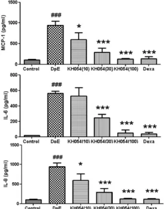
inhibit the production of MCP1, IL6 and IL8 in THP1 and EoL1 cells. Since MCP1 acts as a potent chemoattractant of monocytes and IL8 functions as an essential molecule in the survival, migration and activation of neutrophils, KH054 may inhibit the inflammatory responses by regulation of the immune responses involved in monocytes and neutrophils (Murphy et al., 2000). The secretion of MCP1, IL6 and IL8 increased following treatment with an extract of DpE in THP1 cells (Fig. 2). The cytokines varied from IL-6 ; Control (200.40 ± 28.45 pg /ml), DpE (806.00 ± 54.42 pg /ml), KH054(100) (176.20 ±14.74 pg /ml), Dexamethason (38.20 ± 7.22 pg /ml), IL-8 ; Control (79.50 ± 6.47 pg /ml), DpE (2074.24 ± 76.11 pg /ml), KH054(100) (255.40 ± 120.82 pg /ml), Dexamethason Fig. 2. KH054 inhibits the production of MCP1, IL6 and IL8
increased by DpE or in THP1 cells. THP1 cells were seeded in wells of 24well plates and cultured in RPMI1640 medium containing 0.5% FBS at 37˚C for 24 h. Cells were pretreated in the absence or presence of KH054 at the indicated concentrations. Cells were treated with 10 μg/ml mite extract for 24 h and the supernatant was then collected and analyzed by ELISA. Data are presented as the mean ± SD of three independent experiments. *P<0.05, **P<0.01 and ***P<0.001 was considered to indicate a statistically significant difference between the untreated vs. DpE only treated group or between the DpE only treated vs. KH054 treated group.
MCP1, monocyte chemotactic protein1; IL, interleukin; DpE, Dermatophagoides pteronissinus.
Fig. 3. KH054 inhibits the production of MCP1, IL6 and IL8 increased by DpE in EoL1 cells. EoL1 cells were seeded in 24 well plates and cultured in RPMI1640 medium containing 0.5% FBS at 37˚C for 24 h. Cells were pretreated in the absence or presence of KH054 at the indicated concentrations. Cells were treated with 10 μg/ml DpE for 24 h. The supernatant was collected and analyzed by ELISA.
Data are presented as the mean ± SD of three independent experiments. *P<0.05, and ***P<0.001 were considered to indicate a significant difference between the untreated vs.
DpE only treated group or between the DpE only treated vs.
the KH054treated group. MCP1, monocyte chemotactic protein1; IL, interleukin; DpE, Dermatophagoides pteronissinus.
(160.20 ± 8.87 pg /ml), MCP-1; Control (27.52 ± 6.99 pg /ml), DpE (567.52 ± 118.65 pg /ml), KH054(100) (71.20 ± 14.96 pg /ml), Dexamethason (33.28 ± 6.75 pg /ml) in THP-1 cell, IL-6 ; Control (17.02 ± 1.33 pg / ml), DpE (562.86 ± 28.66 pg /ml), KH054(100) (51.80
± 40.81 pg /ml), Dexamethason (39.60 ± 17.34 pg /ml), IL-8 ; Control (17.02 ± 1.33 pg /ml), DpE (562.86 ± 28.66 pg /ml), KH054(100) (51.80 ± 40.81 pg /ml), Dexamethason (39.60 ± 17.34 pg /ml), MCP-1; Control (17.02 ± 1.33 pg /ml), DpE (562.86 ± 28.66 pg/ml), KH054(100) (51.80 ± 14.96 pg /ml), Dexamethason (39.60 ± 17.34 pg /ml) in EoL-1 cell had the highest amount among these conditions and parts in this study (Fig. 2 and 3). Dexamethason is positive control. KH054 significantly suppressed the production of MCP1 in a dosedependent manner. IL6 expression increased following treatment with a low concentration of KH054, but decreased following treatment with a high concentration when compared with DpE treatment alone.
In EoL1 cells, DpE enhanced the expression of MCP1, IL6 and IL8. MCP1, IL6 and IL8 expression decreased following treatment with KH054 in a dosedependent manner (Fig. 3). Alteration of IL6 by KH054 in EoL1 cells was similar to that in THP1 cells. We first examined whether the protein expression of MCP-1, IL- 6, and IL-8 was affected by DpE in THP-1 cells (Figs.
2 and 3).In THP1 and EoL1 cells, KH054 increased IL6 expression at a low concentration and decreased the expression at a high concentration. The exact mechanism of KH054 remains to be elucidated and is the subject of ongoing studies. Atopic dermatitis is an allergic skin disease characterized by inappropriate epidermalbarrier function, relapsing skin inflammation and IgE mediated sensitization to environmental allergens, including house dust mites. Although drugs for allergy treatment, including atopic dermatitis, are being actively developed, steroids are broadly used as an effective drug for allergy or inflammation therapy. However, steroids elicit a variety of side effects. To investigate a new candidate for allergy treatment, the effect of KH054 was investigated and observed to induce an antiinflammatory effect. In conclusion, KH054 may be promising in the treatment of allergic diseases.
Keratinocytes secrete a number of soluble factors that are capable of upregulating and downregulating immune responses. IL-8, a potent chemoattractant for neutrophils, was reported to be induced from keratinocytes in psoriasis and by the stimulation of other cytokines
(Jiang et al., 1996), IL-1β, IL-6, MCP-1, and tumor necrosis factor (TNF) - α are also important chemical mediators in the acute inflammatory phase of the healing process on skin lesions (Boxman et al., 1996) and are confirmed to be released from activated keratinocytes (Ashbee et al., 1994). To the best of our knowledge, however, there have been no studies on detailed interactions between fungi and keratinocytes, especially the effects of fungi on the production of these cytokines by human keratinocytes IL6 expression increased following treatment with a low concentration of arazyme, but decreased following treatment with a high concentration when compared with mite treatment alone. Alteration of IL6 by arazyme in EoL1 cells was similar to that in THP1 cells (Figs. 2A and 3).
KH054 inhibits the mRNA expression of chemokines and proinflammatory cytokines in LPS-stimulated THP-1 cells
In order to verify the inhibitory effect of KH054 on inflammatory responses stimulated by DpE, the mRNA expression of some proinflammatory cytokines, such as TNF-α, IL-1β and IL-6, and chemokines, such as IL-8, IP-10 and MCP-1, were evaluated using qPCR. The stimulation of THP-1 cells with DpE markedly increased MCP-1, IL-6, and IL-8 mRNA expression. Particularly , KH054 and Dexa(dexamethason) attenuated the expression of these mRNAs (Fig. 4). Of interest, when used at the same concentration of 100μM in DpE- stimulated THP-1 cells, the inhibitory effects of KH054 on the chemokines, MCP-1, IL-6, and IL-8 were almost same with that of dexamethason (p<0.001 for all cytokines).
Although mite antigens have been shown to play an important role in the onset of atopic dermatitis (Furukawa et al., 2004), little is known for the mechanism by which mite antigens induce atopic dermatitis. To investigate the role of a mite antigen on the development of atopic dermatitis, we first evaluated chemokine expressions induced by the mite antigen, extract from D. pteronissinus, in THP-1 cells using RT- PCR analysis. The mite antigen induced DpE expression, a Th2 chemokine, in a dose-dependent manner (Fig. 4). However, it affected the expression of MCP-1, IL-6, and IL-8. These results indicate that the mite antigen specifically upregulates expression in THP- 1 cell, which is considered as the most important mediator for the development of atopy. Using human
keratinocytes, THP-1 cell, we clarified the mechanism by which KH054 prevents a mite antigen-mediated chemokine MCP-1, IL-6, and IL-8 expression, which involves in the pathogenesis of atopic dermatitis.
In summary, the application of KH054 was found to inhibit the development of cytokines induced by DpE.
In addition, the results of the present study shows that KH054 is capable of inhibiting the expression of cytokines, which results from the suppression of DpE activation. Thus, KH054 as an effective new anti- inflammatory agent that may have a potential therapeutic use for preventing the advancement of atopic dermatitis.
Conflicts of Interest: The authors declare that they have no conflicts of interest.
Summary
Recently, oyster mushrooms have been reported for the use of B-glucan-based creams as a complementary therapy for atopic dermatitis. KH054 has been used as a new herbal remedy consisting of oyster mushroom and ginseng. The effect of treatment with atopy on human
monocyte cell lines, THP-1 and EoL-1, was tested for cytokine expression. Ginseng is known to increase the activity of natural killer cells (NK), which plays an important role in innate immunity against infection and tumor development. Monocyte chemotactic protein (MCP-1 / IL-6 / IL) plays an important role in mediating the penetration of various cells into the psoriasis skin. The purpose of this research is to investigate monocyte chemotactic protein (IL-6, IL-8, MCP-1) secretion by dust mites in human acute monocytic leukemia cells (THP-1) and eosinophilic leukemia cells (EoL-1). Dust mite is a pathogen of allergic diseases including atopic dermatitis and asthma.
The inhibitory effect of KH054 on induction of IL-6, IL-8 and MCP-1 secretion by dust mite extract (DpE) in THP-1 and EoL-1 cells was examined. KH054, an extract of oyster mushroom and ginseng, strongly inhibited the production of monocyte chemotactic protein induced by dust mite treatment in monocytic leukemia cells (THP-1) and eosinophilic leukemia cells (EoL-1). Based on the present results, we suggest that KH054 may be useful for developing functional foods Fig. 4. DpE decreased the mRNA expression of MCP-1, IL-6, and IL-8 in THP-1 cells. THP-1 cells seeded into 24 well plates at 1×10⁶ cells/well were cultured in RPMI 1640 containing 0.5% FBS at 37°C for 24 h. Serum starved THP-1 cells were stimulated with 1 lg/ml DpE in the indicated dose-dependent manner for 24 h. Total RNA was extracted from the harvested cells. RNA levels of MCP-1, IL-6, and IL-8 were analyzed by RT- PCR and quantitative results as described.The bands were normalized with GAPDH. data expressed are representative of three individual.
for the treatment of atopic diseases.
Keywords: Monocyte chemotactic protein(MCP-1, IL- 6, IL-8), Oyster mushroom, ginseng, Dust mite
Rererence
Ashbee, HR, Fruin A, Ingham E, Holland WJ. 1994. Cunliffe Humoral immunity to Malassezia furfur serovars A, B and C in patients with pityriasis versicolor, seborrhoeic dermatitis and controls. Exp Dermatol. 3: 106-112.
Bieber T. 2008. Atopic dermatitis N Engl J Med, 358(14):1483–
1494.
Boxman IL, Ruwhof C, Boerman OC, Lowik, CW, Ponec M.
1996. Role of fibroblasts in the regulation of proinflammatory interleukin IL-1, IL-6 and IL-8 levels induced by keratinocyte-derived IL-1. Arch dermatol res. 288: 391-398.’
Brandt EB. Sivaprasad U. 2011. Th2 cytokines and atopic cermatitis. J Clin Cell Immunol. 10: doi:10.4172/2155- 9899.1000110
Furukawa H, Nakamura K, Zheng X, Tojo M, Oyama N, Akiba H. 2004. Enhanced TARC production by dust-mite allergens and its modulation by immunosuppressive drugs in PBMCs from patients with atopic dermatitis J Dermatol Sci. 35:35-42 Gabay, C. 2006. Interleukin-6 and chronic inflammation. Arthritis
res therapy. 8: S3
Heymann PW, Chapman MD, Aalberse RC, Fox JW, Platts-Mills TAE. 1989. Antigenic and structural analysis of group II allergens (Der f II and Der p II) from house dust mites (Dermatophagoides spp.). J aller clinl Immunol. 83: 1055- 1067.
Jesenak M, Urbancek S, Majtan J, Banovcin, P, Hercogova, J.
2016. β-Glucan-based cream (containing pleuran isolated from pleurotus ostreatus) in supportive treatment of mild-to- moderate atopic dermatitis. J Dermatol Treat. 27: 351-354.
Jiang Y, Russell TR, Graves DT, Cheng H, Nong SH, Levitz SM.
1996. Monocyte chemoattractant protein 1 and interleukin-8 production in mononuclear cells stimulated by oral microorganisms. Infect immunity. 64: 4450-4455.
Kim JY, Germolec DR, Luster MI. 1990. Panax ginseng as a potential immunomodulator: studies in mice. Immunopharmacol immunotoxicol. 12: 257-276.
Kim IS, Song GY, Kim DH, Cho SH, Yun CY, Lee JS. 2012. Effect
of (E)-2-(3,4-dimethoxyphenyl)-4-oxo-4H-chrom-en-7yl-3- (3,4dimethoxyphenyl) acrylate on the development of atopic dermatitislike lesions. Life Sci. 91: 338344.
Murphy PM, Baggiolini, M, Charo IF, Hebert, CA, Horuk,R, Matsushima K, Oppenheim JJ, Power CA. 2000. International union of pharmacology. XXII. Nomenclature for chemokine receptors. Pharmacol rev. 52: 145-176.
Le JM, Vilcek J. 1989. Interleukin 6: a multifunctional cytokine regulating immune reactions and the acute phase protein response. Laboratory investigation; j technic meth pathol. 61:
588.
Lee JS, Kim IS, Ryu, JS, Yun, CY. 2008. House dust mite, Dermatophagoides pteronissinus increases expression of MCP-1, IL-6, and IL-8 in human monocytic THP-1 cells.
Cytokine. 42: 365-371.
Lee JS, Kim IS, Ryu JS, Kim JH, Kim JS, Kim DH, Yun,CY. 2012.
The inhibitory effect of Duchesnea chrysantha extract on the development of atopic dermatitislike lesions by regulating IgE and cytokine production in Nc/Nga mice. Phytother Res. 26:
284290.
Liu CF, Chen YL, Chang WT, Shieh, CC, Yu, CK, Reid KBM, Wang JY. 2005. Mite allergen induces nitric oxide production in alveolar macrophage cell lines via CD14/toll‐like receptor 4, and is inhibited by surfactant protein D. Clinic Experi Aller.
35: 1615-1624.
Romagnani S. 1994. Lymphokine production by human T cells in disease states. Ann Rev Immunol 12:227-257
Shakoory B, Fitzgerald SM, Lee SA, Chi DS, Krishnaswamy G.
2004. The role of human mast cell-derived cytokines in eosinophil biology. J interfer cytok res. 24: 271-281.
Smyth MJ, Hayakawa Y, Takeda K, Yagita H. 2002. New aspects of natural-killer-cell surveillance and therapy of cancer. Nat Rev Cancer. 2: 850-861.
Sun XB, Matsumoto T, Kiyohara H, Hirano, M, Yamada H. 1991.
Cytoprotective activity of pectic polysaccharides from the root of Panax ginseng. J Ethnopharmacol 31: 101-107.
Voorhorst T, Spieskma FTM, Varekamp, H, Leupen MJ,Lyklema, AW. 1967. The house dust mite (Dermatophagoides pteronyssinus) and the allergens it produces: Identify with the house dust allergen. J Allergy. 39: 325-339.
Yang EJ, Lee JS, Song BB, Yun CY, Kim DH, Kim IS. 2011.
Antiinflammatory effects of ethanolic extract from Lagerstroemia indica on airway inflammation in mice. J Ethnopharmacol . 136: 422-427.