INTRODUCTION
Eosinophils are major inflammatory effector cells, and blood and tissue eosinophilia is a feature of many allergic disorders, such as bronchial asthma (1). The mechanisms controlling eosinophil-mediated tissue inflammation involve the regu- lation of chemoattractant-dependent interactions with the vascular endothelium and intercellular or cell-extracellular matrix interactions (2). Such interactions are complex pro- cesses largely dependent on the expression of adhesion mole- cules on the eosinophil cell membrane. It has been demon- strated that intercellular adhesion molecule-1 (ICAM-1) is an important factor in many allergic diseases such as asthma (3). ICAM-1 belongs to the immunoglobulin (Ig) superfamily of cellular adhesion molecules and can bind to lymphocyte function-associated antigen-1 (LFA-1), macrophage antigen- 1 (Mac-1), fibrinogen, hyaluronan, and CD43 on leucocytes (4). The upregulation of ICAM-1 expression has been report- ed in activated eosinophils in response to cytokines such as interleukin-3 (IL-3), IL-5, granulocyte macrophage colony- stimulating factor (GM-CSF), and IL-25 (5, 6). Therefore, enhanced expression of ICAM-1 in activated eosinophils at the site of allergic inflammation may allow cell contact-depen-
dent regulation of immune cells in bronchial asthma.
The house dust mite (HDM) Dermatophagoides is the most common indoor allergen and a major cause of perennial asth- ma worldwide (7). In vitro studies suggest that HDM aller- gens directly affect a variety of cell types including bronchial epithelial cells and T cells (8, 9), leading to amplification of allergen-induced bronchial asthma. In addition, it has been reported that extract of HDM D. pteronyssinus can activate eosinophils to generate GM-CSF, TNF- , and IL-8 by acti- vation of NF- B (10). The transcription factor NF- B is a central regulator of the immune system and promotes the transcription of over 150 genes (11). For example, upon stim- ulation with pro-inflammatory cytokines such as TNF or IL-1 , I B kinase is activated to phosphorylate I B, which is then degraded by the proteasome. Degradation of I B allows NF- B to translocate into the nucleus where it can bind to B sequences in the promoters of NF- B-dependent genes to upregulate transcription. Recent studies have report- ed that activation of NF- B mediates the transcription of ICAM-1, vascular cell adhesion molecule-1 (VCAM-1), and cytokines such as GM-CSF and IL-8 in leukocytes (10, 12).
However, no information is available regarding the direct effect of D. farinae extract on the expression of adhesion
Byoung Chul Kwon, Myung Hyun Sohn, Kyung Won Kim, Eun Soo Kim, Kyu-Earn Kim, Myeong Heon Shin*
Department of Pediatrics and Institute of Allergy, Biomolecule Secretion Research Center; Department of Parasitology*, Institute of Tropical Medicine, Brain Korea 21 Project for Medical Science, Yonsei University College of Medicine, Seoul, Korea Byoung Chul Kwon and Myung Hyun Sohn contributed equally to this study.
Address for correspondence Myeong Heon Shin, M.D.
Department of Parasitology, Yonsei University College of Medicine, 134 Sinchon-dong, Seodaemun-gu, Seoul 120-752, Korea
Tel : +82.2-2228-1844, Fax : +82.2-363-8676 E-mail : [email protected]
*This work was supported by the pediatric fund of Yonsei University College of Medicine (2003).
815
House Dust Mite Induces Expression of Intercellular Adhesion Molecule-1 in EoL-1 Human Eosinophilic Leukemic Cells
The house dust mite (HDM) is considered to be the most common indoor allergen associated with bronchial asthma. In this study, we investigated whether crude extract of the HDM Dermatophagoides farinae could activate human eosinophilic leuke- mic cells (EoL-1) to induce upregulation of cell-surface adhesion molecules. When EoL-1 cells were incubated with D. farinae extract, expression of intercellular adhe- sion molecule-1 (ICAM-1) significantly increased on the cell surfaces compared to cells incubated with medium alone. In contrast, surface expression of CD11b and CD49d in EoL-1 cells was not affected by D. farinae extract. In addition, pretreat- ment of cells with NF- B inhibitor (MG-132) or JNK inhibitor (SP600125) significantly inhibited ICAM-1 expression promoted by HDM extract. However, neither p38 MAP kinase inhibitor nor MEK inhibitor prevented HDM-induced ICAM-1 expression in EoL-1 cells. These results suggest that crude extract of D. farinae induces ICAM-1 expression in EoL-1 cells through signaling pathways involving both NF- B and JNK.
Key Words : Pyroglyphidae; EoL-1 Cells; Intercellular Adhesion Molecule-1; NF-KappaB
Received : 1 June 2006 Accepted : 22 January 2007
molecules in human eosinophils.
Human eosinophilic leukemic cells (EoL-1) are a useful in vitro model for studying the functions and regulation of hu- man eosinophils. D. farinae has been shown to be a predom- inant species of HDM in Korea (13). Therefore, in the pre- sent study, we investigated the effect of D. farinae extract on ICAM-1 expression on the surfaces of EoL-1 cells, and the involvement of NF- B in the expression of ICAM-1 induced by D. farinae extract.
MATERIALS AND METHODS
Human eosinophilic leukemic cell line EoL-1 and culture conditions
The human eosinophic leukemic cell line EoL-1 (ECACC 94042252) was maintained in RPMI 1640 medium with 25 mM HEPES (Gibco Laboratories, Grand Island, NY, U.S.A.) supplemented with 10% heat inactivated fetal bo- vine serum (FBS, Gibco Laboratories) in 5% CO2and 95%
humidified air at 37℃. Pharmacologic inhibitors used in this study were MG-132 (NF- B proteasome inhibitor), SB- 203580 (p38 MAPK inhibitor), U0126 (MEK 1/2 inhibi- tor), and SP600125 (JNK inhibitor II), which were pur- chased from Calbiochem Corp (San Diego, CA, U.S.A.) and Cell Signaling (Beverly, MA, U.S.A.). EoL-1 cells were pre- treated with various inhibitors for 1 hr at 37℃before incuba- tion with HDM extract.
Preparation of whole body extract of HDM D. farinae
Live or frozen D. farinae, reared at the Department of Par- asitology, Yonsei University College of Medicine, were pul- verized in liquid nitrogen. The HDM sample was defatted with ethylether and then extracted in 100 mL phosphate- buffered saline (PBS) (137 mM NaCl, 1.8 mM KH2PO4, 10 mM Na2HPO4, 27 mM KCl, pH 7.4) for 72 hr at 4℃under constant stirring. The extract was centrifuged at 10,000 g for 1 hr at 4℃, and the resulting supernatant was dialyzed (cutoff molecular weight 1 kDa; Spectrum, Houston, TX, U.S.A.) against distilled water for 48 hr. The dialyzed super- natant was lyophilized and stored at -20℃until use. Endo- toxin, measured by the E-toxate kit (Sigma Chemical Co., St.
Louis, MO, U.S.A.), was not detected in the HDM extract sample. The kit was sensitive to 0.05-0.1 endotoxin units/
mL. The amount of total protein in the extract was measured by a bicinchoninic acid protein assay kit (Pierce, Rockford, IL, U.S.A.).
Flow cytometry analysis
EoL-1 cells (1×10⁶/mL) were incubated for 1 hr in 48-well flat-bottom tissue culture plates (Costar, Cambridge, MA,
U.S.A.) with or without MG-132, SB203580, U0126 or SP600125 at 37℃in a 5% CO2incubator. Following this preincubation, the cells were incubated in the presence or absence of HDM extract (5-200 g/mL) for 4.5 hr in a 5%
CO2incubator. TNF- (20 ng/mL) (R & D systems, Min- neapolis, MN, U.S.A.) was used as a positive control. After incubation, the cells were washed with cold PBS containing 1% FBS and then incubated for 30 min at 4℃in dark with FITC-conjugated mouse anti-human ICAM-1 mAb (BD Pharmingen, San Diego, CA, U.S.A.), PE-conjugated mouse anti-human CD49d (BD Pharmingen) or PE-labeled mouse anti-human CD11b mAb (BD Pharmingen). FITC- or PE- conjugated mouse IgG1(BD Pharmingen) were used as iso- type control antibodies. After washing, the cells were resus- pended in 200 L of PBS buffer for flow cytometric analysis.
Flow cytometric analysis for fluorescent intensity of ICAM- 1, CD49d or CD11b expression on EoL-1 cells was performed on at least 10,000 cells from each sample with the FACScan instrument (BD Biosciences, San Diego, CA, U.S.A.).
Statistical analysis
Data were expressed as the mean±standard error of the mean (SEM) from three to five independent experiments.
Statistical significance between treatment and control groups was assessed by Student’s t-test. A probability value of less than 0.05 was considered significant.
RESULTS
Crude extract of D. farinae induces cell-surface expres- sion of ICAM-1 in EoL-1 cells
To address whether extract of D. farinae is capable of induc- ing surface expression of ICAM-1 in EoL-1 cells, we incu- bated EoL-1 cells with various concentrations (5-200 g/mL) of HDM extract for 4.5 hr. As shown in Fig. 1, HDM extract induced ICAM-1 expression in EoL-1 cells in a dose-depen- dent fashion. At 25, 50, 100, and 200 g/mL, HDM extract strongly induced ICAM-1 expression (mean±SEM fluores- cent intensity: 195.1±5.8, 221.7±7.4, 241.6±9.3, 255.8
±12.2, respectively). TNF- (20 ng/mL) also significantly increased the fluorescent intensity of ICAM-1 expression on EoL-1 cells (mean±SEM fluorescent intensity: medium 137.6±6.3; TNF- treatment 249.9±13.0).
Next, we investigated the effect of HDM extract on the expression of CD11b or CD49d in EoL-1 cells. Treatment of EoL-1 cells with HDM extract (100 g/mL) for 4.5 hr did not cause a significant increase in CD11b or CD49d expres- sion compared to cells incubated in medium alone (data not shown).
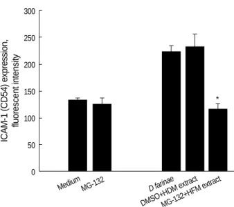
NF- B inhibitor MG-132 suppresses D. farinae extract- induced expression of ICAM-1 in EoL-1 cells
ICAM expression is highly NF- B-dependent (14), and so we investigated the inhibitory effect of NF- B inhibitor MG- 132 on HDM extract-induced upregulation of ICAM-1 in EoL-1 cells. As shown in Fig. 2, pretreatment with 5 M MG-132 resulted in the complete reduction of ICAM exp- ression induced by HDM extract. No cytototoxic effect of MG-132 alone at the concentration tested was observed.
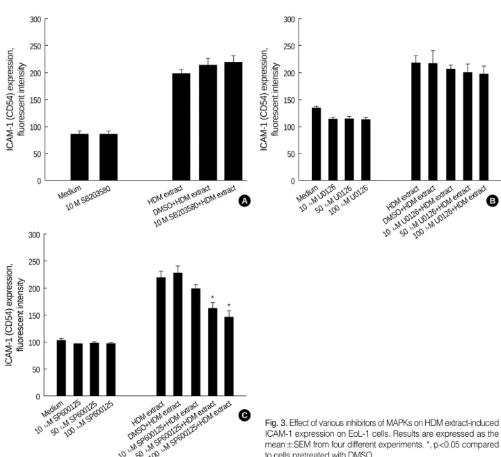
Involvement of JNK, but not p38 or ERK MAPK, in HDM extract-induced expression of ICAM-1 in EoL-1 cells
A recent report demonstrated that MAPKs, including ERK1/2 and JNK, regulate ICAM-1 expression due to stim- ulation by cytokines (15). We examined the role of MAPKs in HDM extract-induced ICAM-1 expression in EoL-1 cells by using p38 MAPK inhibitor (SB203580), MEK 1/2 inhi- bitor (U0126), or JNK inhibitor (SP600125). As shown in Fig. 3A, B, pretreatment with SB203580 (10 M) or U0126 (10-100 M) did not inhibit HDM extract-triggered upreg- ulation of ICAM expression in EoL-1 cells. In contrast, as shown in Fig. 3C, JNK inhibitor SP600125 reduced expres- sion of ICAM-1 by HDM extract in a dose-dependent man- ner.
DISCUSSION
In the present study, we have demonstrated that crude extract of HDM D. farinae activates EoL-1 cells to induce
ICAM-1 expression. Untreated EoL-1 cells expressed low levels of ICAM-1, but HDM extract significantly enhanced the cell-surface expression of ICAM-1 in a dose-dependent manner. In addition, inhibition of NF- B with the proteo- some inhibitor MG-132 almost completely prevented the surface expression of ICAM-1 in EoL-1 cells induced by HDM extract, suggesting a critical role of NF- B in the expres- sion of ICAM-1. This result is in line with previous reports (5, 16) that ICAM-1 expression promoted by cytokines is NF- B dependent in EoL-1 cells and human eosinophils. Mor- eover, it has been reported that MAPKs including ERK1/2, p38, and JNK are involved in ICAM-1 expression in human eosinophils and pulmonary epithelial cells (5, 15). In this study, we found that pretreatment of EoL-1 cells with JNK inhibitor SP600125, but not p38 inhibitor SB203580 or MEK1/2 inhibitor U0126, resulted in significant reduction of ICAM-1 expression stimulated by HDM extract. This suggests that only JNK MAPK is associated with ICAM-1 expression by HDM extract. Taken together, our results suggest that D.
farinae extract induces ICAM-1 expression on EoL-1 cells via signaling pathways of NF- B and JNK MAPK.
ICAM-1 expression on eosinophils is involved in degran- ulation and superoxide anion production (17, 18). A local inc- rease of eosinophils expressing ICAM-1 has been found in inflammatory respiratory diseases such as asthma and idio- pathic eosinophilic pneumonia (19, 20). Eosinophils in the sputum, but not blood, of symptomatic asthmatics not receiv- ing steroid therapy were found to express ICAM-1 (21). There- fore, our findings suggest that D. farinae-induced ICAM-1 ex- pression might contribute to amplification of eosinophil-me- diated tissue inflammation in bronchial asthma.
Evidence indicates that several mite allergens are proteolytic enzymes (serine and cysteine proteases) and that the protease κ
ICAM-1 (CD54) expression, fluorescent intensity 300
250
200
150
100
50
0 Isotype 0 5 10 25 50 100 200 TNF- D. farinae extract ( g/mL)
Fig. 1.Effect of TNF- and HDM extract on the expression of cell surface ICAM-1 on EoL-1 cells. EoL-1 cells were incubated with or without HDM extract (5-200 g/mL) for 4.5 hr. TNF- (20 ng/mL) was used as a positive control. Data are expressed as the mean
±SEM of triplicate experiments. *, p<0.05 compared to cells in- cubated with medium alone.
*
*
* * *
ICAM-1 (CD54) expression, fluorescent intensity 300
250
200
150
100
50
0
Medium MG-132 D farinae
DMSO+HDM extractMG-132+HFM extract Fig. 2.Effect of MG-132 on HDM extract-induced ICAM-1 expres- sion on EoL-1 cells. Results are expressed as the mean±SEM from four different experiments. *, p<0.05 compared to cells pre- treated cells with DMSO.
*
activity of the mite allergens sensitizes immune cells. For example, D. pteronyssinus allergen Der p 1 can induce upreg- ulation of surface expression of ICAM-1 (CD54) on endothe- lial cells (22) and eosinophils (16). In addition, Der p 1 stim- ulates cytokine expression in airway epithelial cells through a protease-activated receptor-2 (PAR-2)-dependent mecha- nism (23). In contrast, human eosinophils are activated by cysteine protease allergen Der f 1 in a PAR-2 driven manner (24). Further studies on this issue will be required to unravel the immunopathological mechanisms of D. farinae-mediat- ed allergic inflammation in bronchial asthma.
In summary, we report that D. farinae extract induces up- regulation of the cell surface adhesion molecule ICAM-1 in EoL-1 cells through JNK and NF- B pathways. Our find- ings may have clinical implications because the exposure to HDM antigens plays an important role in the regulation of eosinophilic inflammation in patients with bronchial asthma.
REFERENCES
1. Bousquet J, Chanez P, Lacoste JY, Barneon G, Ghavanian N, Enan- der I, Venge P, Ahlstedt S, Simony-Lafontaine J, Godard P, et al. Eo- sinophilic inflammation in asthma. N Engl J Med 1990; 323: 1033-9.
2. Jagels MA, Daffern PJ, Zuraw BL, Hugli TE. Mechanisms and reg- ulation of polymorphonuclear leukocyte and eosinophil adherence to human airway epithelial cells. Am J Respir Cell Mol Biol 1999;
21: 418-27.
3. Tang ML, Fiscus LC. Important roles for L-selectin and ICAM-1 in the development of allergic airway inflammation in asthma. Pulm Pharmacol Ther 2001; 14: 203-10.
4. van de Stolpe A, van der Saag PT. Intercellular adhesion molecule- 1. J Mol Med 1996; 74: 13-33.
5. Wong CK, Ip WK, Lam CW. Interleukin-3, -5, and granulocyte macrophage colony-stimulating factor-induced adhesion molecule expression on eosinophils by p38 mitogen-activated protein kinase ICAM-1 (CD54) expression, fluorescent intensity
300
250
200
150
100
50
0
Medium
10 M SB203580 HDM extract DMSO+HDM extract 10 M SB203580+HDM extract
Fig. 3.Effect of various inhibitors of MAPKs on HDM extract-induced ICAM-1 expression on EoL-1 cells. Results are expressed as the mean±SEM from four different experiments. *, p<0.05 compared to cells pretreated with DMSO.
ICAM-1 (CD54) expression, fluorescent intensity 300
250
200
150
100
50
0 Medium
10 M U0126 50 M
U0126 100 M
U0126
HDM extract DMSO+HDM extract 10
M U0126+HDM extract 50
M U0126+HDM extract 100
M U0126+HDM extract
ICAM-1 (CD54) expression, fluorescent intensity 300
250
200
150
100
50
0
Medium 10 M
SP600125 50 M
SP600125 100 M
SP600125 HDM extract DMSO+HDM extract
10
M SP600125+HDM extract 50
M SP600125+HDM extract 100
M SP600125+HDM extract
* *
A
C
B
′
and nuclear factor- B. Am J Respir Cell Mol Biol 2003; 29: 133-47.
6. Cheung PF, Wong CK, Ip WK, Lam CW. IL-25 regulates the ex- pression of adhesion molecules on eosinophils: mechanism of eo- sinophilia in allergic inflammation. Allergy 2006; 61: 878-85.
7. Platts-Mills TA, Vervloet D, Thomas WR, Aalberse RC, Chapman MD. Indoor allergens and asthma: report of the Third International Workshop. J Allergy Clin Immunol 1997; 100: 2-24.
8. Herbert CA, King CM, Ring PC, Holgate ST, Stewart GA, Thomp- son PJ, Robinson C. Augmentation of permeability in the bronchial epithelium by the house dust mite allergen Der p1. Am J Respir Cell Mol Biol 1995; 12: 369-78.
9. Schulz O, Sewell HF, Shakib F. Proteolytic cleavage of CD25, the alpha subunit of the human T cell interleukin 2 receptor, by Der p 1, a major mite allergen with cysteine protease activity. J Exp Med 1998;
187: 271-5.
10. Coward WR, Sagara H, Wilson SJ, Holgate ST, Church MK. Aller- gen activates peripheral blood eosinophil nuclear factor-kB to gen- erate granulocyte macrophage-colony stimulating factor, tumor nec- rosis factor- and interleukin-8. Clin Exp Allergy 2004; 34: 1071-8.
11. Pomerantz JL, Baltimore D. Two pathways to NF- B. Mol Cell 2002;
10: 693-5.
12. Ip WK, Wong CK, Lam CW. Tumour necrosis factor-alpha-induced expression of intercellular adhesion molecule-1 on human eosino- philic leukaemia EoL-1 cells is mediated by the activation of nucle- ar factor-kappaB pathway. Clin Exp Allergy 2003; 33: 241-8.
13. Ree HI, Jeon SH, Lee IY, Hong CS, Lee DK. Fauna and geographi- cal distribution of house dust mites in Korea. Korean J Parasitol 1997; 35: 9-17.
14. Holden NS, Catley MC, Cambridge LM, Barnes PJ, Newton R. IC- AM-1 expression is highly NF-kappaB-dependent in A549 cells. No role for ERK and p38 MAPK. Eur J Biochem 2004; 271: 785-91.
15. Lin FS, Lin CC, Chien CS, Luo SF, Yang CM. Involvement of p42/
p44 MAPK, JNK, and NF- B in IL-1 -induced ICAM-1 expression in human pulmonary epithelial cells. J Cell Physiol 2005; 202: 464- 73.
16. Wong CK, Li ML, Wang CB, Ip WK, Tian YP, Lam CW. House dust mite allergen Der p 1 elevates the release of inflammatory cyto- kines and expression of adhesion molecules in co-culture of human eosinophils and bronchial epithelial cells. Int Immunol 2006; 18:
1327-35.
17. Horie S, Okubo Y, Hossain M, Momose T, Suzuki J, Isobe M, Seki- guchi M. Intercellular adhesion molecule-1 on eosinophils is involved in eosinophil protein X release induced by cytokines. Immunology 1997; 90: 301-7.
18. Takashi S, Okubo Y, Horie S. Contribution of CD54 to human eo- sinophil and neutrophil superoxide production. J Appl Physiol 2001;
91: 613-22.
19. Mengelers HJ, Maikoe T, Brinkman L, Hooibrink B, Lammers JW, Koenderman L. Immunophenotyping of eosinophils recovered from blood and BAL of allergic asthmatics. Am J Respir Crit Care Med 1994; 149: 345-51.
20. Azuma M, Nakamura Y, Sano T, Okano Y, Sone S. Adhesion mo- lecule expression on eosinophils in idiopathic eosinophilic pneumo- nia. Eur Respir J 1996; 9: 2494-500.
21. Hansel TT, Braunstein JB, Walker C, Blaser K, Bruijnzeel PL, Vir- chow JC Jr, Virchow C Sr. Sputum eosinophils from asthmatics ex- press ICAM-1 and HLA-DR. Clin Exp Immunol 1991; 86: 271-7.
22. Mastrandrea F, Nicotra MR, De Vita L, Coradduzza G, Minardi A, Scarcia G, Manelli M, Cadario G, Parmiani S, Natali PG. Mite anti- gens enhance ICAM-1 and induce VCAM-1 expression on human umbilical vein endothelium. Allergol Immunopathol (Madr) 2003;
31: 259-64.
23. Asokananthan N, Graham PT, Stewart DJ, Bakker AJ, Eidne KA, Thompson PJ, Stewart GA. House dust mite allergens induce proin- flammatory cytokines from respiratory epithelial cells: the cysteine protease allergen, Der p 1, activates protease-activated receptor (PAR)-2 and inactivates PAR-1. J Immunol 2002; 169: 4572-8.
24. Miike S, Kita H. Human eosinophils are activated by cysteine pro- tease and release inflammatory mediators. J Allergy Clin Immunol 2003; 111: 704-13.