https://doi.org/10.4174/astr.2017.92.2.55 Annals of Surgical Treatment and Research
A novel panel of serum miR-21/miR-155/miR-365 as a potential diagnostic biomarker for breast cancer
Ji-Guang Han*, Yong-Dong Jiang*, Chun-Hui Zhang1, Yan-Mei Yang2, Da Pang, Yan-Ni Song, Guo-Qiang Zhang
Departments of Breast Surgery and 1Oncology, The Third Affiliated Hospital of Harbin Medical University, Harbin,
2Institute of Cancer Prevention and Treatment, Harbin Medical University, Harbin, China
INTRODUCTION
Breast cancer, one of the most common types of female can
cers, is a highly heterogeneous disease that has multiple sub
types with distinct clinical outcomes and a high fatality rate globally [1]. In clinic, the status/expression level of hormone receptor including estrogen receptor (ER), progesterone re
cep tor (PR), and human EGFlike receptor 2 (HER2) as well as tu mor grade are often used for classification and target ther
apy indicators of breast cancers [1,2]. Although current early detec tion and target therapies based on the measurement of hor mone receptors have remarkably reduced the rate of mor
tality from breast cancer, their application is still limited due to insu fficient sensitivity and specificity as well as the invasive, un pleasant and inconvenient nature of diagnostic procedures.
To identify unique therapeutic targets, it is necessary to develop predictive and prognostic biomarkers that can be conveniently and reliably used in clinic.
Purpose: Insufficient sensitivity and specificity prevent the use of most existing biomarkers for early detection of breast cancer. Recently, it was reported that serum microRNAs (miRNAs) may be potential biomarkers in many cancer diseases.
In this study, we investigated whether serum levels of 5 miRNAs including miR-21, miR-125b, miR-145, miR-155, and miR- 365 could discriminate breast cancer patients and healthy controls.
Methods: Serum levels of miRNAs were measured by using quantitative real-time polymerase chain reaction in 99 breast cancer patients and 21 healthy controls. The abundance change of serum miRNAs were also evaluated following surgical resection in 20 breast cancer patients. Receiver operating characteristic (ROC) curve analysis was performed to assess the sensitivity and specificity of miRNAs as diagnostic biomarkers.
Results: Serum levels of miR-21 and miR-155 was significantly higher, while miR-365 was significantly lower in breast cancer as compared with healthy controls. The serum levels of miR-21 and miR-155 significantly decreased following sur- gical resection. Additionally, the serum level of miR-155 at stages I and II was significantly higher compared to stage III.
The serum miR-145 level was remarkably higher in progesterone receptor (PR)-positive patients than PR-negative. The posi tivity of miR-21, miR-155, and miR-365 was high compared to CA 153 and CEA in breast cancer. ROC curve analyses of a combination of miR-21, miR-155, and miR-365 yielded much higher area under curve and enhanced sensitivity and specificity in comparison to each miRNA alone.
Conclusion: The combination of serum miR-21/miR-155/miR-365 may potentially serve as a sensitive and specific biomarker that enables differentiation of breast cancer from healthy controls.
[Ann Surg Treat Res 2017;92(2):55-66]
Key Words: Serum, MicroRNAs, miRNA-21, miRNA-155, miRNA-365, Breast neoplasms
Received July 5, 2016, Reviewed July 16, 2016, Accepted September 4, 2016
Corresponding Author: Guo-Qiang Zhang
Department of Breast Surgery, The Third Affiliated Hospital of Harbin Medical University, No 150, Haping Road, Harbin 150040, China
Tel: +86-451-86298060, Fax: +86-451-86298115 E-mail: [email protected]
*Ji-Guang Han and Yong-Dong Jiang contributed equally to this study as co- first authors.
Copyright ⓒ 2017, the Korean Surgical Society
cc Annals of Surgical Treatment and Research is an Open Access Journal. All articles are distributed under the terms of the Creative Commons Attribution Non- Commercial License (http://creativecommons.org/licenses/by-nc/4.0/) which permits unrestricted non-commercial use, distribution, and reproduction in any medium, provided the original work is properly cited.
MicroRNAs (miRNAs), a class of naturally occurring, small and noncoding RNA molecules with a length of 19–25 nucleo
tides, can specifically regulate gene mRNA expressions at post
transcriptional levels [3]. The dysregulation of miRNAs may affect some crucial biological processes of cells leading to tumor development by increasing proliferation, decreasing apoptosis, and enhancing the metastatic potential [3,4]. Recently, it has been reported that miRNAs expression level/profile is altered in various cancers exhibiting great potential in improving the diagnosis and prognosis of cancer [5]. Notably, it has been documented that certain secreted miRNAs originating from cancer tissues are protected from endogenous RNase by some unknown mechanisms [6], and thus can be detected in blood and other body fluids [7,8]. In breast cancer tissues, miRNAs have been shown to function as either oncogenes or tu mor suppressors [9], and circulating miRNAs may correlate with disease progression, therapeutic responses and patient sur
vival [10,11], suggesting that the evaluation of miRNAs levels in the blood, either serum or plasma, may be used as non
invasive bloodbased biomarkers. Even though many studies have compared changes of miRNAs levels in blood between breast cancer patients and healthy controls in order to map the profiling of miRNAs and identify some specific miRNAs as potential biomarkers in breast cancer [12], the results are not always consistent due to the differences in study design, such as sample size, patient source and characteristics, and RNA preparation, as well as detection methods or profiling platforms that were used.
In our study, the serum levels of 5 miRNAs including miR
21, miR155, miR125b, miR145, and miR365 that have been indicated as a recurrent presence in breast cancer patients [12,13], were compared between breast cancer patients and healthy controls. We found that the serum level of miR21 was significantly higher, while miR155 and miR365 was signifi
cantly lower in breast cancer than healthy control, and receiver operating curve (ROC) analyses showed that the combination of miR21/miR155/miR365 led to higher sensitivity and specificity in distinguishing breast cancer from healthy controls, indicating a potential as biomarkers for the diagnosis and monitoring of breast cancer.
METHODS
Patients
Blood samples were collected from 99 patients with breast cancer (range, 31–77 years; mean, 48.95 years) and 21 agemat
ched healthy female volunteers (range, 35–59 years; mean, 45.38 years; without current or previous malignancy or inflammatory condition). In addition, the paired blood samples were collected from a 20patient subset both before and 3 weeks after breast cancer surgery. All participants had signed an informed consent
to participate in this study. The study was approved by the Ethics Review Board of Harbin Medical University.
All patients’ breast cancer was histologically confirmed. The surgical patients’ clinicopathological and relevant demographic characteristics were documented in our prospectively main
tained breast cancer database. The clinical stage of breast cancer of all patients was classified according to the TNM classification system of the American Joint Committee on Cancer. The number of various groups’ breast cancer patients is summarized in Table 1.
Serum preparation
A 5mL sample of the whole blood was collected in a Vacu
tainer Serum Separator Tube (Becton Dickinson, Franklin Lakes, NJ, USA). The blood was left to clot at room temperature for 30 minutes, and then centrifuged at 2,000 rpm for 10 minutes at 4oC. The resulting serum was collected, aliquoted, and stored at –80oC.
RNA extraction
Total RNA was extracted from 300 mL of serum using the mirVana PARIS Kit (Ambion, Carlsbad, CA, USA), and finally eluted into 100 mL of preheated (95oC) Elution Solution ac
cording to the manufacturer’s protocol. The eluate was then collected and stored at –20oC. RNA concentration and inte
grity were determined using NanoDrop spectrophotometry
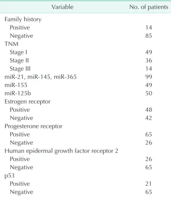
Table 1. The number of patients with breast cancer in va
rious groups (n = 99)
Variable No. of patients
Family history
Positive 14
Negative 85
TNM
Stage I 49
Stage II 36
Stage III 14
miR21, miR145, miR365 99
miR155 49
miR125b 50
Estrogen receptor
Positive 48
Negative 42
Progesterone receptor
Positive 65
Negative 26
Human epidermal growth factor receptor 2
Positive 26
Negative 65
p53
Positive 21
Negative 65
(NanoDrop ND1000; NanoDrop Technologies, Wilmington, DE, USA). The RNA concentration (mg/mL) was calculated by the value of OD260 (OD260 × dilution factor × 40), and adjusted to 0.002 mg/mL. The total RNA was stored at –80oC.
RT and real-time qPCR
The level of miRNAs was quantified in duplicate using quantitative reverse transcription polymerase chain reaction
(RTPCR) and human TaqMan MicroRNA Assay Kit (Applied Biosystems, Foster City, CA, USA). The RT reaction was carried out in a 15mL TaqMan MicroRNA Reverse Transcription System containing 5 mL of RNA extract, 0.15 mL of 100mM dNTPs, 1 mL of Multiscribe Reverse Transcriptase (50 U/mL), 1.5 mL of 10 × RT buffer, 0.19 mL of RNase inhibitor (20 U/mL), 1 mL of gene
specific primer and 4.16 mL of nucleasefree water. For cDNA synthesis, the above mixtures were incubated at 16oC for 30
miRNAlevel(Log2)10 CT
A B
Healthy (n = 21)
P < 0.0001 4
2 0 2
4 Cancer (n = 99)
miRNA21
miRNAlevel(Log2)10 CT
Healthy (n = 21)
P = 0.4414 2
1 0
2
3 Cancer (n = 50)
miRNA125b
1
C D
miRNAlevel(Log2)10 CT
Healthy (n = 21)
P = 0.2129 4
2 0 2
6 Cancer (n = 99)
miRNA145
miRNAlevel(Log2)10 CT
Healthy (n = 21)
P = 0.0005 2
0
Cancer (n = 49) miRNA155
4
2
6 4
E
miRNAlevel(Log2)10 CT
Healthy (n = 21)
P < 0.0001 4
2 0 2
Cancer (n = 99) miRNA365
6 4
Fig. 1. The serum abundance of microRNAs (miRNAs) in breast cancer patients and healthy controls. Total RNA was extracted from serum and quantitative polymerase chain reaction was per formed for evaluation of miRNA abundance.
As compared with healthy controls, the serum levels of miR
21 (A) and miR155 (D) were significantly higher, while miR
365 (E) was significantly lower in breast cancer patients.
The serum con centrations of miR125b (B) and miR365 (C) showed no difference between breast cancer patients and healthy con trols. Data are expressed as mean ± standard deviation.
minutes, 42oC for 30 minutes, and 85oC for 5 minutes.
A 4mL cDNA solution was amplified using 10 mL of TaqMan Gene Expression Master Mix, 1 mL of genespecific primers/
probe and 5 mL of nucleasefree water in a final volume of 20 mL. Quantitative PCR was performed on a 7000 RealTime PCR system (Applied Biosystems) by incubation at 95oC for 10 minutes, and 40 cycles of 95oC for 15 seconds and 60oC for 1 minute. The values of cycle threshold (Ct) were calculated with
SDS 1.4 software (Applied Biosystems).
Measurement of hormone status, p53, CA 153, and CEA
The levels of CA 153 and CEA were measured through electrochemiluminescence assays, and the level of hormone status including ER, PR, HER2, and p53 was measured by immuno
histochemistry assay in the Laboratory Department of the
miRNAlevel(Log2)10 CT
A B
Negative (n = 85)
P = 0.964 4
2
0
2 Positive (n = 14)
miRNA21
miRNAlevel(Log2)10 CT
P = 0.942 2
1 0
2 3
miRNA125b
1
C D
miRNAlevel(Log2)10 CT
P = 0.909 4
2 0 2
6
miRNA145
miRNAlevel(Log2)10 CT
P = 0.760 2
0
miRNA155
4
2
6 4
E
miRNAlevel(Log2)10 CT
P = 0.792 4
2 0 2
miRNA365
6 4
Positive (n = 6) Negative (n = 44)
Negative (n = 85) Positive (n = 14) Negative (n = 41) Positive (n = 8)
Negative (n = 85) Positive (n = 14)
Fig. 2. Effects of family history on the serum microRNAs (miRNAs) level in breast cancer patients. The serum level of miRNAs was assessed in family history positive and negative breast cancer patients. Family history showed no effects on serum miRNAs level in breast cancer patients. Data are expressed as mean ± standard deviation.
Third Affiliated Hospital of Harbin Medical University.
Statistical analysis
The unpaired ttest, a twotailed MannWhitney test, was used to compare the differential expression of serum miRNAs
between breast cancer and normal samples, between family history positive and negative patients, and between the patients with ER (or PR, HER2, p53) positive and negative. The paired ttest was used for comparison of serum miRNAs level between pre and postoperative samples in breast cancer. The oneway
miRNAlevel(Log2)10 CT
A B
4 2 0
4
miRNA21
miRNAlevel(Log2)10 CT
2 1 0
2 3
miRNA125b
1
C D
miRNAlevel(Log2)10 CT
4 2 0 2
6
miRNA145
miRNAlevel(Log2)10 CT
2 0
miRNA155
4
2
6 4
E
miRNAlevel(Log2)10 CT
4 2 0 2
miRNA365
6 4 2
P < 0.01
Healthy
(n = 21) Stage 1
(n = 49) Stage 2
(n = 36) Stage 3 (n = 14) NS
NS
Healthy
(n = 21) Stage 1
(n = 25) Stage 2
(n = 13) Stage 3 (n = 12)
NS
Healthy
(n = 21) Stage 1
(n = 49) Stage 2
(n = 36) Stage 3 (n = 14)
P < 0.05
Healthy
(n = 21) Stage 1
(n = 23) Stage 2
(n = 18) Stage 3 (n = 8) P < 0.05
P < 0.05
Healthy
(n = 21) Stage 1
(n = 49) Stage 2
(n = 36) Stage 3 (n = 14) NS
Fig. 3. Comparisons of the serum microRNAs (miRNAs) level in breast can cer patients across different TNM stages.
The serum levels of miR21 (A), miR125b (B), miR145 (C), and miR365 (E) showed no difference across different TNM stages. Compared to stages I and II, the serum miR
155 level was lower in breast cancer patients at stage III (D).
In comparison with healthy controls, the miR21 level was remarkably higher in breast cancer patients at any TNM stage (A), miR155 was significantly higher at stages I and II (D), whereas miR365 was significantly lower at stages I and III (E).
Data are expressed as mean ± standard deviation. NS, not significant.
A B miRNA125b
C miRNA145 miRNA155
E miRNA365
miRNAlevel(Log2)10 CT
5 4 3 2 1 0
3
miRNA21
2
0.598
ER 42 1
ER+
48 PR
26 PR+
65 HER2
65 HER2+
26 p53
65 p53+
21 (n) 0.272 0.295 0.110 (P)
miRNAlevel(Log2)10 CT
4 3 2 1 0
4 2
0.373
ER 20 1
ER+
26 PR
17 PR+
32 HER2
15 HER2+
26 p53
35 p53+
10 (n) 0.546 0.778 0.117 (P)
miRNAlevel(Log2)10 CT
0.382
ER 42 5.0 2.5 0.0 2.5
5.0
ER+
48 PR
26 PR+
HER2 HER2+ p53 65
p53+
21 (n) 26
65
miRNAlevel(Log2)10 CT
4 2 0
6 4
0.632
ER 42 2
ER+
48 PR
26 PR+
25 HER2
65 HER2+
26 p53
65 p53+
21 (n) 0.471 0.579 0.865 (P)
miRNAlevel(Log2)10 CT
4 2 0
6 4
0.772
ER 22 2
ER+
22 PR
9 PR+
35 HER2
33 HER2+
11 p53
30 p53+
11 (n) 0.905 0.342 0.193 (P)
65
3
0.041 0.750 0.390 (P)
Positiverate(%)
80 60 40 20
0 miR-21 miR-155 miR-365 CEA CA153
F D
Fig. 4. Comparisons of the serum miRNAs levels in breast cancer patients with different hormone status. (AE) The serum levels of miRNAs were compared in various groups of breast cancer patients classified by hormone status including estrogen receptor (ER), progesterone receptor (PR), and human epidermal growth factor receptor 2 (HER2) as well as p53. The serum level of miR145 was significantly higher in PRpositive patients as compared with PR negative. The number for each group was indicated in the images. Data are expressed as mean ± standard deviation. (F) The positive rates of miR21, miR155, and miR365 as well as CEA and CA153 in breast cancer patients. Positivity of the three miRNAs was determined based on the confidence intervals (CI). The value of miR21 and miR155 level ≥ the CI, and the value of miR365 level ≤ the CI, was considered positive.
analysis of variance was used to compare the differential serum miRNAs level between normal and breast cancer patients at different TNM stages. ROCs were generated using logistic regression models, to evaluate the diagnostic performance of various miRNAs and a combination of miRNAs. Area under curve (AUC) was used as the evaluation criteria; the higher AUC, the better diagnostic performance.
All analysis was performed using SAS 9.5 (SAS Institute Inc., Cary, NC, USA) and GraphPad Prism ver. 6 (GraphPad Software Inc., La Jolla, CA, USA). The P < 0.05 was considered as significant difference.
RESULTS
Serum miRNAs levels in breast cancer and healthy samples
It is difficult to extract RNA from serum due to its low abun
dance. In our experiments, the average concentration of the total RNA from 300 mL of serum was 0.118 mg/mL ranging from 0.042 to 0.208 mg/mL. As compared with normal samples, the
level of miR21 and miR155 was significantly higher (miR21:
0.86 ± 0.941 vs. –2.74 ± 1.055, P <0.0001; miR155: –1.24 ± 1.022 vs. –2.01 ± 0.808, P = 0.0005), while miR365 was significantly lower (–0.84 ± 0.873 vs. –0.24 ± 0.317, P < 0.0001) in breast cancer patients’ serum. The level of serum miR125b and miR
145 showed no significant difference between breast cancer and normal samples (Fig. 1).
Effects of family history and TNM stage on serum miRNAs level in breast cancer patients
We compared the serum level of the five miRNAs in breast cancer patients with or without family history. The five miRNAs levels were not significantly different between family history positive and negative patients with breast cancer (Fig. 2).
The serum levels of miR21, miR125b, miR145, miR155, and miR365 in breast cancer patients at different TNM stages were also evaluated to determine if the serum miRNAs could be de tected in earlystage breast cancer (Fig. 3). Across 3 stages, the serum levels of miR21, miR125b, miR145, and miR365 showed no marked difference. In comparison to stages I and II, A
miRNA21level(Log2)10 CT
4 3 2 1 0
4 2 1
miRNA21level(Log2)10 CT
4 2 0
4 2
P = 0.0005 Preoperation
Postoperation
20 3
Preoperation Postoperation 19
18 17 16 15 14 13 12 11 10 9 8 7 6 5 4 3 2 1
B
miRNA21level(Log2)10 CT
2 1 0
5 2 1
miRNA21level(Log2)10 CT
2 0
6 2
P = 0.0011 Preoperation
Postoperation
20 3
Preoperation Postoperation 19
18 17 16 15 14 13 12 11 10 9 8 7 6 5 4 3 2 1 4
4
Fig. 5. Alteration of the serum miR21 and miR155 levels in breast cancer patients receiving surgery. Total serum RNA was ex
tracted preoperation and postoperation in 20 breast cancer patients, and quantitative polymerase chain reaction was performed for evaluation of changes of miRNAs levels. In comparison with preoperation, the serum levels of miR21 (A) and miR155 (B) were significantly reduced 3 weeks postsurgery in breast cancers. Data are expressed as mean ± standard deviation.
the serum miR155 level was remarkably lower (stage III: –2.40
± 1.151 vs. stage I: –1.03 ± 0.790, stage II: –1.00 ± 0.919, P <
0.05) in breast cancer patients at stage III. Notably, compared to normal samples, the serum level of miR21 was significantly higher (stage I: 0.80 ± 0.980, stage II: 0.92 ± 0.994, stage III: 0.87
± 0.674 vs. healthy controls: –0.27 ± 1.055; P < 0.01) in breast can cer at any TNM stage. Nevertheless, in comparison with nor mal controls, the serum level of miR365 was significantly lower (stage I: –0.92 ± 0.753, stage III: –0.97 ± 1.043 vs. healthy con trol: –0.24 ± 0.317; P < 0.05) at both stages I and III.
Comparison of serum miRNAs levels in various groups of breast cancer patients
We compared the serum levels of miR21, miR125b, miR
145, miR155, and miR365 in various groups of breast cancer patients classified by hormone status including ER, PR, and HER2 as well as p53 (Fig. 4). We only detected that the serum level of miR145 was significantly higher (–0.46 ± 0.953 vs.
–0.66 ± 0.777, P = 0.041) in PRpositive patients as compared with PR negative (Fig. 4C). Additionally, in breast cancer pa
tients, the positivity of miR21, miR155 and miR365 was 58%,
0 100
Sensitivity%
100%-Specificity%
0
Sensitivity%
Identity%
100 80 60 40 20
A B
C D
E
80 60
40 20
miRNA21 AUC area: 0.788 Std. Error: 0.055 95% CI: 0.681 to 0.895 P < 0.0001
0 100
Sensitivity%
100%-Specificity%
0 100 80 60 40 20
80 60
40 20
miRNA125b AUC area: 0.559 Std. Error: 0.074 95% CI: 0.413 to 0.705 P = 0.435
0
0
100
100
Sensitivity%Sensitivity%
100%-Specificity%
100%-Specificity%
0
0 100
100 80
80 60
60 40
40 20
20
80
80 60
60 40
40 20
20
miRNA145 AUC area: 0.587 Std. Error: 0.064 95% CI: 0.462 to 0.763 P = 0.210
miRNA365 AUC area: 0.795 Std. Error: 0.044 95% CI: 0.708 to 0.881 P < 0.0001
0 100
Sensitivity%
100%-Specificity%
0 100 80 60 40 20
80 60
40 20
miRNA155 AUC area: 0.749 Std. Error: 0.056 95% CI: 0.639 to 0.858 P = 0.0007
Sensitivity%
Identity%
Sensitivity%
Identity%
Sensitivity%
Identity%
Sensitivity%
Identity%
Fig. 6. Area under curve (AUCs) of receiver operating char
ac teristic (ROC) for serum miRNAs levels. The three miRNAs in cluding miR21 (A), miR155 (D), and miR365 (E) show higher sensitivity and specificity than miR125b (B) and miR
145 (C). The AUC area, standard (Std.) error, 95% confidence in terval (CI), and the Pvalues are indicated in the images.
48%, and 59%, respectively. Nevertheless, the positivity of CA 153 and CEA was 33% and 20%, respectively (Fig. 4F).
Comparison of serum miRNAs levels before and after surgery in breast cancer patients
The serum level of miRNAs was analyzed in the paired pre
and postoperative samples from 20 patients with breast cancer.
As compared with the preoperative samples, the level of miR
21 and miR155 decreased significantly (miR21: –0.01 ± 1.318 vs. 1.31 ± 1.082, P = 0.0005; miR155: –1.98 ± 0.944 vs. –0.89 ± 0.921, P = 0.0011) in the postoperative samples (Fig. 5).
ROC curve analysis
ROC curve analysis was performed to evaluate the diagnostic value for the five miRNAs. The AUCs closer to 1 reflect more substantial differences between breast cancer and normal sam
ples. ROC curve analysis revealed that the three miRNAs, miR
21, miR155, and miR365, had significantly higher AUCs with
values of 0.788, 0.749, and 0.795, respectively (Fig. 6, Table 2). At the optimal cutoff value of –0.089, with the values of sensitivity plus specificity considered to be maximal for miR21, the sensitivity and specificity were 66.67% and 88.89%, respectively.
At the optimal cutoff value of –1.171 for miR155, the sensitivity and specificity were 100% and 51.02%, respectively. At the optimal cutoff value of –0.4722 for miR365, the sensitivity and specificity were 85.71% and 72.73%, respectively.
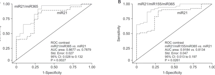
We further performed ROC curve analyses for combinations of miR21, miR155, and miR365. Compared to miR21 alone, the combination of miR21/miR365 yielded higher AUC (0.8677 vs. 0.7879, P = 0.0027), and better sensitivity (96.99%) and specificity (66.67%) (Fig. 7A). A combination of the three miRNs also created higher AUC (0.9184 vs. 0.8134; P = 0.0261), and better sensitivity (85.71%) and specificity (85.72%) (Fig. 7B).
DISCUSSION
Endocrine treatment is conventionally used for ER+ patients, and trastuzumab for HER2+ patients [14,15]. Although these biomarkers are commonly used in clinic for target therapy and management of breast cancer, there is still room for improve
ment, such as early diagnosis of tumor lesions, identification of highrisk patients, development of predictive biomarkers for monitoring progress and therapy effects, etc. By mapping the circulating miRNAs signature and profiling, some miRNAs have been found differentially expressed in breast cancer and normal tissues [16]. Although the clinical application of serum miRNAs as a noninvasive diagnostic strategy is promising, the miRNA signatures should be further investigated and validated for different subtypes of breast cancers. In the current study, we analyzed the serum level of five miRNAs including miR21, miR125b, miR145, miR155, and miR365 in breast cancers and
1.00
Sensitivity
A B
0
1-Specificity 0
1.00 0.75 0.50 0.25
miR21
ROC contrast
miR21/miR365 miR21 AUC area: 0.8677 0.7879 Std. Error: 0.027
95% CI: 0.028 to 0.132 P = 0.0027
vs.
vs.
0.75 0.50
0.25 miR21/miR365
1.00
Sensitivity
0
1-Specificity 0
1.00 0.75 0.50 0.25
miR21
ROC contrast
miR21/miR155/miR365 miR21 AUC area: 0.9184 0.8134 Std. Error: 0.047
95% CI: 0.013 to 0.197 P = 0.0261
vs.
vs.
0.75 0.50
0.25
miR21/miR155/miR365
Fig. 7. Area under curve (AUCs) of receiver operating characteristic (ROC) for combination of microRNAs (miRNAs). The combination of miR21 and miR365, as well as miR21, miR155 and miR365, shows higher sensitivity and specificity than each miRNA alone, e.g. miR21. The AUC area, standard (Std.) error, 95% confidence interval (CI), and the Pvalues are indicated in the images.
Table 2. The area under curve, sensitivity and specificity of miRNAs
miRNAs AUCs Sensitivity
(%) Specificity
(%) Pvalue
miR21 0.788 66.67 88.89 <0.001*
miR125b 0.559 0.435
miR145 0.587 0.210
miR155 0.749 100 51.02 <0.001*
miR365 0.795 85.71 72.73 <0.001*
miR21/miR155 0.868 96.99 66.67 0.003*
miR21/miR155/
miR365 0.918 85.71 85.72 0.026*
AUC, area under curve; miRNA, microRNA.
*P < 0.05, statistically significant.
healthy controls, followed by construction of ROC curves to determine the sensitivity and specificity of circulating miRNAs as potential biomarkers for breast cancer.
Our results showed that the serum concentrations of miR
21 and miR155 were significantly elevated, while miR365 was significantly downregulated in patients with breast cancer compared with healthy controls (Fig. 1). MiR21 is involved in regulating the expression of multiple tumor suppressor genes, and plays a crucial role in the occurrence and development of a variety of cancer diseases [17]. In breast cancer, several studies have shown that upregulation of miR21 was detected in both serum and tissues [18], and that serum miR21 could be used as an noninvasive biomarker for diagnosis and prognosis of breast cancer as well as an indicator for the invasiveness of the tumor [19]. MiR155 plays a role in various physiological and pathological processes, and oversilencing by miR155 may result in apoptotic resistance and thus triggering oncogenic cascades.
It has been reported that the amount of both circulating miR
155 in serum and noncirculating miR155 in tissue were elevated in patients with breast cancer [20,21]. Therefore, miR
21 and miR155 may act as oncogenic factors, and thus may be potential targets for breast cancer therapy. Additionally, we found that miR155 level was remarkably higher at stages I and II compared to stage III in breast cancer patients (Fig. 3).
Similarly, it has been shown that miR7 was associated with breast cancer grades [22]. Reduction of tissue miR365 has been reported in different types of cancers such as hepatocellular carcinoma (HCC) [23] and lung cancer [24]. An in vitro experi
ment showed that overexpression of miR365 remarkably sup
pressed proliferation and migration capacities of HCC cell [23].
Kodahl et al. [25] also showed that the serum miR365 was downregulated in early stage breast cancer patients. Family history is an important predisposing factor in breast cancer.
How ever, we did not find differential expression of the five miRNAs between family history positive and negative breast cancer patients (Fig. 2).
In this study, the serum abundance of miR125b and miR145 displayed no remarked difference between breast cancer and healthy control (Fig. 1). Nevertheless, by comparing the miRNAs profile between 61 breast cancers and 10 healthy controls in the Mexican population, it was found that the serum levels of miR
145 and miR125b were significantly higher in patients with breast cancer than healthy controls [26]. The current analysis just found that serum miR145 abundance was significantly higher in PRpositive breast cancer patients compared to the PRnegative (Fig. 4C), indicating that serum miR145 level may separate the PRpositive subtypes of breast cancer. A previous study also examined the association of miRNAs expression in serum with different tumor hormone status, and they found 7 miRNAs with differential expression for women whose breast cancer differed by HER2 expression [27]. Notably, it was
reported that the level of miR145 was downregulated both in the in vitro cultured breast cancer cell lines [28] and in the serum from early stage breast cancer patients [25]. CA153 and CEA are the most widely used circulating biomarkers in moni
toring patients with breast cancer. Nevertheless, we found that the positivity of miR21, miR155 and miR365 was higher in comparison with CEA and CA 153 in breast cancer patients (Fig. 4F). Similarly, serum miR21 and miR30a has a higher sensitivity in diagnosis of breast cancer compared with CEA and CA153 [29,30].
The serum level of miR21 and miR155 was further analyzed in 20 patients with breast cancer before surgery and 3 weeks after tumor removal. The abundance of serum miR21 and miR155 was significantly reduced in the postoperative sam
ples as compared with the paired preoperative samples (Fig.
5). Consistently, Sochor et al. [20] found that early breast cancer patients significantly overexpressed several oncogenic miRNAs such as miR155, miR181b, miR19a, and miR24, which dramatically decreased following surgical resection.
Kodahl et al. [25] reported that circulating miR3383p, miR223, and miR148a exhibited lower, and miR107 exhibited higher levels postoperatively than the preoperative samples from 24 postmenopausal women with ER+ earlystage breast cancer.
These findings suggest that serum oncogenic miRNAs may be potentially used for diagnostic purpose and relapse judgement.
To determine the diagnostic performance of miRNAs for breast cancer, we performed ROC curve analysis, and the AUC was used as the evaluation criteria; the higher AUC, the better diag nostic performance. Our data revealed that the 3 miRNAs miR21, miR155, and miR365 had significantly higher AUC with the values of 0.788, 0.749, and 0.795, respectively (Fig.
6), suggesting that we were able to discriminate breast cancer from healthy controls. The sensitivity for the three miRNAs (miR21, miR155, and miR365) was 66.7%, 100%, and 85.7%, respec tively, and the specificity was 88.9%, 51.02%, and 72.7%, respectively (Fig. 6, Table 2). It has been reported that a miRNA panel could accurately distinguish cancers from normal subjects [4,7,8,11,12]. A combination of ROC curve analyses of miR145, miR155, and miR382 exhibited much better sensitivity and specificity than each miRNA alone [30]. In this study, ROC curve was analyzed in a panel of 3 miRNAs (miR21, miR155, and miR365). Our data demonstrate that a combination of miR21 and miR365 yielded a significantly high AUC (0.868), sensitivity (96.97%), and specificity (66.67%). The combination of miR21, miR155, and miR365 further generated much higher AUC (0.918), accompanied by relatively higher sensitivity (85.71) and specificity (85.72) (Fig. 7, Table 2). Accordingly, our results implied that a panel of miRNAs (e.g., a combination of miR
21/miR155/miR365) remarkably enhanced the diagnostic performance with high sensitivity and specificity, and thus may provide an improved indicator for breast cancer diagnosis and
screening.
Taken together, our study provides evidence that evaluation of serum miRNAs level can be used as biomarkers for diagnosis of breast cancer, and that a combination of miR21/miR155/miR
365 may potentially serve as a sensitive and specific biomarker that enables the differentiation of breast cancer from healthy controls.
CONFLICTS OF INTEREST
No potential conflict of interest relevant to this article was reported.
1. Schettini F, Buono G, Cardalesi C, Desideri I, De Placido S, Del Mastro L. Hormone Receptor/Human Epidermal Growth Factor Receptor 2positive breast cancer:
Where we are now and where we are going. Cancer Treat Rev 2016;46:206.
2. Duffy MJ. Role of tumor markers in pa
tients with solid cancers: a critical review.
Eur J Intern Med 2007;18:17584.
3. Pileczki V, CojocneanuPetric R, Maralani M, Neagoe IB, Sandulescu R. MicroRNAs as regulators of apoptosis mechanisms in cancer. Clujul Med 2016;89:505.
4. Solomides CC, Evans BJ, Navenot JM, Vadigepalli R, Peiper SC, Wang ZX. Micro
RNA profiling in lung cancer reveals new mole cular markers for diagnosis. Acta Cytol 2012;56:64554.
5. Markou A, Sourvinou I, Vorkas PA, Yousef GM, Lianidou E. Clinical evaluation of microRNA expression profiling in non small cell lung cancer. Lung Cancer 2013;
81:38896.
6. Toiyama Y, Takahashi M, Hur K, Nagasaka T, Tanaka K, Inoue Y, et al. Serum miR21 as a diagnostic and prognostic biomarker in colorectal cancer. J Natl Cancer Inst 2013;105:84959.
7. Liu R, Chen X, Du Y, Yao W, Shen L, Wang C, et al. Serum microRNA expression pro
file as a biomarker in the diagnosis and prognosis of pancreatic cancer. Clin Chem 2012;58:6108.
8. MarAguilar F, LunaAguirre CM, Moreno
Rocha JC, AraizaChavez J, Trevino V, RodriguezPadilla C, et al. Differential ex pres sion of miR21, miR125b and miR
191 in breast cancer tissue. Asia Pac J Clin
Oncol 2013;9:539.
9. Yan LX, Huang XF, Shao Q, Huang MY, Deng L, Wu QL, et al. MicroRNA miR21 overexpression in human breast cancer is associated with advanced clinical stage, lymph node metastasis and patient poor prognosis. RNA 2008;14:234860.
10. Iorio MV, Ferracin M, Liu CG, Veronese A, Spizzo R, Sabbioni S, et al. MicroRNA gene ex pression deregulation in human breast cancer. Cancer Res 2005;65:706570.
11. Bertoli G, Cava C, Castiglioni I. Micro
RNAs: new biomarkers for diagnosis, pro
g nosis, therapy prediction and therapeutic tools for breast cancer. Theranostics 2015;
5:112243.
12. Ravelli A, Reuben JM, Lanza F, Anfossi S, Cappelletti MR, Zanotti L, et al. Breast can cer circulating biomarkers: advantages, draw backs, and new insights. Tumour Biol 2015;36:665365.
13. AlKhanbashi M, AlMoundhri M. Micro
ribo nucleic acid and carcinogenesis:
breast cancer as an example. Oncol Rev 2015;9:279.
14. Fatima S, Faridi N, Gill S. Breast cancer:
steroid receptors and other prognostic in
di cators. J Coll Physicians Surg Pak 2005;
15:2303.
15. Stark AT, Claud S, Kapke A, Lu M, Linden M, Griggs J. Race modifies the asso ciation bet ween breast carcinoma path ologic pro
g nostic indicators and the posi tive sta tus for HER2/neu. Cancer 2005;104:218996.
16. Graveel CR, Calderone HM, Westerhuis JJ, Winn ME, Sempere LF. Critical analysis of the potential for microRNA biomarkers in breast cancer management. Breast Cancer
(Dove Med Press) 2015;7:5979.
17. Huang Y, Yang YB, Zhang XH, Yu XL, Wang ZB, Cheng XC. MicroRNA21 gene and cancer. Med Oncol 2013;30:376.
18. Savad S, Mehdipour P, Miryounesi M, Shirkoohi R, Fereidooni F, Mansouri F, et al. Expression analysis of MiR21, MiR
205, and MiR342 in breast cancer in Iran.
Asian Pac J Cancer Prev 2012;13:8737.
19. Heneghan HM, Miller N, Lowery AJ, Sweeney KJ, Newell J, Kerin MJ. Cir
culating microRNAs as novel mini mally inva sive biomarkers for breast can cer.
Ann Surg 2010;251:499505.
20. Sochor M, Basova P, Pesta M, Dusilkova N, Bartos J, Burda P, et al. Oncogenic microRNAs: miR155, miR19a, miR181b, and miR24 enable monitoring of early breast cancer in serum. BMC Cancer 2014;
14:448.
21. Mattiske S, Suetani RJ, Neilsen PM, Callen DF. The oncogenic role of miR
155 in breast cancer. Cancer Epidemiol Biomarkers Prev 2012;21:123643.
22. Lyng MB, Lænkholm AV, Sokilde R, Gravgaard KH, Litman T, Ditzel HJ. Global microRNA expression profiling of high
risk ER+ breast cancers from pa tients re ceiving adjuvant tamoxifen mono
the rapy: a DBCG study. PLoS One 2012;7:
e36170.
23. Chen Z, Huang Z, Ye Q, Ming Y, Zhang S, Zhao Y, et al. Prognostic significance and antiproliferation effect of microRNA365 in hepatocellular carcinoma. Int J Clin Exp Pathol 2015;8:170511.
24. Kang SM, Lee HJ, Cho JY. MicroRNA365 regulates NKX21, a key mediator of lung
REFERENCES
cancer. Cancer Lett 2013;335:48794.
25. Kodahl AR, Lyng MB, Binder H, Cold S, Gravgaard K, Knoop AS, et al. Novel cir
culating microRNA signature as a po ten
tial noninvasive multimarker test in ER
positive earlystage breast cancer: a case control study. Mol Oncol 2014;8:87483.
26. MarAguilar F, MendozaRamirez JA, MalagonSantiago I, EspinoSilva PK, SantuarioFacio SK, RuizFlores P, et al.
Serum circulating microRNA profiling for
iden tification of potential breast cancer biomarkers. Dis Markers 2013;34:1639.
27. Godfrey AC, Xu Z, Weinberg CR, Getts RC, Wade PA, DeRoo LA, et al. Serum microRNA expression as an early marker for breast cancer risk in prospectively collected samples from the Sister Study cohort. Breast Cancer Res 2013;15:R42.
28. Polytarchou C, Iliopoulos D, Struhl K.
An integrated transcriptional regulatory circuit that reinforces the breast cancer
stem cell state. Proc Natl Acad Sci U S A 2012;109:144705.
29. Zeng RC, Zhang W, Yan XQ, Ye ZQ, Chen ED, Huang DP, et al. Downregulation of miRNA30a in human plasma is a novel marker for breast cancer. Med Oncol 2013;
30:477.
30. Gao J, Zhang Q, Xu J, Guo L, Li X. Clinical significance of serum miR21 in breast can cer compared with CA153 and CEA.
Chin J Cancer Res 2013;25:7438.