Original Article
Diagnostic and clinicopathological significance of Ki67
mRNA expression in cervical cancer tissue
Dawn Chung1,2, Sunyoung Park3, Geehyuk Kim3, Hye-Young Wang4, Kwang Hwa Park5, Sang Wun Kim6, Sunghoon Kim6, Young Tae Kim6, Hyeyoung Lee3, Eun Ji Nam6
1Department of Obstetrics and Gynecology, Yonsei University Wonju College of Medicine, Wonju, Korea; 2Department of Obstetrics and Gynecology, Yonsei University Graduate School of Medicine, Seoul, Korea; 3Department of Biomedical Laboratory Science, College of Health Sciences, Yonsei University, Wonju, Korea; 4Optipharm M&D Inc., Wonju Eco Environmental Technology Center, Wonju, Korea; 5Department of Pathology,
Yonsei University Wonju College of Medicine, Wonju, Korea; 6Department of Obstetrics and Gynecology, Institute of
Women’s Life Medical Science, Yonsei University College of Medicine, Seoul, Korea
Received January 15, 2017; Accepted February 21, 2017; Epub June 1, 2017; Published June 15, 2017
Abstract: Ki67 is a key biomarker associated with cancer cell proliferation and poor prognosis. We previously evalu-ated the diagnostic potential of quantitatively measured Ki67 mRNA levels in formalin-fixed paraffin-embedded (FFPE) cervical cancer tissue samples. In the present study, we continued this avenue of research using quantita-tive reverse transcriptase PCR (RT-qPCR) to measure Ki67 mRNA levels in FFPE cervical tissues and performed an assessment with each clinical prognostic factor of patients. We obtained 190 FFPE cervical tissue samples that comprised of 80 squamous cell carcinoma (SCC), 10 adenocarcinoma (ADC), 30 HSIL, 30 LSIL, and 40 normal cervical tissue samples. And using this assay, we also evaluated the predictive value of Ki67 in cases with a low-grade squamous intraepithelial lesion (LSIL) and those with a high-low-grade squamous intraepithelial lesion (HSIL). As a result, Ki67 mRNA levels were increased in SCC and ADC cervical cancer tissues (n = 90) compared to those in normal cervical tissues (n = 40) (P < 0.001). The diagnostic validity of the Ki67 mRNA assay was evaluated and demonstrated a sensitivity of 93.3% (95% confidence interval (CI) = 86.1 to 97.5) and a specificity of 97.5% (95% CI = 86.8 to 99.9). Ki67 mRNA positivity was 93.3% for cervical cancer, 40.0% for HSIL, 13.3% for LSIL, and 2.5% for normal tissue samples. Furthermore, we found that high levels of Ki67 mRNA expression in cervical cancer were associated with lymph node status (P = 0.01). In conclusion, Ki67 mRNA assay can provide an additional accurate approach for molecular diagnosing cervical cancer, and also predict prognosis of cervical cancer depending on LSIL and/or HSIL status.
Keywords: Cervical cancer, Ki67 mRNA, HPV E6/E7, RT-qPCR, molecular diagnosis
Introduction
Cervical cancer is the third most common malignancy in women globally and one of the leading causes of morbidity and mortality in women worldwide. The World Health Orga- nization estimates that approximately 527,600 women are newly diagnosed and there are 265,700 deaths from cervical cancer every year [1]. Human papillomavirus (HPV) is a major cause of cervical cancer and is the most com-mon sexually transmitted pathogen acom-mong women and men [2]. Therefore, the detection of HPV is routinely performed in exfoliated infect-ed cervical cells or tissues of patients with cervical cancer or patients with precancerous lesion [3-7].
However, most high-risk HPV infections resolve spontaneously within 1 to 2 years [2]. Several studies have investigated the molecular mech-anisms underlying cervical cancer carcinogen-esis to identify potential diagnostic or prognos-tic biomarkers for cervical cancer [8-11]. For example, putative molecular markers such as p16, p53, and Ki67 have been identified in cer-vical carcinogenesis. Their respective coding genes and proteins have been characterized, and their roles in the process have been stud-ied with the aim of improving diagnosis and treatment of cervical cancer.
Several studies found that among these differ-ent markers, immunohistochemical (IHC) stain-ing of Ki67 was an effective method for the
prognosis of different tumor types [12-15]. Ki67 is associated with cell cycle activity and is expressed at varying levels during G1, S, G2, and M phases, but is not expressed in G0 [10, 16]. In our previous study, we demonstrated the potential diagnostic value of Ki67 in cervi-cal cancer by quantitatively measuring Ki67 mRNA levels in formalin-fixed paraffin-embed-ded (FFPE) cervical cancer tissue samples [17]. A purpose of the current study is to investigate the relationship between Ki67 mRNA level and clinicopathological measures of patients with cervical cancer.
Low-grade squamous intraepithelial lesion (LSIL) and high-grade squamous intraepithelial lesion (HSIL) are important precancerous courses in the development of cervical cancer, but their carcinogenesis is not well-known [16, 18, 19]. Cox et al, performed a meta-analysis and concluded that the likelihood of progres-sion from LSIL to HSIL was approxima- tely 10% within 2 years [20]. However, there is
went pathological testing at the Department of Pathology, Yonsei University Wonju Severance Christian Hospital between January 2010 and December 2014. This study was approved by the Institutional Ethics Committee of Yonsei University Wonju College of Medicine (approval no. CR315052), and all subjects were provided written informed consent. Of the 190 FFPE tissue samples collected, 40 (21.1%) were normal, 30 (15.8%) were LSIL, 30 (15.8%) were HSIL, 10 (5.3%) were adenocarcinoma (ADC), and 80 (42.1%) were squamous cell carcinoma (SCC) (Table 1). Using a prior grading system for diagnosis, we categorized grade 1 cervical intraepithelial neoplasia (CIN 1) tissue as LSIL and grade 3 cervical intraepithelial neoplasia (CIN 3) tissue as HSIL. FFPE normal tissue samples included 40 chronic cervicitis speci-mens obtained from patients who underwent a hysterectomy for other benign gynecological diseases such as leiomyoma and adeno- myosis.
Table 1. Clinical characteristics of patients Cancer (n = 90)
n (%) HSIL (n = 30)n (%) LSIL (n = 30)n (%) Normal (n = 40)n (%) Age ≤50 years 41 (45.6) 23 (76.7) 20 (66.7) 24 (60.0) >50 years 49 (54.4) 7 (23.3) 10 (33.3) 16 (40.0) HPV DNA chip Negative 7 (7.8) 1 (3.3) 11 (36.7) Positive 66 (73.3) 21 (70.0) 10 (33.3) Unknown* 17 (18.9) 8 (26.7) 9 (30.0) Histology SCC 80 (88.9) ADC 10 (11.1) FIGO stage < IIB 34 (37.8) ≥ IIB 43 (47.8) Unknown 13 (14.4) Lymph nodes Negative 37 (41.1) Positive 37 (41.1) Unknown 16 (17.8) Tumor size ≤4 cm 43 (47.8) >4 cm 33 (36.7) Unknown 14 (15.5)
ADC, adenocarcinoma; LSIL, low-grade squamous intraepithelial lesion; HSIL, high-grade squamous intraepithelial lesion; HPV, human papillomavirus; SCC, squamous cell carcinoma. *Unknown stands for cases referred to other institution after biopsy without any baseline study.
no accurate method or approach to identify which patients with LSIL will progress to HSIL. Therefore, a better understanding of cervical cancer progression is necessary for improved management of patients with either LSIL or HSIL. In this study, we evaluated the discriminatory power of a Ki67 mRNA assay using quantitative reverse tran-scriptase PCR (RT-qPCR). We determined threshold cutoff for the diagnosis of cervical cancer using one hundred and ninety FFPE cervical cancer tissues and studied their clinical rele-vance. Additionally, the pre-dictive prognostic value of Ki67 mRNA was assessed in cases with LSIL or HSIL. Materials and methods
Patients and samples
We retrospectively obtained 190 FFPE cervical tissues from patients who
under-Deparaffinization of FFPE tissue and total RNA extraction
Three 10-μm sections from each paraffin block of cervical tissue were used for total RNA extraction. Extractions were performed using the Qiagen RNeasy FFPE mini kit (Qiagen, Hilden, Germany) according to the manufactur-er’s protocol. RNA purity and concentration were determined by measuring absorbance at 260 nm and 280 nm using a spectrophotome-ter (Infinite 200, Tecan, Salzburg, Austria). All RNA preparation and handling was performed in a laminar flow hood under RNase-free condi-tions. Isolated RNA was stored at -70°C.
cDNA synthesis
Complementary DNA (cDNA) was synthesized using the M-MLV Reverse Transcriptase kit (Invitrogen, Carlsbad, CA, USA) and random hexamers (Invitrogen) according to the manu-facturer’s recommendations. Briefly, 10 μL of total RNA was added to a master mix contain-ing 10 mM dNTPs at neutral pH, 0.25 μg ran-dom hexamers, and 5-μL DEPC-treated water. Reactions were incubated at 65°C for 5 min and chilled on ice. A mixture of 4 μL 5× First-Strand Buffer, 2 μL 0.1 M dithiothreitol, and 1 μL M-MLV reverse transcriptase (RT) was
added, and cDNA synthesis was synthesized at 25°C for 10 min, followed by 37°C for 50 min, and 70°C for 15 min.
Ki67 mRNA RT-qPCR assay
Quantitative real-time PCR amplification of the OPTIMYGENE Ki67 mRNA assay (Optipharm, Osong, Republic of Korea) was performed in 10 μL 2× Thunderbird probe qPCR mix (Toyobo, Osaka, Japan), 3 μL primer and TaqMan probe mixture, 2 μL template cDNA, and distilled water (DW) to a final volume of 20 μL per sample. Positive and negative controls were included. No-template controls were included in each run and consisted of sterile DW instead of template DNA. PCR cycling was 95°C for 3 min, followed by 40 cycles of 95°C for 3 sec-onds, and 55°C for 30 seconds. mRNA levels were quantified by determining the cycle thre- shold (CT), which is defined as the number of PCR cycles required for fluorescence to exceed a value significantly higher than that of the background fluorescence. To avoid false nega-tives because of mRNA degradation, glyce- raldehyde-3-phosphate dehydrogenase (GAP- DH) was used as an internal control. The amount of Ki67 mRNA was determined using the comparative CT method (ΔΔCT method) [21], Table 2. Ki67 mRNA expression in matched non-cancerous and cancerous lesions
Age BMI Cytology Histology Ki67 mRNA expression P value
Non-cancerous lesion Cancerous lesion Fold change
P001 62 33.3 ADC ADC 0.06 0.22 3.67 P = 0.0005* P002 39 22.1 ASCUS SCC 0.26 22.32 85.85 P003 41 25.8 SCC SCC 0.04 7.01 175.25 P004 49 20.5 SCC SCC 0.04 5.9 162.10 P005 36 22.8 ADC ADC 2.62 38.85 14.82 P006 30 21.6 HSIL SCC 0.64 1.18 1.84 P007 40 16.4 SCC SCC 3.12 35.02 11.22 P008 57 21.4 ADC ADC 0.19 11.79 62.05 P009 30 19.6 ADC ADC 0.01 4.35 435.00 P010 57 22.7 SCC SCC 0.19 53.34 280.74 P011 46 20.7 SCC SCC 3.43 131.6 38.37 P012 35 27.6 HSIL SCC 0.25 24.59 98.36 P013 42 21.9 SCC SCC 1.19 39.95 33.57 P014 57 26.2 ADC ADC 0.2 34.54 172.70 P015 67 26.6 HSIL SCC 0.03 35.02 1167.33 P016 65 27.9 SCC SCC 0.28 29.86 106.64
*Ki67 mRNA levels were higher in canceroustissuelesion than that found in matched Non-cancerous tissuelesion (Wilcoxon matched-pairs test). ADC, adenocarcinoma; ASCUS, Atypical squamous cells of undetermined significance; BMI, body mass index; HSIL, high-grade squamous intraepithelial lesion; SCC, squamous cell carcinoma.
was normalized to the internal housekeeping gene GAPDH using the following equation: ΔΔCT = (ΔCT [target sample] - ΔCT [normal sample]).
Statistical analysis
Statistical analyses were performed using GraphPad Prism v5.02 (GraphPad, La Jolla, CA, USA) and SPSS (Statistical Package for the Social Sciences) v23.0 (SPSS Inc., Chicago, IL, USA). The Wilcoxon matched-pairs test was used to compare nonparametric-matched sam-ples, and the Student’s t-test and 95% confi-dence interval (CI) were used to determine sta-tistical significance. Receiver operating charac-teristic (ROC) curves were used to predict cut-off values of the marker. Sensitivity and speci-ficity were calculated using MedCalc v12.5 (MedCalc software, Ostend, Belgium). The Pearson’s chi-square test was used to analyze associations between the positivity of Ki67 mRNA expression and histologically diagnosed samples. For all tests, a P value < 0.05 was considered statistically significant.
Results
Patient characteristics
Patient characteristics are summarized in Table 1. One hundred and ninety FFPE tissue sam-ples were used in this study of which 90 (47.3%) were cancer, 30 (15.8%) were HSIL, 30 (15.8%) were LSIL, and 40 (21.1%) were normal sam-ples. Especially, for cervical cancer cases, data on histology, FIGO stage, tumor size, and lymph node positivity were retrospectively reviewed from patient medical records (Table 1). Among 90 cases of cervical cancer, 80 (88.9%) cases were diagnosed as SCC, 43 (47.8%) cases were more than FIGO stage IIB, 33 (36.7%) cases had more than 4 cm of tumor size, and 37 (41.1%) cases showed positive lymph nodes.
Ki67 mRNA levels for matched FFPE cancer-ous and non-cancercancer-ous tissues
To evaluate the effectiveness of the Ki67 mRNA assay, matched FFPE cancerous and non-can-cerous tissues from 16 cervical cancer patients were tested. Using ∆∆CT to determine mRNA levels in matched tissue samples, we found that levels of Ki67 mRNA in cancer tissues were higher than those in matched normal tis-sues (P = 0.0005). The Ki67 mRNA levels in
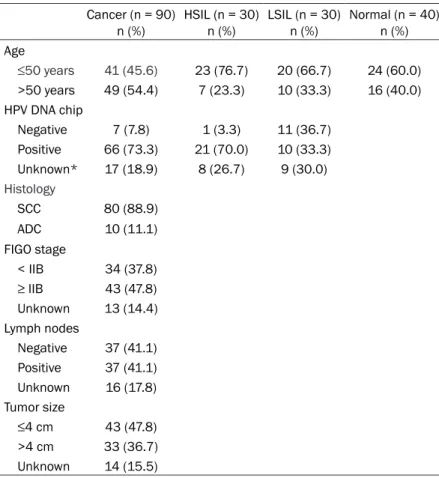
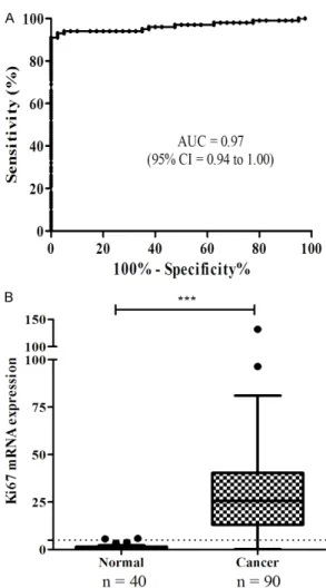
Figure 1. Receiver operating characteristics (ROC) curve analysis. A. The area under the ROC curve (AUC) was 0.97 (95% CI = 0.94 to 1.00, P < 0.001). B. Ki67 mRNA levels were significantly higher in FFPE cancer tissues compared to that found in FFPE normal tissues (t-test, P < 0.001) using a diagnostic threshold of 5 (shown as a horizontal dotted line). ***P < 0.001.
Table 3. Sensitivity, specificity, NPV, and PPV of Ki67 mRNA levels in cervical cancer and normal tissues Ki67 mRNA RT-qPCR assay (n = 130) 95% CI Sensitivity 93.3% 86.1-97.5 Specificity 97.5% 86.8-99.9 PPV 98.8% 93.6-100.0 NPV 86.7% 73.2-95.0
PPV, positive predictive value; NPV, negative predictive value; CI, confidence interval.
measuring mRNA relative to a reference gene using CFX Manager Software v1.6 (Bio-Rad, Hercules, CA, USA). The amount of Ki67 mRNA
normal tissues ranged from 0.01 to 3.43 com-pared to those in cancer tissues ranged from 0.22 to 131.60. The Ki67 mRNA levels in cervi-cal cancer tissues were 1.84- to 1167.33-fold higher compared to levels found in normal tis-sues (Table 2).
Diagnostic value of Ki67 for cervical cancer
ROC curve analysis was performed to deter-mine the optimal diagnostic cutoff value for the assay to discriminate normal tissues from those with cervical cancer. Ki67 mRNA levels were analyzed in 90 FFPE cancer tissues and
40 FFPE normal tissues, and found that the area under the ROC curve (AUC) was 0.97 (95% CI = 0.94 to 1.00, P < 0.001, Figure 1A). We also found that the levels of Ki67 mRNA in cer-vical cancer tissues were significantly increased compared to that found in normal cervical tis-sues. Based on these findings, we set a diag-nostic cutoff (threshold) of 5 (P < 0.001, Figure 1B). Using a threshold of 5, the assay had a sensitivity of 93.3% (95% CI = 86.1 to 97.5), a specificity of 97.5% (95% CI = 86.8 to 99.9), a positive predictive value of 98.8% (95% CI = 93.6 to 100.0), and a negative predictive value of 86.7% (95% CI = 73.2 to 95.0) (Table 3).
Figure 2. Box and whisker plots of Ki67 mRNA levels in histologically diagnosed FFPE cervical tissues. Ki67 mRNA levels in FFPE normal tissues were significantly lower than that found in LSIL, HSIL, and cancer tissues (t-test, P < 0.0001). ***P < 0.001, *P < 0.05.
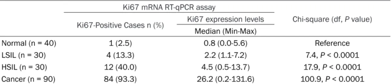
Table 4. Association between Ki67 mRNA expression levels and histologically diagnosed cervical grades
Ki67 mRNA RT-qPCR assay
Chi-square (df, P value) Ki67-Positive Cases n (%) Ki67 expression levels
Median (Min-Max)
Normal (n = 40) 1 (2.5) 0.8 (0.0-5.6) Reference
LSIL (n = 30) 4 (13.3) 2.2 (1.1-7.2) 7.4, P < 0.0001
HSIL (n = 30) 12 (40.0) 4.5 (0.5-13.7) 17.9, P < 0.0001
Cancer (n = 90) 84 (93.3) 26.2 (0.2-131.6) 100.9, P < 0.0001
Ki67 mRNA levels in histologically diagnosed FFPE cervical tissues
Ki67 mRNA levels were evaluated with histo-logically diagnosed cervical tissues to deter-mine whether Ki67 mRNA levels discriminated between normal, LSIL, HSIL, and cancer tis-sues. Ki67 mRNA levels were ranged from 0.0 to 5.6 (median 0.8) in 40 normal tissues, from 1.1 to 7.2 (2.2) in 30 LSIL tissues, from 0.5 to 13.7 (4.5) in 30 HSIL tissues, and from 0.2 to 131.6 (26.2) in 90 cancer tissues. The highest Ki67 mRNA levels were expressed in cancer tis-sues, with progressively lower but still elevated levels were checked in HSIL and LSIL tissues compared to the levels in normal tissues (P < 0.0001) (Figure 2 and Table 4). Using a cutoff of 5, positivity for Ki67 was 2.5% (1/40 cases) for normal, 13.3% (4/30 cases) for LSIL, 40.0% (12/30 cases) for HSIL, and 93.3% (84/90 cases) for cancer. We found that this Ki67 mRNA assay using a diagnostic threshold of 5 discriminated between normal and abnormal cervical lesions (P < 0.001) (Table 4).
Ki67 mRNA expression in relation to clinical prognostic parameters in cervical cancer tis-sues
The median value of Ki67 mRNA levels in cervi-cal cancer tissues was 26.2. To determine
whether there was an association between Ki67 mRNA levels in cervical cancer tissues and clinical prognostic parameters, specifically, age, HPV status, FIGO stage, lymph node posi-tivity, and tumor size, the Ki67 mRNA levels-were divided into two groups: a low Ki67 (below the median Ki67 mRNA level) group and a high Ki67 (above the median Ki67 mRNA level) group. Among the clinical prognostic parame-ters of cervical cancer,lymph node metastasis showed statistically significant relation with a high Ki67 group (P = 0.01) (Table 5).
Discussion
Cervical cancer is a leading cause of cancer mortality in 35-55 year old women worldwide. To avoid unnecessary treatment of transient HPV infections and related benign lesions, the optimal screening strategy for cervical cancer should efficiently and accurately identify pre-cursor lesions that will progress to an invasive cancer [22]. The purpose of the present study was to evaluate Ki67 mRNA expression levels with histological grades to identify and under-stand the relationship between the Ki67 mRNA assay and clinicopathological parameters of cervical cancer, and assess the performance evaluation of the assay as a diagnostic test for the detection of cervical cancer.
Our study compared Ki67 mRNA levels between matched cancerous and noncancerous tissues from 16 patients diagnosed with cervical SCC or ADC, and found that there were significant differences in mRNA levels between matched FFPE tissue samples (P = 0.0005) (Figure 1). In fact, comparing to normal tissues, Ki67 mRNA expression levels in cancer tissues enabled their discrimination regardless of whether they were ADC or SCC. Yamamoto et al found that Ki67 mRNA expression levels were informative as a Ki67-labeling index in patients with breast cancer [8]. Taking a cue from this, in cervical cancer, this Ki67 mRNA assay was validated as adiscriminative marker.
Using 90 cervical cancer and 40 normal FFPE tissue samples, we studied this Ki67 mRNA assay accurately discriminated between cervi-cal cancer and normal tissues with a high sen-sitivity of 93.3% (95% CI 86.1 to 97.5) and a high specificity of 97.5% (95% CI 86.8 to 99.9) (Table 3). Several studies have demonstrated that Ki67 expression using immunoquantifica-tion can provide greater discriminaimmunoquantifica-tion not only Table 5. Ki67 mRNA expression correlated
with clinical parameters in 90 cervical cancer patients
Ki67 mRNA expression Number
of cases Low (Ki67 < 26.2) High (Ki67 ≥ 26.2) valueP Age (years) ≤50 years 41 18 23 0.92 >50 years 49 21 28 HPV DNA chip Negative 7 2 5 0.48 Positive 66 28 38 FIGO stage < IIB 34 18 16 0.68 ≥ IIB 43 17 26 Lymph node Negative 37 21 16 0.01 Positive 37 12 25 Tumor size < 4 cm 43 23 22 0.30 ≥ 4 cm 33 13 20
between normal and cancer tissues but also between LSIL and HSIL. And the reported aver-age positivity rates found in normal, LSIL, and HSIL were 7.9%, 49%, and 90%, respectively [11, 23]. Similarly, in our study, positivity rates of Ki67 mRNA expression were 2.5%, 13.3%, and 40.0% in normal, LSIL, and HSIL respec-tively. Moreover, the positivity rates of Ki67 mRNA expression in cancer was 93.3%, and these were statistically significant (P < 0.001) (Table 4).
We also found that increased Ki67 mRNA expression in cancer samples was significantly higher than that found in normal, LSIL, and HSIL samples (Figure 2). Since about 10% of cases with LSIL progress to HSIL, cytological and/or histological follow-up are more fre-quently needed in LSIL patients with tendency of disease progression. Chen et al and Zhou et al were attempt to predict progression using Ki67 immunocytochemistry and immunohisto-chemistry tests and showed similar results that distinguishing LSIL and HSIL [24, 25]. In addi-tion to burdening the healthcare system, the challenge of using such a broad screening approach is that it reduces overtreatment for follow-up in patients with LSIL, the majority of who do not progress to HSIL.
Through clinicopathological prognostic param-eter analysis separating patients into high Ki67 expression (median ≥ 26.2) and low Ki67 expression (median < 26.2) groups, lymph node positivity was associated with Ki67 mRNA levels (Table 5). Shokouh studied that Ki67 IHC was correlated to lymph node status in breast cancer [9]. Yang et al showed lymph node metastasis and immunohistochemical markers Ki67 correlated for the predicting lymph node metastasis in endometrial cancer [26]. Cervical cancer in this study as well as breast and endo-metrial cancer in other studies also showed the interrelation of high Ki67 could be associated with lymph node positivity.
Previous studies demonstrated that IHC stain-ing of Ki67 may be used to complement HPV testing [27, 28]. Because of HPV screening pro-grams, HPV infection status is widely tested, but there are no predictors to determine the risk of high-risk HPV infection causing progres-sion to cancer. We demonstrated that Ki67 mRNA assay can provide an additional accu-rate approach for molecular diagnosing
cal cancer, and also predict prognosis of cervi-cal cancerdepending on LSIL and/or HSIL status.
Acknowledgements
This research was supported by the Basic Science Research Program through the National Research Foundation of Korea (NRF) funded by the Ministry of Science, ICT, and Future Planning (2015R1A2A2A04004455). Disclosure of conflict of interest
None.
Address correspondence to: Dr. Hyeyoung Lee, Department of Biomedical Laboratory Science, College of Health Sciences, Yonsei University, 1 Yonseidae-gil, Wonju, Gangwon, 220-710, Korea. Tel: +82-33-760-2740; Fax: +82-33-760-2561; E-mail: [email protected]; Dr. Eun Ji Nam, De- partment of Obstetrics and Gynecology, Institute of Women’s Life Medical Science, Yonsei University College of Medicine, 50-1, Yonsei-ro, Seodaemun-gu, Seoul 03722, Korea. Tel: +82-2-2228-2250; Fax: +82-2-313-8350; E-mail: [email protected]
References
[1] Torre LA, Bray F, Siegel RL, Ferlay J, Lortet-Tieu-lent J and Jemal A. Global cancer statistics, 2012. CA Cancer J Clin 2015; 65: 87-108. [2] Schiffman M, Castle PE, Jeronimo J, Rodriguez
AC and Wacholder S. Human papillomavirus and cervical cancer. Lancet 2007; 370: 890-907.
[3] Salimovic-Besic I, Tomic-Cica A, Smailji A and Hukic M. Comparison of the detection of HPV-16, 18, 31, 33, and 45 by type-specific DNA- and E6/E7 mRNA-based assays of HPV DNA positive women with abnormal Pap smears. J Virol Methods 2013; 194: 222-228.
[4] Munkhdelger J, Choi Y, Lee D, Kim S, Kim G, Park S, Choi E, Jin H, Jeon BY, Lee H and Park KH. Comparison of the performance of the Nu-cliSENS EasyQ HPV E6/E7 mRNA assay and HPV DNA chip for testing squamous cell le-sions of the uterine cervix. Diagn Microbiol In-fect Dis 2014; 79: 422-427.
[5] Castro FA, Koshiol J, Quint W, Wheeler CM, Gil-lison ML, Vaughan LM, Kleter B, van Doorn LJ, Chaturvedi AK, Hildesheim A, Schiffman M, Wang SS, Zuna RE, Walker JL, Dunn ST and Wentzensen N. Detection of HPV DNA in paraf-fin-embedded cervical samples: a comparison of four genotyping methods. BMC Infect Dis 2015; 15: 544.
[6] Brink AA, Snijders PJ and Meijer CJ. HPV detec-tion methods. Dis Markers 2007; 23: 273-281. [7] Eide ML and Debaque H. HPV detection meth-ods and genotyping techniques in screening for cervical cancer. Ann Pathol 2012; 32: e15-23, 401-409.
[8] Yamamoto S, Ibusuki M, Yamamoto Y, Fu P, Fu-jiwara S, Murakami K and Iwase H. Clinical rel-evance of Ki67 gene expression analysis using formalin-fixed paraffin-embedded breast can-cer specimens. Breast Cancan-cer 2013; 20: 262-270.
[9] Shokouh TZ, Ezatollah A and Barand P. Inter-relationships Between Ki67, HER2/neu, p53, ER, and PR status and their associations with tumor grade and lymph node involvement in breast carcinoma subtypes retrospective- observational analytical study. Medicine (Baltimore) 2015; 94: e1359.
[10] Li LT, Jiang G, Chen Q and Zheng JN. Ki67 is a promising molecular target in the diagnosis of cancer. Mol Med Rep 2015; 11: 1566-1572. [11] Calil LN, Edelweiss MI, Meurer L, Igansi CN and
Bozzetti MC. p16 INK4a and Ki67 expression in normal, dysplastic and neoplastic uterine cervical epithelium and human papillomavirus (HPV) infection. Pathol Res Pract 2014; 210: 482-487.
[12] Luttmer R, Dijkstra MG, Snijders PJ, Berkhof J, van Kemenade FJ, Rozendaal L, Helmerhorst TJ, Verheijen RH, Ter Harmsel WA, van Baal WM, Graziosi PG, Quint WG, Spruijt JW, van Di-jken DK, Heideman DA and Meijer CJ. p16/Ki-67 dual-stained cytology for detecting cervical (pre)cancer in a HPV-positive gynecologic out-patient population. Mod Pathol 2016; 29: 870-878.
[13] Pan D, Wei K, Ling Y, Su S, Zhu M and Chen G. The prognostic role of Ki-67/MIB-1 in cervical cancer: a systematic review with meta-analy-sis. Med Sci Monit 2015; 21: 882-889. [14] Piri R, Ghaffari A, Azami-Aghdash S, Ali-Akbar
YP, Saleh P and Naghavi-Behzad M. Ki-67/ MIB-1 as a prognostic marker in cervical can-cer-a systematic review with meta-analysis. Asian Pac J Cancer Prev 2015; 16: 6997-7002. [15] Ancuta E, Ancuta C, Cozma LG, Iordache C,
Anghelache-Lupascu I, Anton E, Carasevici E and Chirieac R. Tumor biomarkers in cervical cancer: focus on Ki-67 proliferation factor and E-cadherin expression. Rom J Morphol Embryol 2009; 50: 413-418.
[16] Scholzen T and Gerdes J. The Ki-67 protein: from the known and the unknown. J Cell Physi-ol 2000; 182: 311-322.
[17] Wang HY, Kim G, Cho H, Kim S, Lee D, Park S, Park KH and Lee H. Diagnostic performance of HPV E6/E7, hTERT, and Ki67 mRNA RT-qPCR assays on formalin-fixed paraffin-embedded
cervical tissue specimens from women with cervical cancer. Exp Mol Pathol 2015; 98: 510-516.
[18] Weinstein LC, Buchanan EM, Hillson C and Chambers CV. Screening and prevention: cervi-cal cancer. Prim Care 2009; 36: 559-74. [19] Safaeian M, Solomon D and Castle PE.
Cervi-cal cancer prevention-cerviCervi-cal screening: sci-ence in evolution. ObstetGynecol Clin North Am 2007; 34: 739-60.
[20] Cox JT. The development of cervical cancer and its precursors: what is the role of human papillomavirus infection? Curr Opin Obstet Gy-necol 2006; 18 Suppl 1: s5-s13.
[21] Livak KJ and Schmittgen TD. Analysis of rela-tive gene expression data using real-time quantitative PCR and the 2(T)(-Delta Delta C) method. Methods 2001; 25: 402-408. [22] Saslow D, Solomon D, Lawson HW, Killackey
M, Kulasingam SL, Cain J, Garcia FA, Moriarty AT, Waxman AG, Wilbur DC, Wentzensen N, Downs LS Jr, Spitzer M, Moscicki AB, Franco EL, Stoler MH, Schiffman M, Castle PE, Myers ER; ACS-ASCCP-ASCP Cervical Cancer Guide-line Committee. American Cancer Society, American Society for Colposcopy and Cervical Pathology, and American Society for Clinical Pathology screening guidelines for the preven-tion and early detecpreven-tion of cervical cancer. CA Cancer J Clin 2012; 62: 147-172.
[23] Pacchiarotti A, Ferrari F, Bellardini P, Chini F, Collina G, Dalla Palma P, Ghiringhello B, Mac-callini V, Musolino F, Negri G, Pisa R, Sabatucci I and Giorgi Rossi P. Prognostic value of p16-INK4A protein in women with negative or CIN1 histology result: a follow-up study. Int J Cancer 2014; 134: 897-904.
[24] Chen CC, Huang LW, Bai CH and Lee CC. Pre-dictive value of p16/Ki67 immunocytochemis-try for triage of women with abnormal Papani-colaou test in cervical cancer screening: a systematic review and meta-analysis. Ann Saudi Med 2016; 36: 245-251.
[25] Zhou WQ, Sheng QY, Sheng YH, Hou WJ, Xu GX, Wu YM and Lu H. Expressions of survivin, P16(INK4a), COX-2, and Ki-67 in cervical can-cer progression reveal the potential clinical ap-plication. Eur J Gynaecol Oncol 2015; 36: 62-68.
[26] Yang B, Shan B, Xue X, Wang H, Shan W, Ning C, Zhou Q, Chen X and Luo X. Predicting lymph node metastasis in endometrial cancer using serum CA125 combined with immunohisto-chemical markers PR and Ki67, and a com-parison with other prediction models. PLoS One 2016; 11: e0155145.
[27] Agoff SN, Lin P, Morihara J, Mao C, Kiviat NB and Koutsky LA. p16(INK4a) expression corre-lates with degree of cervical neoplasia: A
com-p16/Ki67 dual staining on formalin-fixed par-affin-embedded cervical specimens: correla-tion with HPV-DNA test, E6/E7 mRNA test, and potential clinical applications. Biomed Res Int 2013; 2013: 453606.
parison detection of high-risk with Ki-67 ex-pression and HPV types. Mod Pathol 2003; 16: 665-673.
[28] Zappacosta R, Colasante A, Viola P, D’Antuono T, Lattanzio G, Capanna S, Gatta DMP and Rosini S. Chromogenic in situ hybridization and