*Corresponding author : Jun-Sang Ham, National Institute of Animal Science, RDA, Suwon 441-350, Korea. Tel : 82-31-290-1692, Fax : 82-31-290-1697, E-mail : [email protected]
산양유의 특성
-
유지방, 체세포, 그리고 산양취 -정석근․이승규․김동훈․함준상*
농촌진흥청 축산과학원
Characteristics of Goat Milk
- Milk Fat, Somatic Cell Count, and Goaty Flavor -
Seok-Geun Jeong, Seung-Gyu Lee, Dong-Hun Kim and Jun-Sang Ham*
National Institute of Animal Science, RDA
---
ABSTRACT
Since goat milk infant formula has been increased, it is expected that goat milk consumption would be increased. This review summarizes the characteristics of goat milk especially, milk fat, somatic cell count, and goaty flavor. Average milk fat content for one year of twelve goat milk farms was 3.6%, but 2.9~3.1% in summer, which means summer goat milk could not meet the 'Processing and Ingredient Standard for Animal Products'. More than 3.2% for goat milk fat content in 'Processing and Ingredient Standard for Animal Products' should be amended. In addition to, hygienic standard for goat milk should be newly established because goat milk has naturally higher somatic cell count with noninfectious factors. It is thought that 6-trans nonenal and some branched fatty acids are responsible for the goaty flavor. It is necessary to minimize goaty flavor from farm to table because goaty flavor is the most important factor for the promotion of goat milk industry .
(Key words : goat milk, allergenicity, somatic cell count, goaty flavor)
---
서 론
산양유 제품은 산양의 산간 방목지에서 천연의 산야초를 먹는 먹이 습성으로 인하여 웰빙 식품의 개념으로 뿐만 아 니라 산양유 조제식의 형태로 다시 소비되기 시작하고 있다 (Park, 2006). 산양유 제품의 공급은 대규모 유가공 회사에서 산양 조제 분유 생산을, 소규모 산양유가공 회사에서 신선 유와 발효유를 생산하는 형태로 이루어지고 있다. 대규모 유가공 회사에 의해 주도되는 유아용 조제식으로의 소비 증 가는 미래에 산양유의 소비가 증가할 것이라는 예상을 가능 하게 하며, 국내 산양유 산업의 육성이 필요함을 말해주고 있다. 산양유 산업의 육성을 위해서는 산양유의 홍보 및 산 양유 특유취 저감 등의 기술 개발이 필요하다. 본 고에서는 산양유 유지방 함량의 계절적 변이, 산양유의 체세포 수가 높은 점, 그리고 산양취의 원인 물질 등 산양유의 특성에 대
하여 고찰하였다.
본 론
1. 유지방
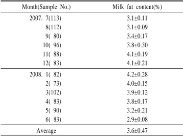
Devendra(1972)와 Mba 등(1975)은 British Alpines, Anglo- Nubians, 그리고 Saanens이 온대 기후에 비해 열대 기후에 서 유지방 함량이 낮은 산양유를 생산한다는 점에 주목했 으며, 미국에서 Saanen의 평균 유지방 함량은 2005년에 3.3%, 2006년에 3.2%, 2007년에 3.3%로 보고되고 있다(American Dairy Goat Association, 2008). 산양유 가공업체에 납유 중 인 12개 농가의 1년간 유지방 함량을 Table 1에 표시하였 다. Saanen이 대부분인 산양 목장의 평균 유지방 함량은 3.6%로 미국 Saanen의 평균보다 높게 나타났으나, 계절별 변이가 심하여 여름철인 6, 7, 8월에는 각각 2.9, 3.1, 3.1%
로 나타났다. 이는 여름철에 농후 사료를 급여하지 않기 때 문인 것으로 설명된다. 그런데, 우리나라 ‘축산물의 가공기 준 및 성분규격’(2007)에서는 산양유의 유지방 함량을 3.2%
Table 1. Monthly variation of goat milk fat from 12 goat farms Month(Sample No.) Milk fat content(%)
2007. 7(113) 8(112) 9( 80) 10( 96) 11( 88) 12( 83)
3.1±0.11 3.1±0.09 3.4±0.17 3.8±0.30 4.1±0.19 4.1±0.21 2008. 1( 82)
2( 73) 3(102) 4( 83) 5( 90) 6( 83)
4.2±0.28 4.0±0.15 3.9±0.12 3.8±0.17 3.2±0.21 2.9±0.08
Average 3.6±0.47
이상으로 규정하고 있으며, 행정기관에서는 지방 함량 미 달에 대해 행정 처분을 하게 된다. 유지방은 크림, 버터 등 유제품 제조의 원료가 되므로 유대 결정의 중요한 기준 으로 사용되어 왔으며, ‘축산물의 가공기준 및 성분규격’
(2007)에서는 우유류, 유당 분해 우유, 가공유, 크림 발효 유와 농후 크림 발효유의 지방 함량을 각각 3.0, 3.0, 2.7, 그리고 8.0% 이상으로 규정하고 있다. 하지만 최근에는 소 비자들의 지방 섭취 기피와 함께 저지방 제품의 인기가 높 아지고 있으며 ‘축산물의 가공기준 및 성분규격’(2007)에 서 저지방 우유류, 저지방 유당 분해 우유, 저지방 가공유 의 지방 함량을 2.0% 이하로 규정하고 있다. 따라서, 산양 유의 지방 함량이 3.2%가 미달되기 때문에 영업정지 처분 을 받는 것은 제도적인 결함으로 생각되며 이에 대한 조속 한 개선이 필요하며, 가수 등 부정유의 단속을 위해서는 무지고형분 기준을 적용하는 것이 바람직할 것으로 생각 된다.
2. 체세포
체세포 수는 목장 관리 수단으로서 유질 평가를 위한 지표 로 인정되고 있다. 하지만 우유와 양유(sheep milk)에서 인정 되는 평가방법이기는 하지만 산양유에는 적용할 수 없다 (Paape 등, 2007). 이는 유방이 감염되지 않아도 산양의 체세 포 수가 소나 양보다 높게 나타나기 때문이다(Table 2). 감 염되지 않은 유방에서 채취한 산양유에서도 체세포 수는 27 만에서 200만으로 나타났으며, 감염된 유방에서는 65만에 서 420만의 체세포 수를 나타내었다. 유방내 감염 이외에 관리 방법, 비유기, 산차 및 CAEV(caprine arthritis-encephalitis virus) 감염이 산양유의 체세포 수 증가에 영향을 미친다. 유 선이 감염되지 않아도 체세포 수는 비유기와 산차의 증가에 따라 증가된다(Dulin et al., 1983; Luengo et al., 2004). 비유 기와 산차의 증가에 따른 체세포 수 증가는 유방내 감염도
원인이 될 수 있지만 많은 부분이 비감염적 요인 때문이다 (Paape와 Contreras, 1997). Wilson 등(1995)은 산양에서 체세 포 수 변이의 90% 이상은 유방내 감염때문이 아니라고 주 장하였으며, 착유일수 증가와 몇 월인가가 체세포 수 증가 에 가장 큰 원인이라고 하였다. 산차와 유생산 감소도 체 세포 수의 증가에 유의적인 원인이 되며, 체세포 수 변이 의 75%는 설명되지 못한다. 설명되지 못하는 부분은 혐기 성 박테리아인 Mycoplasma나 CAEV가 원인이 될 수 있다 (Paape et al., 2001). 대부분의 연구자는 비유기에 따른 체세 포 수 증가는 희석 효과로 설명될 수 있음을 지적한다. 왜냐 하면 비유기 증가에 따라 유생산이 감소하고 비유기동안 체 세포 수가 직선적으로 증가하기 때문이다. 게다가, 비유기가 비슷한 경우 체세포 수에 미치는 계절의 효과는 유생산과 관련이 있다(Sanchez et al., 1998). 산양유에서 체세포 수 증 가의 다른 요인들은 발정기, 백신, 사료 변화 및 착유과정 변 화이다(Paape와 Contreras, 1997). 이러한 요인들은 산양에 신 체적 스트레스로 작용하므로 우유 생산 감소 때문에 체세포 가 증가하는 것으로 설명될 수 있다.
우유 및 양유와 달리 산양유는 다형핵호중구(Polymorpho- nuclear neutrophilic leukocytes, PMN)가 유선 감염에 관계없 이 대부분을 차지한다(Dulin et al., 1983). PMN은 감염이 되 지 않은 산양유에서 체세포의 45~74%를 차지하고, 유선 감 염시에는 71~86%를 차지한다. 산양유에서 상피세포는 차 지하는 비율이 적으며 세포질 입자(cytoplasmic particles)의 존재 때문에 광학현미경에 의한 구별은 쉽지 않다. 초기 연구 에서는 상피세포가 전체 세포의 1% 미만이라고 보고(Dulin et al., 1982)되었으나, 최근에는 유선이 감염되지 않았을 때 상피세포는 6%에 달한다고 보고되었다(Contreras et al., 1998). 산양에서 유즙 분비는 주로 아포크린(apocrine)이기 때 문에(Wooding et al., 1970), 세포질 입자가 유선 분비 세포 말단 부위에서 젖으로 들어간다. 감염되지 않은 유선내 젖 의 세포질 입자수는 71~306×103/mL이고, 감염된 경우는 98~231×103/mL이다(Dulin et al., 1983). 비록 이들 입자의 대부분 핵이 없지만, 약 1% 정도는 핵 절편을 가지는 것으 로 관찰되었다(Dulin et al., 1982). 따라서, 산양유의 체세포 수 측정에는 다른 방법이 필요하며(Table 3), DNA 특이적 세포 계수 방법(fluoro-optical electronic cell counter, DNA 특 이 염색법을 이용한 체세포 수 측정)만이 사용되어야 한다.
Dulin 등(1982)의 연구는 pyronin Y-methyl green 염색과 Fosso- matic(fluoro-optical electronic counter)에 의한 체세포 수가 유 의적 차이가 없음을 가리킨다. DNA 특이적 방법인 Wisconsin mastitis test도 Fossomatic 체세포 수와 유의적 차이를 보이 지 않았다. Coulter electronic 세포 측정과 Levowitz-Weber로 염색한 직접검경법은 유의적으로 높은 수치를 나타내었다.
우유에서 체세포 수의 간접측정법으로 많이 사용되고 있는
Table 2. Results of milk SCC scores for healthy and infected goat udder halves1,2)
Reference Type Cells×103/mL
Uninfected Infected
Dulin et al., 1982 Dulin et al., 1983
Poutrel and Lerondelle, 1983 Timms and Schultz, 1985 Lerondelle et al., 1992
Kalogridou-Vassiliadou et al., 1992 Wilson et al., 1995
Contreras et al., 1996
Corrales et al., 1996 De Cremoux et al., 1996
Ferrer et al., 1996 Le Mens et al., 1996 Poutrel et al., 1996
Sanchez et al., 1996
Vihan, 1996 Contreras et al., 1997
AM GM AM GM AM AM LS GM AM AM GM AM AM GM GM AM GM AM AM GM
280 481 614 337 520 270 303~650
396 1115 1235 493 973 349~571
1059 272 687 341 938 332 2000
1690 1778 1293 659 1040CNS
3800 373~800
873 1909 2186 1078CNS~2731MP 1764CNS~3591MP
1900~3393 2511 932CNS~2443MP 1462CNS~4213MP
1218 2147 708 2500
1) Adapted from Sanchez et al.(1998).
2) AM = Arithmetic mean; GM = geometric mean; LS = linear score; CNS = udder halves infected with coagulase-negative staphylococci; MP = udder halves infected with major pathogens.
Table 3. Comparison of methods for estimating SCC in goat milk1) Method No. of cells(×105/mL)2) Pyronin Y-methyl green stain
Fossomatic cell counter Wisconsin mastitis test Coulter Counter Levowitz-Weber stain Significance level Standard error
3.40a 3.65ab 4.94bc 6.44cd 7.92d p<0.01
±1.1324
1) Adapted from Dulin et al.(1982).
2) Each value represents the mean of 24 determinations run in duplicate.
Means with the same subscript letter in common are not significantly different(p<0.05).
CMT(California Mastitis Test)도 산양유에서 스크리닝법으로 사용될 수 있다(Schalm et al., 1971; Schalm과 Noorlander, 1957). CMT 반응은 체세포에서 유래한 DNA에 기초한다 (Carroll과 Schalm, 1962; Paape et al., 1962). CMT에 사용되 는 시약은 sodium alkylarylsulfonate, sodium hydroxide 및 pH 지시제이다. 시약이 체세포를 분해하여 DNA가 sodium hy- droxide 용액으로 방출되어 젤의 형성을 일으킨다. Coulter electronic cell counter 같은 입자 계수기나 DNA 특이적이 아
닌 염색을 이용한 직접검경법은 산양유에 사용되어서는 안 된다(Dulin et al., 1982). 양유는 세포질 입자가 산양유의 1/10인 15×103/mL이므로 전자 입자계수기와 DNA 특이적이 아닌 염색법도 체세포 수 측정에 사용될 수 있다. Fosso- matic cell counter 같은 fluor-optical electronic 세포계수에서 시료는 40℃에서 1~5일 후에 분석하여야 한다. 신선한 시 료는 DNA를 ethidium bromide로 염색하기 위해 60℃에서 15 분 가열이 필요하다(Miller et al., 1986). 산양유에는 세포질 입 자가 많기 때문에 pyronin Y-methyl green 염색법이 미국에서 공식적 염색법으로 사용되고 있다(Packard et al., 1992).
산양에서는 감염되지 않아도 자연적으로 체세포 수가 높고 비감염적 요인으로 체세포 수가 증가하기 때문에 체 세포 수로 유방염을 예측하는 것은 적절하지 못하다(Haenlein 과 Hinckly, 1995). Poutrel과 Lerondelle(1983)은 체세포 수 1×10⁶/ mL 이상의 85%는 주요 병원균에 의한 감염으로 분 류할 수 있으나, 모든 병원균 감염을 포함하였을 때는 분류 의 정확도가 떨어짐을 발견했다. 비유기와 다른 요인을 고 려한 기준이 최근에 제안되었다(Sanchez et al., 1998). 미국 에서 산양유 체세포의 법적 한계는 1×10⁶/mL(Pateurized Milk Ordinance, 1995)이고, EU에서는 아직 법적 한계가 마련되지 않고 있다. 미국의 경우는 너무 엄격하며 계절적 요인이 고
려될 필요가 있다. 우리나라의 경우도 산양유의 체세포 수 에 대해 규정되어 있지 않다. 축산물가공처리법 제2조 4항 에서 “원유”라 함은 판매 또는 판매를 위한 처리․가공을 목 적으로 하는 착유상태의 우유와 양유를 말하고, 제4조 2항 의 규정에 의거 “원유의 위생등급기준”이 고시되어 있다(농 림부 수의과학검역원, 2002). 일반적으로 산양유를 양유에 준하여 원유의 위생등급 기준을 적용하기 쉬우나, 상기한 바와 같은 이유로 산양유는 별도로 체세포 수 등급기준을 마련해야 할 것으로 생각된다.
3. 산양취
산양유의 품질을 심각하게 위협하는 외부적 요인으로 숫 산양이 산양취의 원인이 되며, 숫 산양은 착유장이나 착유 산양으로부터 격리된 곳에서 사육하도록 권장되고 있다. 발 정기의 숫 산양은 뇨를 볼과 목 부위에 분사하고, 이것이 특 징적인 냄새를 갖고 있긴 하지만 주요 숫 산양 냄새는 피지 선의 분비 때문이다. 피지선은 뿔의 기저에 위치하고 성장 기 동안 비대해지며, 암컷에도 있기는 하지만 보통 활성화 되지 않는다. Smith 등(1984)은 발정기에 산양목장 수 백미 터 밖에서도 검출될 수 있는 냄새 중 하나가 nonenal이라고 주장하였으며 5-, 6-, 7-trans, 그리고 6-cis nonenals가 농장에 서 검출되는 냄새와 비슷하며, 6-trans nonenal을 가장 주요 한 물질로 추정하였다. 또한, nonenal이 없는 숫 산양 지방 시 료에서 6-trans nonenal을 합성하여 분비선 지방(gland lipid)에 6-trans nonenal의 전구물질이 있음을 확인하였다. Keppler 등(1965)도 nonenals 이성체의 냄새를 평가한 바, 비슷한 냄 새를 가진 5-trans나 7-trans nonenals보다 6-trans nonenal이 100~200배 강력하다고 하였다.
전통적으로 C-6, C-8 및 C-10 단쇄지방산이 숫 산양 냄새 와 관련이 있다고 알려졌으나, Sugiyama 등(1981)은 일본 재 래 숫 산양에서 분비선 냄새의 주요 물질로 4-ethyl 분지지방 산(branched fatty acid)을 분리하였다. Wong 등(1975)은 야생 산양육에 산양취가 있으며, 이는 4-methyl octanoic acid 때문 이라고 보고하였다. 스위스의 Givaudan 회사에서는 산양유 내 산양취의 원인 물질로 4-ethyl oct-2-enoic acid를 분리하였 다. 분지지방산은 분비선 지방내 전체 지방산의 55% 이상을 차지하며 조성이 복잡하기 때문에 다른 결과가 보고되고 있 다. 미량의 6-trans nonenal과 분지지방산은 각기 또는 복합 적으로 우유의 산패취와는 다른 산양취를 발생한다.
유제품의 품질 평가에 있어서 관능적 특성 특히, 향기와 맛(풍미)은 가장 중요한 평가 요인 중의 하나이다. 산양유 제품의 산양취를 줄이기 위해서는 산양취 원인 물질에 대한 구명이 필요하며, 산양 사육 농가의 착유 및 저장 그리고 산 양유 가공업체에서 집유 및 가공․유통 중에 발생될 수 있 는 지방 산화 및 분해에 의한 휘발성 저급 유리지방산 생성
이 최소화될 수 있도록 주의를 기울여야 하겠다.
결 론
대규모 유가공 회사에 의해 주도되는 유아용 조제식으로 의 소비 증가는 미래에 산양유의 소비가 증가할 것이라는 예상을 가능하게 하며, 국내 산양유 산업의 육성이 필요함 을 말해주고 있다. 산양유 가공업체에 납유중인 12개 목장 의 1년 평균 유지방 함량은 3.6%로 나타났으나, 농후사료를 급여하지 않는 여름철에는 2.9~3.1%로 나타나 ‘축산물의 가공기준 및 성분규격’의 3.2% 이상을 충족시키지 못하는 것으로 나타나 이에 대한 개선이 필요하다. 또한, 산양에서 는 감염되지 않아도 자연적으로 체세포 수가 높고 비감염적 요인으로 체세포 수가 증가하기 때문에 “원유의 위생등급기 준”을 따르는 것은 무리가 있으며, 산양유의 체세포 수 등급 기준을 별도로 마련해야 할 것으로 생각된다. 6-trans nonenal 과 분지지방산이 산양취의 주요 원인 물질로 보고되고 있으 며, 산양취의 관리는 산양유 산업의 성패를 결정할 수 있는 주요한 요인이 될 수 있으므로 목장에서부터 유통까지 세심 한 관리가 필요하다.
참고문헌
1. Carroll, E. J. and Schalm, O. W. 1962. Effect of deoxyri- bonuclease on the California test for mastitis. J. Dairy Sci.
45:1094-1097.
2. Contreras, A., Sierra, D., Corrales, J. C., Sanchez, A. and Marco, J. 1996. Physiological threshold of somatic-cell count and California Mastitis Test for diagnosis of caprine subclinical mastitis. Small Ruminant Res. 21:259-264.
3. Contreras, A., Paape, M. J., DiCarlo, A. L., Miller, R. H.
and Rainard, P. 1997. Evaluation of selected antibiotic residue screening test for milk from individual goats. J.
Dairy Sci. 80:1113-1119.
4.. Contreras, A., Sierra, D., Corrales, J. C., Sanchez, A. and Gon- zalo, C. 1998. Diagnostico indirecto de las mamitis caprinas.
Mamitis caprina II. Ovis. 54:25-36.
5. Corrales, J. C., Sanchez, A., Sierra, D., Marco, J. C. and Contreras, A. 1996. Relationship between somatic cell counts and intramammary pathogens goats. pp.35-39 In Somatic Cells and Milk of Small Ruminants. R. Rubino ed. Wagen- ingen, the Nethelands.
6. De Cremoux, Poutrel, R. B., Pillet, R., Perrin, G., Ducellier, M., and Heuchel, V. 1996. Cell counts for diagnosing caprine bacterial mammary infections. pp.35-39 In Somatic Cells
and Milk of Small Ruminants. R. Rubino ed. Wageningen, the Nethelands.
7. Devendra, C. 1972. The composition of milk of Britsh Alpine and Anglo-Nubian goats imported into Trinidad. J.
Dariy Res. 39:381-385.
8. Dulin, A. M., Paape, M. J. and Wergin, W. P. 1982.
Differentiation and enumeration of somatic cells in goat milk. J. Food Prot. 45:435-439.
9. Dulin, A. M., Paape, M. J., Schultze, W. D. and Weinland, B. T. 1983. Effect of parity, stage of lactation, and intra- mammary infection on concentration of somatic cells and cytoplasmic particles in goat milk. J. Dairy Sci. 66:2426- 2433.
10. Ferrer, O., Real, F, Acosta, B. and Molina, J. M. 1996.
Physiological cellular values in Canary(Agrupacion caprina canaria) Goat Milk. pp.81-84. In Somatic cells and milk of small ruminants. R. Rubino ed. Wageningen, the Nethe- lands.
11. Haenlein, G. F. W. and Hinckley, L. S. 1995. Goat milk somatic cell count situation in USA. Int. J. Anim. Sci.
10:305-310.
12. http://adga.org/facts.htm
13. Kalogridou-Vasiliadou, D., Manolkidis, K. and Tsigoida, A.
1992. Somatic cell counts in relation to infection status of the goat udder. J. Dairy Res. 59:21-28.
14. Keppler, J. G., Schols, J. A., Feenstra, W. H. and Meij- boom, P. W. 1965. Components of the hardening flavor present in hardened linseed oil and soybean oil. J. Am. Oil Chem. Soc. 42:246.
15. Le Mens, P., Dalmas, S. and Humbert, G. 1996. Relations entre l'activite de la N-acetyl-glucosaminidale(NAG-ase), le nombre de cellules, l'apititude a la coagulation du lait et le statut infectieux mammarie chez la chevre. pp.311-317. In Somatic cells and milk of small ruminants. R. Rubino ed.
Wageningen, the Nethelands.
16. Lerondelle, C., Richard, Y. and Issartial, J. 1992. Factors affecting somatic cell counts in goat milk. Small Ruminant Res. 8:129-139.
17. Luengo, C., Sanchez, A., Corrales, J. C., Fernandez, C. and Contreras, A. 2004. Influence of intramammary infection and non-infection factors on somatic cell counts in dairy goats. J. Dairy Res. 71:169-174.
18. Mba, A. U., Boyo, B. S. and Oyenuga, V. A. 1975. Studies on the milk composition of West African dwarf, Red Soloto and Saanen goats at different stages of lactation. I. Total
solids, butterfat, solids-not fat protein, lactose and energy contents of milk. J. Dairy Res. 42:217-226.
19. Miller, R. H., Paape, M. J. and Action, J. C. 1986. Com- parison of milk somatic cell counts by Coulter and Fosso- matic Counters. J. Dairy Sci. 69:1942-1946.
20. Paape, M. J., Wiggans, G. R., Bannerman, D. D., Thomas, D. L., Sanders, A. H., Contreras, A., Moroni, P. and Miller, R. H. 2007. Monitoring goat and sheep milk somatic cell counts. Small Ruminant Res. 68:114-125.
21. Paape, M. J., Poutrel, B., Contreras, A., Marco, J. C. and Capuco, A. V. 2001. Milk somatic cells and lactation in small ruminants. J. Dairy Sci. 75:556-565.
22. Paape, M. J. and Contreras, A. 1997. Historical perspective on the evolution of the milk somatic cell count. Flemish Vet. J. 66(suppl.):93-105.
23. Paape, M. J., Hafs, H. D. and Snyder, W. W. 1962. Feulgen DNA in milk as a measure of udder irritation. J. Anim. Sci.
21(Suppl.):1028.
24. Packard Jr., V. S., Tatini, S., Fugua, R., Heady, J. and Gilman, C. 1992. Direct microscopic methods for bacteria or somatic cells. In: Methods for the examination of dairy products, R.
T. Marshall, ed., 16th ed. Am. Public Health Assoc., Wa- shington, DC. pp. 309-321.
25. Park, S. Y. 2006. Production and consumption of goat milk products in Korea. 2006. J. Korean Dairy Technol. Sci.
24(2):39-45.
26. Pasteurized Milk Ordinance (PMO). 1995. Grade "A" Pas- teurized milk ordinance. U.S. Dept. of Health and Human Services, Washington, DC.
27. Poutrel, B. and Lerondelle, C. 1983. Cell content of goat milk: California Mastitis Test, Coulter Counter, and Fosso- matic for predicting half infection. J. Dairy Sci. 66:2575- 2579.
28. Poutrel, B., De Cremoux, R., Pillet, R., Heuchel, V. and Duceilliez, M. 1996. Caprine mammary infections with respect to cell counts in milk. pp.61-64. In Somatic Cells and Milk of Small Ruminants. R. Rubino ed. Wageningen, the Nethelands.
29. Sanchez, A. Contreras, A., Corrales, J. C., Sierra, D. and Marco, J. 1996. El recuento de celulas somaticas como instumento de control de mamitis subclinicas in las cabra Murciano-Granadina. pp.205-212. In XXI Jornadas Cientificas de la SEOC.
30. Sanchez, A. Corrales, J. C., Marco, J. and Contreras, A.
1998. Use of somatic cell counts for control of mastitis in
goats. In: Mamitis caprinas II. Ovis 54:37-51.
31. Schalm, O. W. and Noorlander. 1957. Experiments and obser- vations leading to development of the California mastitis test. JAVMA. 130:199-204.
32. Schalm, O. W., Carroll, E. J. and Jain, N. C. 1971. Physical and chemical tests for detection of mastitis. In: Bovine Mastitis, Lea & Febiger, Philadelphia, PA. pp.150-155.
33. Smith, P. W., Parks, O. W. and Schwartz, D. P. 1984. Cha- racterization of male goat odors: 6-Trans nonenal. J. Dairy Sci. 67:794-801.
34. Sugiyama, T., Sasada, H., Masaki, J. and Yamashia, K. 1981.
Unusual fatty acids with specific odor from mature male goat. J. Agric. Biol. Chem. 45:2655.
35. Timms, L. L. and Schulttz, H. 1985. N-acetyl-B-D-gluco- saminidase activity and somatic cells in goat milk. J. Dairy Sci. 68:3363-3366.
36. Vihan, V. S. 1996. Determination of lysosomal-enzyme activity,
somatic cells, percent fat and protein in subclinical caprine mastitis. pp.31-34. In Somatic cells and milk of small ruminants. R. Rubino ed. Wageningen, the Nethelands.
37. Wilson, D. J., Stewart, K. N. and Sears, P. M. 1995. Effects of stage of lactation, production, parity and season on somatic cell counts in infected and uninfected dairy goats.
Small Ruminant Res. 16:165-169.
38. Wong, E., Johnson, C. B. and Nixon, L. N. 1975. The contri- bution of 4-methyl octanoic acid to mutton and goat meat flavor. N. Z. J. Agri. Res. 18:261.
39. Wooding, F. B. P., Peaker, M. and Linzell, J. L. 1970.
Theories of milk secretion: Evidence from the electron microscopic examination of milk. Nature. 226:762-764.
40. 농림부 국립수의과학검역원. 2002. 원유의위생등급기준 (검역원고시 2002-4).
41. 농림부 국립수의과학검역원. 2007. 축산물의가공기준및 성분규격(검역원고시 2007-20).