만성 점액낭염의 합병증으로 다발성의 작은 쌀알 모양의 하 얀 결절이 발생된다2 , 4 , 8 , 9 ). 비록 미립체의 형성은 대부분 만성 류마토이드 관절염에서 관찰 되지만, 전신적인 어떤 기저 질환 없이도 발생할 수 있다2 ). 미립체를 동반할 수 있는 이미 알려 진 전신적인 관절증인 색소 융모 결절성 활액막염과 활막 연골 종증과 같은 활막 증식증을 의심함으로써 감별 진단하여야 한 다7 ). 또한 회전근 개 파열 관절증 환자에게서 회전근 개 전층 파열과 함께 인회석 결정(apatite crystal)으로 인해 염증을 동반하는 Milwaukee shoulder도 감별해야 한다1 ). 그러므로 미립체의 수술 전 감별 진단과 정확한 진단을 위해 단순 방사 선과 자기 공명 영상 소견이 필요하다2 , 4 , 7 , 8 , 1 0 )
.
저자들은 류마토이드 관절염 환자에서 견관절과 견봉하-삼 각근하 점액낭에 동시에 다발성으로 생긴 미립체의 자기 공명 영상 소견과 함께 미립체의 제거 및 회전근 개 전층 파열에 대 해 관절경적 치료를 보고하는 바이다.
증 례
5 5세의 여자 환자로 5개월전 부터 우측 견관절 연부 조직 종 창과 간헐적인 통증을 호소하였다. 과거력상 전신적으로 대칭 적인 다발성 관절염으로 전형적 류마토이드 관절염을 1 0년전부 터 진단받고 치료 중이었다. 류마토이드 인자 양성이었고, C- 반응성 단백은 89.7 mg/dl, 적혈구 침강속도는 62 mm/h으 로 증가하였다. 외상 또는 다른 전신 질환의 과거력은 없었다.
진찰 소견상 우측 삼각근 주위에 큰 종창과 경도의 견관절 운동 제한이 있었다. 즉 우측 견관절의 운동 범위는 외전 7 0 도, 굴곡 1 0 0도, 내회전 2 0도였으며 외회전시 통증을 호소하 였다.
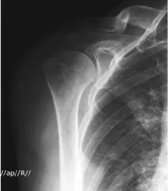
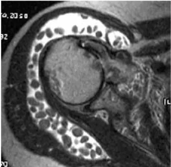
단순 방사선 소견상 상완골 근위부의 석회화 되지 않은 연 부 조직 종괴를 보였다(Fig. 1). 그리고 우측 견관절뿐만 아 니라 손목 관절 부위에 피질골이 얇아진 골다공증과 관절 주변 이 골파괴 되어 관절 간격이 좁아진 전형적인 류마토이드 관절 염의 소견이 관찰 되었다(Fig. 2). 10-MHz의 탐촉자를 가진 초음파(Philips, Ultramark 400C, Best, Netherlands)의 소견상 견봉하-삼각근하 전체에 다발성의 저에코 종괴가 보였 다(Fig. 3). 또한, 자기 공명 영상(Philips, Intera 1.5T, Best, Netherlands)에서는 T1 강조 영상에서 등신호 ( i s o t e n s e )를 나타냈고(Fig. 4), T2 강조 영상에서는 저신호
류마토이드 관절염에서 회전근 개 전층 파열과 동반되어 견관절과 견봉하 및 삼각근하 점액낭에 동시에 발생된 다발성 미립체:
관절경적 치료 및 자기 공명 영상 소견 -1례 보고-
노규철・정영기・유정한・나경수 한림대학교의과대학강남성심병원정형외과
류마토이드관절염이나혈청학적음성의여러관절병증에서작은크기의섬유소성미립체의형성은흔히나타나는소견이다.
그러나 류마토이드 관절염에서 견관절의 견봉하-삼각근하 점액낭염과 연관되어 생기는 다발성의 미립체는 굉장히 드물다.
지금까지, 문헌상만성적인류마토이드관절염환자에서견관절의회전근개전층파열과동반되어견관절과견봉하-삼각근 하점액낭에동시에발생된다발성미립체를관혈적이아닌관절경적으로치료한예는없는것으로사료되어이에보고하고자 한다. 또한, 견봉하-삼각근하점액낭에발생한미립체에대한자기공명영상소견도최근까지단지몇몇보고에만언급되어 왔다. 그래서 다발성 미립체와 연관된 여러가지의 활액막 증식성 질환들의감별진단을위해 견봉하-삼각근하점액낭에 생 긴다발성미립체의자기공명영상소견을기술하고자한다.
색인단어: 류마토이드관절염, 미립체, 견봉하-삼각근하점액낭염, 회전근개전층파열, 관절경, 자기공명영상
통신저자: 유 정 한
서울특별시 영등포구 대림1동 9 4 8 - 1
한림대학교 의과대학 강남성심병원 정형외과학교실 TEL: 02) 829-5165・FAX: 02) 834-1728 E-mail: jhbyoo@kornet.net
65
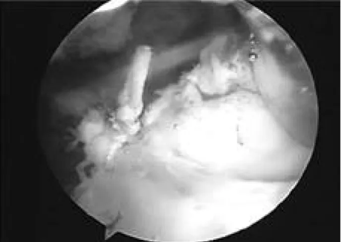
( h y p o t e n s e )의 골격근에 비해 오히려 경도의 고신호( h y p e r- t e n s e )를 갖는 다양한 크기의 다발성 결절로 채워진 견봉하- 삼각근하 점액낭의 현저한 팽창이 보였다(Fig. 5). 관절경 수 술 소견상 약 3 0 0개 이상의 작은 쌀 모양의 유리체가 견봉하- 삼각근하 점액낭과 동시에 견관절낭에서 관찰되었다(Fig. 6).
관절경을 이용한 수술시 필터가 달린 흡입관을 통해 미립체를 제거하였으며, 견관절과 견봉하-삼각근하 점액낭의 활막 제거 술을 동시에 시행하였다. 미립체의 크기 범위는 약 5~9 mm 였다(Fig. 7). 관절경 소견상 회전근 개의 전층 파열이 있었 다(Fig. 8). 파열된 회전근 개는 약 1×4 cm 정도의 종파열 로 관절경을 이용해 흡수성 봉합사인 P D S와 비흡수성 봉합사 인 E t h i b o n d를 이용하여 건과 건의 봉합을 시행하였다( F i g . 9). 병리학적으로 점액낭 벽은 융모성 세포로 덮여 있었으며 림프성 결절이 존재하였다. 미립체 자체는 조밀한 섬유성 물질 로 구성 되어 있었다(Fig. 10).
Fig. 1. Radiographs showed soft tissue mass in the region of the proximal humerus without soft tissue calcifications.
Fig. 2. The right shoulder and other involved joints showed evidence of rheumatoid arthritis.
Fig. 3. Ultrasound showed multiple hypoechoic mass with almost completely subacromial-subdeltoid bursae.
Fig. 4. MR imaging showed a markedly distended subacromi- al-subdeltoid bursae, filled with multiple nodules of varying sizes that were isointense on T1-weighted.
수술 후 첫째날 부터 현저한 동통의 감소와 운동 범위의 향 상이 보였다. 수술 후 둘째날 에 능동적 견관절 운동을 시작하 였다. 수술 후 2달째 추시 관찰시 환자는 외전 9 0도, 굴곡 1 2 0도, 내회전 및 외회전 4 5도의 운동범위를 얻었다. 수술 후 7개월에 환자는 견관절의 완전한 운동 범위를 얻었다. 동통, 종창은 없었으며 견관절의 잠김 증상도 없었다.
고 찰
미립체는 교원질과 섬유소에 둘러싸인 호산성( a c i d o p h i l i c ) 물질로 내측에 무정형의 중심체를 갖는다. 그것들은 특히 혈청 검사 양성을 보이는 슬관절의 류마토이드 관절염과 관련이 있
으나, 질환 자체의 기간이나 중증도, 방사선학적 변화와는 관 련이 없다2 , 7 ). 저자들의 증례에서는 방사선적, 혈청학적 그리고 병리학적 결과들을 통해 다발성 관절의 류마토이드 관절염의 진단을 확진할 수 있었다.
특히, 점진적으로 증가하는 활액막의 비후와 간헐적인 악화 가 동반된 삼출이 수개월 또는 수년에 걸쳐 발현된다. 병리학 적으로 점액낭 벽은 치밀한 림프 형질 세포성 침투가 동반된 융모성 증식 활액막으로 둘러싸여 있다2 , 9 , 1 1 )
. 병인은 불분명하 지만, 몇몇 저자들은 활액막의 미세 경색으로 인한 활액막 박 리와 활액막액으로부터 생겨난 섬유소에 덮여 발생한다고 주
장한다2 , 3 ). 또 다른 저자들은 활액막 구성 요소와는 별개로 초
기의 미립체가 활막액에서 형성된 후 섬유소의 응집으로 점차 커지게 된다고 제안한다2 , 7 ).
자기 공명 영상 소견으로 활막 연골종증에서 보이는 유리 체, 미립체 그리고 색소성 융모결절 활액막염을 구별할 수 있 다. 미립체의 자기 공명 영상에 대한 이전 보고들은 T 1과 T 2
Fig. 5. MR imaging showed slightly hyperintense on T2- weighted spin-echo images relative to the hypointense skeletal muscle.
Fig. 6. During arthroscopic surgery, more than 300 small fine loose rice-like bodies were detected in the subacromial and gleno-humeral spaces.
Fig. 7. These bodies ranged in size from 5 to 9 mm.
Fig. 8. There was communication between the subacromial bursa and the joint, implying full-thickness rotator cuff tear.
강조 영상에서 골격근과 비교할 때 등신호 또는 저신호를 보인 다고 말하고 있다. 본 증례에서는 미립체가 T2 강조 영상에서 약간의 고신호를 보이나, 주변의 점액낭 액과는 명확히 구분되 었다2 , 4 , 7 , 8 , 1 0 )
.
미립체의 우선 감별 진단은 활액막으로 둘러싸인 점액낭을 거의 포함하지 않고 무기화 되지 않은 화생성 연골( m e t a- plastic cartilage)을 가지는 활막 골연골종증이다5 ). 무기질화 가 진행하면, 방사선 검사상 이상 소견이 관찰된다. 특히, 단 순 방사선 촬영이 정상 소견이라면 비석회화 또는 석회화 결절 을 보이는 경우에는 더욱이 자기 공명 영상이 필요하다. 무기 질화 되지 않고, 연골이 함유된 활막 연골종증의 종물은 저신 호의 골격근과 비슷하게 보여진다2 ). 본 증례에서 미립체들은 T1 강조 영상에서 등신호를 보였고, T2 강조 영상에서 미세 하게 증가된 신호를 보여 사실상 식별이 불가능하였다. 그러나 미립체들과는 다르게 일반적으로 T1, T2 강조 영상에서 약간
고신호를 보이는 활막 연골종증은 T1 강조 영상에서 식별될 수 있으나, T2 강조 영상에서는 점액낭 액체와 구별되지 않을 수 있어 이런 차이점을 이용해 감별할 수 있다. 또한 회전근 개 전층 파열과 동반되어 인회석 결정(apatite crystal)으로 인해 염증을 동반하는 Milwaukee shoulder도 감별해야 할 질환이나 본 증례에서는 회전근 개 파열 관절증은 없었으며 인 회석 결정도 없었다1 ).
활액막 절제술은 활액막 증식 질환의 치료로 권장되고 있 다. 또한, 미립체들과 동반된 증식성 활액막에서 관혈적이든 관절경적이든 유리체 제거술과 활액막 절제술로 치료 될 수 있 다. 관절경적 변연 절제술 및 활액막 절제술은 류마토이드 질 환 환자에게 있어서 통증, 종창의 조절 및 관절 기능 향상에 큰 도움이 된다.
과거력상 전신적인 관절병증이 있어 미립체를 형성하는 여 러가지의 활액막 증식성 질환인 색소 융모 결절성 활액막염과 활막 연골종증 등의 질환을 감별 진단하는 것은 무엇보다 수술 전에 꼭 행해져야 한다. 특히 단순 방사선과 자기 공명 영상 소견은 미립체 를 형성하는 상기 질환들의 술전 감별 진단에 도움이 되므로 세밀한 관찰이 필요하다. 또한 관절경적 치료가 관혈적 방법에 비해 넓은 시야를 가지기 때문에 관절경적 미립 체 제거술은 다른 연관된 관절내 병변을 치료할 수 있을뿐 아 니라 결과적으로 적은 이환율로 치료 후 빠른 재활 운동과 환 자를 가능한 빨리 정상 일상 생활로 복귀 시킬 수 있어 유용한 술식으로 사료된다.
참고문헌
01. Antoniou J, Tsai A, Baker D, Schumacher R, Williams GR and Iannotti JP: Milwaukee shoulder: correlating possible etiologic variables. Clin Orthop Relat Res, 407:79-85, 2003.
02. Chen A, Wong LY, Sheu CY and Chen BF : Distinguishing multiple rice body formation in chronic subacromial-subdeltoid bursitis from synovial chondro - matosis. Skeletal Radiol, 31:119-121, 2002.
03. Cheung HS, Ryan LM, Kozin F and McCarty DJ:
Synovial origins of Rice bodies in joint fluid. Arthritis Rheum, 23:72-76, 1980.
04. Griffith JF, Peh WC, Evans NS, Smallman LA, Wong RW and Thomas AM: Multiple rice body formation in chronic subacromial/subdeltoid bursitis: MR appear - ances. Clin Radiol, 51:511-514, 1996.
05. Milgram JW : Synovial osteochondromatosis: a histopathological study of thirty cases. J Bone Joint Surg, 59(A):792-801, 1977.
06. Popert AJ, Scott DL, Wainwright AC, Walton KW, Williamson N and Chapman JH: Frequency of occur - Fig. 9. Tear was longitudinal shape so we performed side by
side suture with PDS absorbable sutures and Ethibond non-absorbable sutures.
Fig. 10. Pathologically, the bursal wall was lined with villous cells and lymphoid nodules were present. Rice bodies were noted to consist of dense fibrinous material.
rence, mode of development, and significance of rice bod - ies in rheumatoid joints. Ann Rheum Dis, 41:109-117, 1982.
07. Spence LD, Adams J, Gibbons D, Mason MD and Eustace S: Rice body formation in bicipito-radial bursitis:
ultrasound, CT, and MRI findings. Skeletal Radiol, 27:30- 32, 1998.
08. Stein AJ, Case JL, Berman J and Levy H: Case report 770. Chronic subacromial bursitis with massive formation of rices bodies. Skeletal Radiol, 22:71-73, 1993.
09. Steinfeld R, Rock MG, Younge DA and Cofield RH:
Massive subacromial bursitis with rice bodies: Report of three cases, one of which was bilateral. Clin Orthop Relat Res, 301:185-190, 1994.
10. Tan CH, Rai SB and Chandy J: MRI appearances of multiple rice body formation in chronic subacromial and subdeltoid bursitis, in association with synovial chodro - matosis. Clin Radiol, 59:753-757, 2004.
11. Witwity T, Uhlmann R, Nagy MH, Bhasin VB, Bahgat MM and Singh AK: Shoulder rheumatoid arthritis asso - ciated with chondromatosis, treated by arthroscopy.
Arthroscopy, 7:233-236, 1991.
Multiple Rice Body in both Glenohumeral Joint and Subacromial & Subdeltoid Bursa Simultaneously combined with Full Thickness Cuff Tear in Rheumatoid
Arthritis: Arthroscopic Treatment & MR Appearance -A Case Report-
Kyu Cheol Noh, M.D., Yung Khee Chung, M.D., Jung Han Yoo, M.D., Kyong Soo Nah, M.D.
Department of Orthopaedic Surgery, Kangnam Sacred Heart Hospital, Hallym University School of Medicine, Seoul, Korea
Numerous small fibrinous rice bodies are a common finding in joints afflicted with rheumatoid arthri- tis(RA) or seronegative arthropathy. Subacromial and subdeltoid bursitis of the shoulder associated with multiple rice body formation is a rare occurrence. To our knowledge, this is the first report to describe the arthroscopic treatment of massive rice bodies in both glenohumeral joint and subacromi- al-subdeltoid bursae combined with full thickness of rotator cuff in RA. Besides, the MR appearance of subacromial-subdeltoid bursal rice bodies have been previously described in only few recent reports. Therefore, we also describe the MR appearances subacromial-subdeltoid bursae associated with multiple rice bodies in RA.
Key Words: Rheumatoid arthritis, Rice body, Subacromial and subdeltoid bursitis, Full thickness cuff tear, Arthroscopy, MRI
Address reprint requests to Jung-Han Yoo, M.D.
Department of Orthopaedic Surgery, Kangnam Sacred Heart Hospital,
Hallym University School of Medicine 948-1 Daerim 1 Dong, Youngdeungpo-gu, 150-950, Seoul, Korea.
TEL: 82-2-829-5165, FAX: 82-2-834-1728, E-mail: jhbyoo@kornet.net
= ABSTRACT =