https://doi.org/10.4174/astr.2020.98.2.89 Annals of Surgical Treatment and Research
Is platelet-rich plasma improves the anastomotic healing in hyperthermic intraperitoneal chemotherapy with
oxaliplatin: an experimental rat study
Omer Faruk Buk1, Sonmez Ocak1, Bugra Genc2, Bahattin Avcı3, Hatice Olger Uzuner4
1Department of General Surgery, University of Healthy Sciences, Samsun Research and Training Hospital, Samsun, Turkey
2Department of Animal Nutrition and Nutritional Diseases, Faculty of Veterinary Medicine, Ondokuz Mayis University, Samsun, Turkey
3Department of Biochemistry, Faculty of Medicine, Ondokuz Mayis University, Samsun, Turkey
4Department of Pathology, University of Healthy Sciences, Samsun Research and Training Hospital, Samsun, Turkey
INTRODUCTION
Despite developing diagnostic tools and screening programs, approximately 4%–7% of patients with colon cancer have peritoneal carcinomatosis (PC) at the time of diagnosis [1].
Metachronous peritoneal metastasis can also be seen in 4% to 50% of patients during follow-up [2-4]. In the past, PC was accepted as the terminal stage. However, recently a new treatment modality called ‘hyperthermic intraperitoneal chemotherapy (HIPEC) combined with cytoreductive surgery
Received August 1, 2019, Revised October 30, 2019, Accepted January 4, 2020
Corresponding Author: Sonmez Ocak
Department of General Surgery, University of Healthy Sciences, Samsun Research and Training Hospital, 199 Barış Bulvarı Kadıköy Mahallesi, İlkadım/Samsun 55090, Turkey
Tel: +90-506-531-48-29, Fax: +90-362-277-88-65 E-mail: [email protected]
ORCID: https://orcid.org/0000-0001-9550-4268
Copyright ⓒ 2020, the Korean Surgical Society
cc Annals of Surgical Treatment and Research is an Open Access Journal. All articles are distributed under the terms of the Creative Commons Attribution Non- Commercial License (http://creativecommons.org/licenses/by-nc/4.0/) which permits unrestricted non-commercial use, distribution, and reproduction in any medium, provided the original work is properly cited.
Purpose: Hyperthermic intraperitoneal chemotherapy (HIPEC) is a novel treatment option for peritoneal surface malignancies. Due to cytotoxic effects of chemotherapeutic agents, anastomosis healing can be impaired and lead to leakage rates higher than conventional intestinal surgery. In this experimental study, we aimed to investigate the effects of platelet-rich plasma (PRP) on colonic anastomosis in rats that received HIPEC with oxaliplatin.
Methods: Thirty rats were divided into 3 groups. Group 1 was determined as control group and hyperthermic saline perfusion was performed after colon anastomosis. In group 2, colon anastomosis then hyperthermic oxaliplatin perfusion was performed. In the last group, the colonic anastomosis was enhanced by PRP gel and then hyperthermic oxaliplatin perfusion was performed. All the rats were reoperated on postoperative day 7 and anastomotic bursting pressure values were recorded. Tissue samples were taken for hydroxyproline assay and histopathological examination.
Results: Control group had higher anastomotic bursting pressure value than group 2 and group 3 (P < 0.001). There were significant differences in anastomotic bursting pressure between groups 2 and 3 (P < 0.001). Group 2 had significantly lower hydroxyproline levels than group 3 and control group (P < 0.001). Histopathological examination revealed that PRP application reduced inflammatory response.
Conclusion: PRP application on colonic anastomosis improves anastomotic healing and can reduce anastomosis related complications and stoma creation; though further clinical studies are needed.
[Ann Surg Treat Res 2020;98(2):89-95]
Key Words: Anastomotic leak, Colonic neoplasms, Hyperthermia, Oxaliplatin, Platelet-rich plasma
(CRS)’ has provided prolonged survival rates with acceptable mortality and morbidity rates [5,6]. Anastomotic leakage rates have been found to be higher in CRS plus HIPEC compared to conventional colorectal surgery ranging between 10%–25% in various studies, which is why stomal diversion is required in such patients [7-11].
Platelet-rich plasma (PRP) contains numerous platelet- derived growth factors which improve wound healing and have been widely used in various surgical procedures. Promising experimental studies have been reported regarding PRP intestinal anastomotic healing and PRP’s application [12-14].
In this experimental animal study, we aimed to investigate whether PRP application has favorable effects on colonic anastomosis in rats when performed with oxaliplatin-based HIPEC.
METHODS
This study was conducted at the Experimental Animal Application and Research Center of Ondokuz Mayıs University upon the approval of the Local Ethical Committee of Ex- perimental Animal of Ondokuz Mayıs University (approval number: 2017/51). Thirty-five male Wistar-Albino rats were used.
Rats were maintained at 22℃ ± 2℃ with a 12-hour light/dark cycle at 40%–50% humidity. Rats were allowed free access to water and standard laboratory rat pellets.
PRP preparation
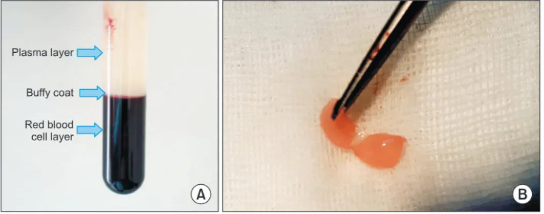
Five rats were selected as donors and used for PRP pre- paration. Blood samples were obtained by cardiac puncture and 8.5 mL of blood from each donor was drawn into the acid- citrate-dextrose tubes (BD Vacutainer, BD, Franklin Lakes, NJ, USA). The tubes were then centrifuged at 2,500 rpm and 22℃ for 10 minutes. After the first spin, 3 layers were seen as follows; (1) light yellow plasma layer, (2) buffy coat layer, and (3) red blood cells layer (Fig. 1A). Plasma and buffy coat layers were taken carefully and drawn into another tube, then centrifuged at 3,500 rpm and 22℃ for 15 minutes. Finally, 1-mL PRP was harvested and mixed with 1-mL thrombine (Human Thrombin Lyophilized, Baxter, Cherry Hill, NJ, USA) and 0.5-mL calcium chloride in order to obtain the gel form of PRP (Fig. 1B).
Study groups and surgical procedure
Rats were divided equally into 3 groups (Fig. 2): Group 1:
Control group; left colon was transected and end-to-end colonic anastomosis was performed, then hyperthermic (42℃) saline
Plasma layer
Buffy coat
Red blood cell layer
A B
Fig. 1. (A) Three layers were seen after first spin; plasma, buffy coat, and red blood cells layers.
(B) Gel form of platelet-rich pla- sma.
Fig. 2. Study protocol. PRP, pla- telet-rich plasma; HIPEC, hyper- thermic intraperitoneal che mo- therapy. Group 1, control group;
group 2, oxaliplatin alone group;
group 3, oxaliplatin and platelet- rich plasma group.
Sacrified for PRP preperation
n = 5
Group 1 n = 10
Group 2 n = 10
Group 3 n = 10
Control group Left colon resection and colocolonic anastomosis then hyperthermic saline perfusion
HIPEC group Left colon resection and colocolonic anastomosis then HIPEC with oxaliplatin
HIPEC + PRP group Left colon resection and colocolonic anastomosis, PRP application then HIPEC
with oxaliplatin 35 Wistar albino
rats
were perfused for 60 minutes. Group 2: Oxaliplatin alone group;
left colon was transected and end-to-end colonic anastomosis was performed followed by HIPEC with oxaliplatin (77.5 mg/
kg) at 42℃ and 5 mg/kg for 60 minutes. Group 3: Oxaliplatin and PRP group; left colon was transected, end-to-end colonic anastomosis was performed and PRP gel was applied to the anastomosis site, then HIPEC was performed with oxaliplatin (77.5 mg/kg) at 42℃ and 5 mg/kg for 60 minutes.
Surgical procedure
Ketamine hydrochloride (Ketalar, Park Devis, Istanbul, Turkey) at a dose of 50 mg/kg, and xylazine (Rompun, Bayer, Istanbul, Turkey) at a dose of 5 mg/kg were used for anesthesia.
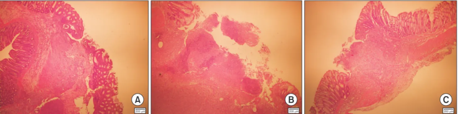
After skin shaving and skin preparation with 10% povidone iodine, a laparotomy was performed and the left colon was identified and transected. End-to-end anastomosis was per- formed with 5/0 polypropylene sutures (Prolene, Ethicon, Cincinnati, OH, USA). In group 3, PRP gel was applied all around the anastomosis line (Fig. 3). Inflow and outflow catheters were placed to the right supradiaphragmatic space and pelvis, respectively, then the abdomen was closed with separate 3/0 silk sutures. After closure of the abdomen oxaliplatin in 5%
dextrose solution was perfused for 60 minutes by a roller pump in groups 2 and 3. The perfusate was heated by a heater plate and the temperature of the abdomen was monitored continuously with a digital thermometer. The animals were taken back to their cages and fed chow and water after the operation.
Anastomotic bursting pressure measurement
On the seventh day following surgery, the animals were reoperated on and both abdomen and colonic anastomosis were evaluated. The colon segment involving the anastomosis was removed and 2 rubber catheters were placed into the proximal and distal lumen of the excised segment. The catheters were fixed with 3/0 silk sutures and catheters were connected to a syringe pump and a manual sphygmomanometer to measure the anastomotic bursting pressure (ABP). The colon segment was insufflated with air by syringe pump and when the first air bubble was detected, the pressure value was recorded as ABP.
After recording ABP, the colon segment was opened and divided into 2 pieces, one piece was to be used for histopathological analysis and therefore put into 10% formalin solution, and the other was stored at -20℃ for hydroxyproline assay. The rats were euthanized with a high dose of anesthetic agent.
Histologic examination
All materials were fixed in formalin and embedded in paraffin. Haematoxylin-eosin stained sections were evaluated by one pathologist without knowledge of the group. An Olympus (BX51) light microscope was used. Verhofstad scale was used to evaluate the anastomotic healing by scoring the degrees of necrosis, neutrophils, macrophages, lymphocytes, edema, and mucosal epithelium [15].
Hydroxyproline assessment
Tissues were homogenized with liquid nitrogen and were soaked in a phosphate buffer solution (10 mM; pH, 7.2). Tis- sue samples were sonicated for 10 minutes at +4°C with 220 V (METU electromechanical, Serial No. 30607, Berlin, Ger many) before they were stored at -80°C until assay. The homogenates, which were defrosted at room temperature, were later centrifuged at +4°C for 5 minutes with 15,000 g.
The supernatants were taken for hydroxyproline analysis.
The concentrations of hydroxyproline in the sample were measured using a commercially available enzyme-linked immunosorbent Hydroxyproline Assay Kit (CEA621Ge, Cat.
No.l180307228; Cloud-Clone Corp., TX, USA). This assay employs the competitive inhibition enzyme immunoassay technique. A monoclonal antibody specific to hydroxyproline was precoated onto a microplate. A competitive inhibition reaction was launched between biotin labeled hydroxyproline and unlabeled hydroxyproline (standards or samples) with the precoated antibody specific to hydroxyproline. After incubation the unbound conjugate was washed off. Next, avidin conjugated to horseradish peroxidase (HRP) was added to each microplate and incubated. The amount of bound HRP conjugate was in reverse proportion to the concentration of hydroxyproline in the sample. After addition of the substrate solution, the intensity of color developed was in reverse proportion to the Fig. 3. The gel form of platelet-rich plasma was applied
around the colonic anastomosis. It adheres quickly to the anastomotic site and was not affected by hyperthermic oxaliplatin perfusion.
concentration of hydroxyproline in the sample. The protein amount of each sample of the tissue homogenates was calculated with the Lowry method [16]. Tissue hydroxyproline values were expressed as ng per mg protein. All assays were conducted according to the manufacturer’s instructions. The samples, which showed higher concentrations, were diluted and measured in duplicate.
Statistical analysis
Sample size was calculated by the statistical package (G*Power software) from previous experimental rat studies data on intestinal anastomosis healing with a power of 80% α-error and 5% β-error, and it is estimated that at least 10 samples were needed for each group. Numerical data were presented as mean ± standard deviation. One-way analysis of variance test was used for analyzing the differences between the mean values of the parameters (ABP, tissue hydroxyproline level, and histological scores) and either Tukey honestly significant difference or Tamhane post hoc tests were used to analyze the differences between the groups. A P-value below 0.05 was considered statistically significant.
RESULTS
All the animals survived until the end of the study. No wound complications, including wound infection, seroma, or
wound dehiscence, occurred. No local or systemic complications related to PRP were observed. In group 3, it was observed that hyperthermic perfusion did not remove the PRP gel due to PRP gel adhering strongly to the anastomosis line.
Anastomotic bursting pressure
The ABP values of the groups are given in Table 1. In variant analysis, as expected, group 1 had higher ABP value than group 2 and 3 (P < 0.001 and P < 0.001, respectively). Group 2 had lower ABP than group 3 and the difference was significant (P <
0.001). These findings indicate that PRP gel application has a positive effect on ABP.
Hydroxyproline levels
Table 1 shows the hydroxyproline levels of the groups. Group 2 had significantly lower hydroxyproline levels than groups 1 and 3 (P < 0.001). The difference between groups 1 and 3 was significant (P < 0.001). These data show that PRP application improved wound healing.
Histopathological examination
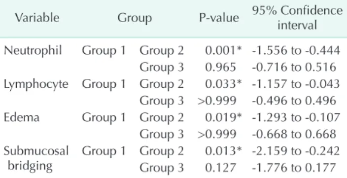
Verhofstad scale is a good guideline for evaluating anasto- motic wound healing and is widely used in experimental studies [13,15,17,18]. In our study, histopathological analysis demonstrated that PRP application improved the anastomotic healing on a cellular basis by decreasing neutrophils and
Table 1. The mean values of anastomotic bursting pressure and hydroxyproline levels of the study groups
Variable Group 1 Group 2 Group 3 P-value
Anastomotic bursting pressure (mmHg) 180 ± 9.14 94.90 ± 9.90 125.7 ± 15.63 <0.001*
Hydroyproline level (ng/mg protein) 314.69 ± 75.57 92.00 ± 26.97 280.92 ± 45.85 <0.001*
Values are presented as mean ± standard deviation.
Group 1, control group; group 2, oxaliplatin alone group; group 3, oxaliplatin and platelet-rich plasma group.
*P < 0.05, statistically significant difference.
A B C
200 m 200 m 200 m
Fig. 4. (A) Group 1: There is marked submucosal bringing, less edema, and inflammation (H&E, ×40). (B) Group 2: There is huge necrotic exudate on the luminal surface and between the anastomosis sides (H&E, ×40). (C) Group 3: There is less inflammation, edema and more bringing (H&E, ×40). Group 1, control group; group 2, oxaliplatin alone group; group 3, oxaliplatin and platelet-rich plasma group.
lym pho cytes amounts, as well as degree of edema. Also, sub- mucosal bridging was significantly better in group 1 and group 3 than in group 2 (Fig. 4) Detailed data analysis is shown in Tables 2 and 3.
DISCUSSION
Despite the survival advantages of CRS combined with HIPEC, this treatment modality has higher morbidity rates when compared with conventional surgical procedures. It is believed that morbidity would be caused by either major surgery or drug toxicity [19]. Anastomosis leakage can occur in up to 10%–25% of cases in those whom have undergone colorectal resections. For this reason, the majority of surgeons prefer to perform stomas (either ileostomy or colostomy) to prevent fecal peritonitis and its fatal results [20]. However, stoma closure requires additional surgical intervention, and reduced quality of life in patients with stomas has been reported [21]. Doud et al. [22] reported that stoma closure surgery is associated with a higher rate of morbidity and mortality for patients on whom HIPEC was performed.
PRP is an autologous concentrate that contains numerous growth factors, and several experimental studies have shown that PRP application improves wound healing as well as intestinal anastomosis healing [12,13,23,24]. In this experimental study, our data showed that PRP reduced tissue inflammation and increased hydroxyproline levels and ABP.
These data suggest that PRP gel improves impaired anastomotic healing due to hyperthermic oxaliplatin perfusion. In light of these findings, we propose to use PRP gel reinforcement to colonic anastomosis to prevent stoma and stoma related morbidity and mortality by reducing anastomotic leakage rates.
Our proposal has several strengths outlined as follows; PRP gel application is safe and has no reported adverse effects to date. It
will also not cause any allergic events due to being autologous.
PRP gel application is easy to prepare and use, can be obtained in about an hour in the operating room, and has a lower cost compared to other reinforcement materials [25]. Furthermore, we evaluated the anastomosis strength on postoperative day 7. Both experimental and clinical studies have shown that collagen lysis started in the first 3 to 5 days and anastomosis strength reached the lowest value on the seventh day, thus most of the anastomosis leakage occurred within those days [12,26- 29]. Median ABP value, as well as the median hydroxyproline level, was significantly higher in the PRP applied group (group 3) than in group 2. These data suggest that applying PRP gel has a positive impact on impaired anastomosis healing due to hyperthermic oxaliplatin perfusion.
This study has several limitations. First of all; this ex- periment was based on an animal model, thus the effects of this application on humans are unknown. Although there are numerous experimental studies suggesting favorable effects of PRP on intestinal anastomosis healing, interestingly, to date there are no clinical studies regarding this issue except one. Recently, Casella et al. [25] used PRP gel as a stapler line reinforcement material in patients who had received laparoscopic sleeve gastrectomy. They followed 20 patients and no leakage was observed during the 1-year follow-up period.
Despite the small number of patients, the authors encourage us to use PRP on gastrointestinal anastomosis; of course, further clinical studies including a larger number of patients are needed. Secondly, the rats were reoperated at postoperative day 7 in our study. Therefore, any late complications could not be evaluated including abscess, ileus, etc., as well as postoperative mortality. Thirdly, and perhaps most importantly, concern of cancer progression has been considered by some authors [30].
PRP contains numerous growth factors, which improve wound healing, but could also increase the proliferation of tumor cells, and are unacceptable for cancer patients. However, the latter is Table 2. Comparison of histopathological parameters of the
groups according to the Verhofstad scale
Variable Group 1 Group 2 Group 3 P-value Necrosis 2.3 ± 0.82 2.5 ± 0.52 2.7 ± 0.48 0.378 Neutrophil 1.9 ± 0.56 2.9 ± 0.52 2.0 ± 0.47 <0.001*
Lymphocyte 1.8 ± 0.42 2.4 ± 0.51 1.8 ± 0.42 0.008*
Macrophages 2.0 ± 0.47 2.5 ± 0.52 2.3 ± 0.67 0.156 Edema 2.1 ± 0.56 2.8 ± 0.42 2.1 ± 0.56 0.007*
Mucosal
epithelium 2.6 ± 0.966 2.6 ± 0.516 2.5 ± 0.84 0.949 Submucosal
layer 0.8 ± 0.63 2.0 ± 0.94 1.6 ± 0.96 0.014*
Values are presented as mean ± standard deviation (point).
Group 1, control group; group 2, oxaliplatin alone group; group 3, oxaliplatin and platelet-rich plasma group.
*P < 0.05, statistically significant difference.
Table 3. Post hoc analysis of the histopathological para- meters between the groups those with significant differences Variable Group P-value 95% Confidence interval
Neutrophil Group 1 Group 2 0.001* -1.556 to -0.444 Group 3 0.965 -0.716 to 0.516 Lymphocyte Group 1 Group 2 0.033* -1.157 to -0.043
Group 3 >0.999 -0.496 to 0.496 Edema Group 1 Group 2 0.019* -1.293 to -0.107
Group 3 >0.999 -0.668 to 0.668 Submucosal
bridging Group 1 Group 2 0.013* -2.159 to -0.242 Group 3 0.127 -1.776 to 0.177 Group 1, control group; group 2, oxaliplatin alone group; group 3, oxaliplatin and platelet-rich plasma group.
*P < 0.05, statistically significant difference.
not proven yet and still remains as a theory.
In conclusion, our data suggest the use of PRP gel on colonic anastomosis improves anastomotic wound healing in the cellular base and these findings will encourage the surgeons performing anastomosis without creation of stomas in patients who have received HIPEC with oxaliplatin. Further human
studies are needed to support our suggestion.
CONFLICTS OF INTEREST
No potential conflict of interest relevant to this article was reported.
REFERENCES
1. Aoyagi T, Terracina KP, Raza A, Takabe K. Current treatment options for colon cancer peritoneal carcinomatosis. World J Gastroenterol 2014;20:12493-500.
2. Nadler A, McCart JA, Govindarajan A.
Peritoneal carcinomatosis from colon cancer: a systematic review of the data for cytoreduction and intraperitoneal chemotherapy. Clin Colon Rectal Surg 2015;28:234-46.
3. Nagata H, Ishihara S, Hata K, Murono K, Kaneko M, Yasuda K, et al. Survival and prognostic factors for metachronous peritoneal metastasis in patients with colon cancer. Ann Surg Oncol 2017;24:
1269-80.
4. Bhatt A, Bhamre R, Rohila J, Kalikar V, Desouza A, Saklani A. Patients with extensive regional lymph node involve- ment (pN2) following potentially cura- tive surgery for colorectal cancer are at increased risk for developing peri toneal metastases: a retrospective single-insti- tution study. Colorectal Dis 2019;21:287- 96.
5. Glehen O, Kwiatkowski F, Sugarbaker PH, Elias D, Levine EA, De Simone M, et al. Cytoreductive surgery combined with perioperative intraperitoneal chemot- herapy for the management of peritoneal carcinomatosis from colorectal cancer:
a multi-institutional study. J Clin Oncol 2004;22:3284-92.
6. Verwaal VJ, van Ruth S, Witkamp A, Boot H, van Slooten G, Zoetmulder FA.
Long-term survival of peritoneal carcino- matosis of colorectal origin. Ann Surg Oncol 2005;12:65-71.
7. Casado-Adam A, Alderman R, Stuart OA,
Chang D, Sugarbaker PH. Gastrointestinal complications in 147 consecutive patients with peritoneal surface malignancy treated by cytoreductive surgery and peri- operative intraperitoneal chemo therapy.
Int J Surg Oncol 2011;2011:468698.
8. Hompes D, D’Hoore A, Van Cutsem E, Fieuws S, Ceelen W, Peeters M, et al. The treatment of peritoneal carcinomatosis of colorectal cancer with complete cyto- reduc tive surgery and hyperthermic intra- peritoneal peroperative chemotherapy (HIPEC) with oxaliplatin: a Belgian multi- centre prospective phase II clinical study.
Ann Surg Oncol 2012;19:2186-94.
9. Gervais MK, Dube P, McConnell Y, Drolet P, Mitchell A, Sideris L. Cytoreductive surgery plus hyperthermic intraperitoneal chemo therapy with oxaliplatin for peri- toneal carcinomatosis arising from colo- rectal cancer. J Surg Oncol 2013;108:438- 43.
10. Riss S, Chandrakumaran K, Dayal S, Cecil TD, Mohamed F, Moran BJ. Risk of definitive stoma after surgery for peri- toneal malignancy in 958 patients: com- parative study between complete cyto- reductive surgery and maximal tumor debulking. Eur J Surg Oncol 2015;41:392-5.
11. Whealon MD, Gahagan JV, Sujatha- Bhaskar S, O’Leary MP, Selleck M, Dumitra S, et al. Is fecal diversion needed in pelvic anastomoses during hyper- thermic intraperitoneal chemo therapy (HIPEC)? Ann Surg Oncol 2017;24:2122-8.
12. Yamaguchi R, Terashima H, Yoneyama S, Tadano S, Ohkohchi N. Effects of platelet- rich plasma on intestinal anastomotic healing in rats: PRP concentration is a key
factor. J Surg Res 2012;173:258-66.
13. Sozutek A, Colak T, Cetinkunar S, Reyhan E, Irkorucu O, Polat G, et al. The effect of platelet-rich-plasma on the healing of left colonic anastomosis in a rat model of intra-abdominal sepsis. J Invest Surg 2016;29:294-301.
14. Alves R, Grimalt R. A review of platelet- rich plasma: history, biology, mechanism of action, and classification. Skin Appen- dage Disord 2018;4:18-24.
15. Verhofstad MH, Lange WP, van der Laak JA, Verhofstad AA, Hendriks T. Micro- scopic analysis of anastomotic healing in the intestine of normal and diabetic rats.
Dis Colon Rectum 2001;44:423-31.
16. Lowry OH, Rosebrough NJ, Farr AL, Randall RJ. Protein measurement with the Folin phenol reagent. J Biol Chem 1951;193:265-75.
17. Strebel K, Nielsen SR, Biagini M, Qvist N.
Effect of Humira® on intestinal anasto- motic response in rabbits. J Invest Surg 2015;28:167-72.
18. Aghayeva A, Benlice C, Bilgin IA, Atukeren P, Dogusoy G, Demir F, et al. The effects of hyperthermic intraperitoneal chemo- perfusion on colonic anastomosis: an ex- peri mental study in a rat model. Tumori 2017;103:307-13.
19. von Breitenbuch P, Piso P, Schlitt HJ.
Safety of rectum anastomosis after cyto- reduc tive surgery and hyperthermic intra- peritoneal chemotherapy. J Surg Oncol 2018;118:551-6.
20. Deraco M, Glehen O, Helm CW, Morris DL, van der Speeten K. Cytoreductive sur- gery & perioperative chemotherapy for peri toneal surface malignancy: textbook
and video atlas. Woodbury (CT): Cine- Med Publishing Inc.; 2013.
21. Braumann C, Mul ler V, K nies M, Aufmesser B, Schwenk W, Koplin G. Qua- lity of life and need for care in pa tients with an ostomy: a survey of 2647 patients of the Berlin OStomy-Study (BOSS).
Langenbecks Arch Surg 2016;401:1191-201.
22. Doud AN, Levine EA, Fino NF, Stewart JH, Shen P, Votanopoulos KI. Stoma creation and reversal after cytoreductive surgery with hyperthermic intraperitoneal che- mo therapy. Ann Surg Oncol 2016;23:503- 10.
23. Yol S, Tekin A, Yilmaz H, Kucukkartallar T, Esen H, Caglayan O, et al. Effects of platelet rich plasma on colonic anasto- mosis. J Surg Res 2008;146:190-4.
24. Esat Duymus M, Temel S, Ozer H, Kemal Urhan M, Kaya F, Aslan F, et al. Com pa- rison of the effects of plateletrich pla s ma pre pared in various forms on the healing of dermal wounds in rats. Wounds 2016;
28:99-108.
25. Casella G, Soricelli E, Genco A, Ferrazza G, Basso N, Redler A. Use of platelet-rich plasma to reinforce the staple line du ring laparoscopic sleeve gastrectomy: fea si- bility study and preliminary out come. J Laparoendosc Adv Surg Tech A 2015;25:
222-7.
26. Hyman N, Manchester TL, Osler T, Burns B, Cataldo PA. Anastomotic leaks after intestinal anastomosis: it’s later than you think. Ann Surg 2007;245:254-8.
27. Zhou B, Ren J, Ding C, Wu Y, Chen J, Wang
G, et al. Protection of colonic anastomosis with platelet-rich plasma gel in the open abdomen. Injury 2014;45:864-8.
28. Schiff A, Roy S, Pignot M, Ghosh SK, Fegelman EJ. Diagnosis and management of intraoperative colorectal anastomotic leaks: a global retrospective patient chart review study. Surg Res Pract 2017;2017:
3852731.
29. Daglioglu YK, Duzgun O, Sarici IS, Ulutas KT. Comparison of platelet rich plasma versus fibrin glue on colonic anastomoses in rats. Acta Cir Bras 2018;33:333-40.
30. Spartalis E, Prodromidou A, Spartalis M, Machairas N. Comment on ‘Role of platelet-rich fibrin on intestinal anasto- mosis wound healing in a rat’. Biomed Mater 2018;13:068001.