J Korean Surg Soc 2012;83:21-29
http://dx.doi.org/10.4174/jkss.2012.83.1.21
ORIGINAL ARTICLE
Journal of the Korean Surgical Society
JKSS
pISSN 2233-7903ㆍeISSN 2093-0488
Received January 9, 2012, Revised March 8, 2012, Accepted March 22, 2012 Correspondence to: Jin Cheon Kim
Department of Surgery, University of Ulsan College of Medicine, 86 Asanbyeongwon-gil, Songpa-gu, Seoul 138-736, Korea Tel: +82-2-3010-3489, Fax: +82-2-474-9027, E-mail: jckim@amc.seoul.kr
cc Journal of the Korean Surgical Society is an Open Access Journal. All articles are distributed under the terms of the Creative Commons Attribution Non-Commercial License (http://creativecommons.org/licenses/by-nc/3.0/) which permits unrestricted non-commercial use, distribution, and reproduction in any medium, provided the original work is properly cited.
Characterization of biological responses of colorectal cancer cells to anticancer regimens
Seon Ae Roh
1,2, Eun Young Choi
1,2, Dong Hyung Cho
3, Yong Sik Yoon
1,2, Tae Won Kim
4, Yong Sung Kim
5, Jin Cheon Kim
1,21Department of Surgery, Asan Medical Center, University of Ulsan College of Medicine, Seoul, 2Laboratory of Cancer Biology and Genetics, Asan Institute for Life Sciences, Seoul, 3Graduate School of East-West Medical Science, Kyung Hee University, Global Campus, Yongin, 4Department of Oncology, Asan Medical Center, University of Ulsan College of Medicine, Seoul, 5Division of Medical Genetics, Korea Research Institute of Bioscience & Biotechnology, Daejeon, Korea
Purpose: Identification of subgroups of patients who differ in their response to treatment could help to establish which of the best available chemotherapeutic options are best, based on biological activity. In metastatic colorectal cancer (CRC), novel molecular-targeted agents that act on pathways that regulate cell growth, the cell cycle, apoptosis, angiogenesis, and in- vasion are being developed. Here, we employed an in vitro chemosensitivity assay to evaluate the biological efficacy of con- ventional monotherapies and combination chemotherapy with targeted drugs. Methods: The chemosensitivities of 12 CRC cell lines to the established regimens FOLFOX (5-fluorouracil [5-FU] + leucovorin + oxaliplatin) and FOLFIRI (5-FU + leu- covorin + irinotecan) and to therapy with these regimens in combination with the biologically targeted drugs bevacizumab or cetuximab were comparatively evaluated for their effects on apoptotic and autophagic cell death processes, angiogenesis, and invasion. Results: Each of the chemotherapeutic regimens promoted apoptotic cell death and invasion. All drug regi- mens caused significantly greater apoptotic cell death with activation of caspase-3 in SW480 cells compared to other cells, ef- fects that were associated with a remarkable reduction in matrix metalloproteinase-9 activity. The FOLFOX regimen more ef- fectively promoted apoptotic cell death, angiogenesis, and invasion than the FOLFIRI regimen. Combination therapy with FOLFOX/FOLFIRI regimen and bevacizumab produced a moderate angiogenesis-blocking effect in most cell lines.
Conclusion: The results validate our in vitro chemosensitivity assay, and suggest that it may be applied to help determine ad- equate regimens in individual CRC patients based on the biological characteristics of their tumors.
Key Words: Colorectal neoplasms, Chemotherapy, Pharmacological biomarkers
INTRODUCTION
Colorectal cancer (CRC) is the third most common can- cer and the fourth most frequent cause of cancer deaths worldwide. Surgical resection remains the cornerstone of
management for patients with stage I to III disease; how- ever, a considerable number of patients will ultimately re- lapse and die from their disease. The main prognostic fac- tor for survival or relapse after surgery of localized disease is tumor stage. Accordingly, whereas stage I CRC is usu-
ally cured by surgery alone, adjuvant chemotherapy is currently recommended for stage III and high risk stage II cancers [1,2].
In a metastatic setting, patients are treated with two standard first- and second-line chemotherapy regimens (in either order): 1) FOLFOX, consisting of 5-fluorouracil (5-FU), which blocks thymidylic acid formation and there- fore DNA biosynthesis, with leucovorin (LV; a folate that stabilizes binding of 5-FU to thymidylate synthase) and oxaliplatin; and 2) FOLFIRI, consisting of 5-FU/LV with irinotecan [3]. Oxaliplatin is a platinum derivative that ex- erts antitumor activity against colon carcinomas by virtue of its cytotoxic effects, and irinotecan is a topoisomerase inhibitor that ultimately leads to inhibition of both DNA replication and transcription. Recently, the molecularly targeted drugs bevacizumab and cetuximab have been used in combination with FOLFOX and FOLFIRI in meta- static CRC, providing novel targets for cancer therapy.
Bevacizumab is a monoclonal antibody that binds and sequesters vascular endothelial growth factor (VEGF); ce- tuximab, also a monoclonal antibody, blocks the ligand- binding site of the epidermal growth factor receptor (EGFR) thereby inhibiting EGFR-mediated intracellular signal transduction.
In metastatic CRC, these molecularly targeted agents are being developed for their effects on pathways charac- teristic of tumors, including those that regulate cell growth, the cell cycle, apoptosis, angiogenesis, and in- vasion [4,5]. Angiogenesis -the formation of new capil- laries from the preexisting one- is an essential require- ment of solid tumor growth and metastasis. Accordingly, inhibition of angiogenesis has been explored as an anti- tumor treatment strategy, reflecting the expectation that poorly vascularized tumors would have limited growth potential and restricted metastatic capacity. VEGF, a spe- cific mitogen for endothelial cell proliferation is probably the key mediator of tumor angiogenesis [6,7].
The extracellular matrix (ECM) acts as both a physical scaffold for cells and a repository for growth factors.
Structural changes in ECM proteins accomplished by a complex process controlling the expression and activities of matrix metalloproteinases (MMPs), are a prerequisite for cell migration during tissue remodeling. Overexpres-
sion of MMPs leads to degradation of the ECM, an essen- tial step for tumor invasion and metastasis. Particular groups of MMPs, notably gelatinases A and B, also known as 72 and 92 kDa type IV collagenases or MMP-2 and MMP-9, respectively, are of particular interest with respect to their roles in the development and progression of CRC [8-10].
Cell death can be divided into two categories: apoptosis and necrosis. Apoptosis, or programmed cell death, repre- sents a cell-intrinsic “suicide” mechanism that is regulated by a variety of cellular signaling pathways. In many cases, tumors can be difficult to eliminate because cancer treat- ments often act by damaging cells with radiation or chem- icals, and cause mutations in the apoptotic pathway.
Understanding how apoptosis is regulated in cancer is therefore a major issue in the development of treatments for this disease [11,12]. Autophagy, a lysosome-based mechanism responsible for the degradation of cellular components, including organelles, might act as a balanc- ing mechanism between cell survival and cell death.
Caspases and autophagy are involved in complementary death pathways in cells: in cases where caspases are in- hibited, autophagy is required for cell death. In addition, signals such as steroids in flies and tumor necrosis factor- related apoptosis-inducing ligand (TRAIL) in human cells activate an autophagic cell death process that involves both active caspases and autophagy [13,14].
There is a need for a clinically useful chemosensitivity assay that helps to individualize the treatment delivered to cancer patients. Collagen sponge-gel supported histo- culture, which maintains cellular heterogeneity and an in- tact cyto-architecture, has been utilized to develop the his- toculture drug response assay (HDRA) for individualized chemotherapy. The three-dimensional collagen-based HDRA uses the 3-(4,5-dimethylthiazolyl-2) 2,5-diphe- nyltetrazolium bromide (MTT) assay as a readout for the effectiveness of chemotherapeutic agents [15]. The results obtained using HDRA are highly correlated with clinical drug response, resistance, and survival in advanced CRC.
However, because HDRA uses only MTT assay as an addi- tional means to objectively validate drug efficacy are needed.
The goal of the current study was to examine the effi-
cacy of conventional monotherapy regimens and combi- nation therapies incorporating molecular-targeted drugs, using effects on various biological behaviors of CRC cells as endpoints.
METHODS
Cell lines
Twelve human CRC cell lines, specifically RKO, SW48, HT29, SW480, and HCT116 from the American Type Culture Collection (Rockville, MD, USA); KM12c, WiDr, DLD1, HCT15, Caco2 and LoVo from the Korean Cell Line Bank (Seoul, Korea); and AMC5, established in our labo- ratory, were used in these studies. Cell lines were main- tained in their respective media supplemented with 10%
fetal bovine serum and antibiotics using standard protocols.
Anti-cancer drugs and combinations
The regimens examined included FOLFOX and FOLFIRI, established for colorectal cancer, and their com- binations with biologically targeted drugs. The targeted drugs used were bevacizumab (Avastin; Genetech Inc., San Francisco, CA, USA), a VEGF receptor antibody, and cetuximab (Erbitux; Merck, Darmstadt, Germany), a chi- meric monoclonal antibody that competitively inhibits li- gand binding to the EGFR.
Drug concentrations
Cytotoxicity was assessed using the CellTiter 96 Non- Radioactive Cell Proliferation Assay (Promega, Madison, WI, USA), which is based on the metabolic reduction of the tetrazolium compound MTT by viable cells. Drug concen- trations were initially established to largely coincide with clinical doses and results of empirical assays reported previously. Concentrations for drug combination studies were based on IC50 values (i.e., concentrations that inhibit cell growth by 50%). The final concentrations of single agents and combinations used to distinguish in vitro sensi- tivity from resistance were 50 μg/mL for 5-FU, 10 μg/mL for leucovorin, 20 μg/mL for irinotecan, 40 μg/mL for ox- aliplatin, and 20 μg/mL-for bevacizumab and cetuximab.
These final values were obtained by selecting median val- ues close to the clinical and preclinical doses that pro- duced minimal toxicity in vivo.
Detection of apoptosis by flow cytometry
The apoptotic effects of the respective drug regimens were studied using an Annexin V and propidium iodide (PI) double-labeling technique. In the early stages of apop- tosis, the cell membrane remains intact and is imper- meable to the DNA binding dye, PI. At this same stage, the phosphatidylserine residue to which Annexin V specifi- cally binds is translocated to the extra cellular leaflet of the membrane, making it accessible to Annexin V. In contrast, during necrosis, cells take up PI because the cell mem- brane is ruptured. Thus, cells which take up both fluo- rochromes are a mixture of apoptotic and necrotic cells, whereas cells that exclude PI but bind Annexin V are (early) apoptotic cells. Apoptosis of CRC cells was studied by plating 5 × 10⁵ cells on 6-well plates and culturing with chemotherapeutic regimens for 4 hours. The medium was then removed and replaced with fresh medium, and cells were cultured for 24 hours. After removing the medium and washing with phosphate-buffered saline, cells were resuspended in 200 μL binding buffer and incubated with 5 μL Annexin V-FITC (fluorescein isothiocyanate) and 5 μL of PI (BD Annexin V-FITC Apoptosis Detection Kit 1, Becton Dickinson, Franklin Lakes, NJ, USA) and kept in the dark for 15 minutes. Cells were then analyzed using a FACScan flow cytometer (Becton Dickinson).
Caspase-3 activity
Caspase-3 is responsible for the cleavage of key cellular proteins, such as cytoskeletal proteins, that leads to the typical morphological changes observed in cells under- going apoptosis. As such, it is a critical executer of apoptosis. Activation of caspase-3 requires proteolytic processing of its inactive zymogen into activated p17 and p12 fragments. Cleavage of caspase-3 was measured by Western blot analysis. Samples were extracted in 2 × Laemmli sample buffer (62.5 mM Tris-HCl, 25% glycerol, 2% sodium dodecyl sulfate [SDS], 5% 2-mercaptoethanol, 0.01% Bromophenol Blue; Bio-Rad Laboratories Inc., Hercules, CA, USA), separated by SDS-polyacrylamide
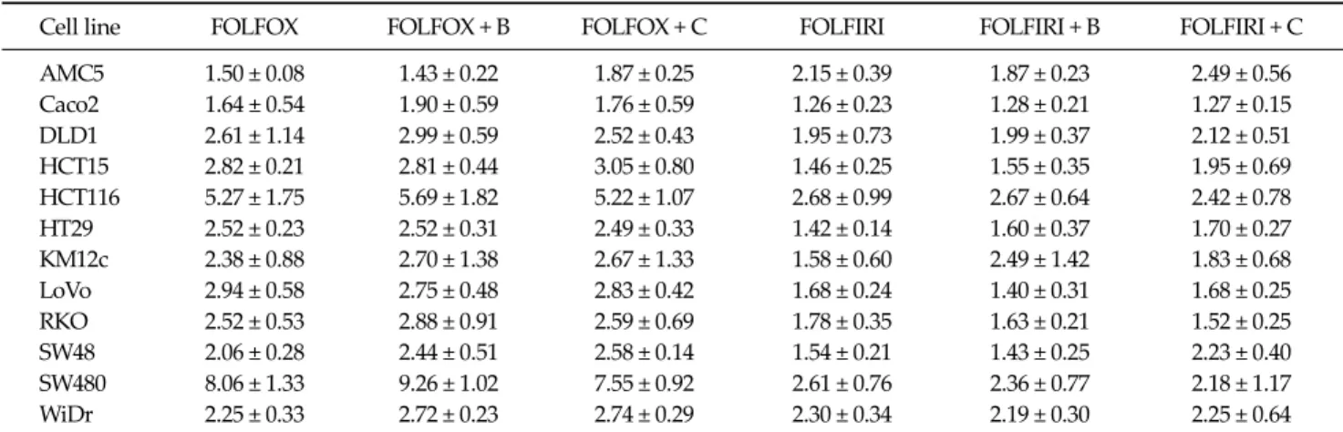
Table 1. Apoptotic cell death in colorectal cancer cell lines following treatment with the indicated drug regimens
Cell line FOLFOX FOLFOX + B FOLFOX + C FOLFIRI FOLFIRI + B FOLFIRI + C
AMC5 1.50 ± 0.08 1.43 ± 0.22 1.87 ± 0.25 2.15 ± 0.39 1.87 ± 0.23 2.49 ± 0.56 Caco2 1.64 ± 0.54 1.90 ± 0.59 1.76 ± 0.59 1.26 ± 0.23 1.28 ± 0.21 1.27 ± 0.15 DLD1 2.61 ± 1.14 2.99 ± 0.59 2.52 ± 0.43 1.95 ± 0.73 1.99 ± 0.37 2.12 ± 0.51 HCT15 2.82 ± 0.21 2.81 ± 0.44 3.05 ± 0.80 1.46 ± 0.25 1.55 ± 0.35 1.95 ± 0.69 HCT116 5.27 ± 1.75 5.69 ± 1.82 5.22 ± 1.07 2.68 ± 0.99 2.67 ± 0.64 2.42 ± 0.78 HT29 2.52 ± 0.23 2.52 ± 0.31 2.49 ± 0.33 1.42 ± 0.14 1.60 ± 0.37 1.70 ± 0.27 KM12c 2.38 ± 0.88 2.70 ± 1.38 2.67 ± 1.33 1.58 ± 0.60 2.49 ± 1.42 1.83 ± 0.68 LoVo 2.94 ± 0.58 2.75 ± 0.48 2.83 ± 0.42 1.68 ± 0.24 1.40 ± 0.31 1.68 ± 0.25
RKO 2.52 ± 0.53 2.88 ± 0.91 2.59 ± 0.69 1.78 ± 0.35 1.63 ± 0.21 1.52 ± 0.25
SW48 2.06 ± 0.28 2.44 ± 0.51 2.58 ± 0.14 1.54 ± 0.21 1.43 ± 0.25 2.23 ± 0.40 SW480 8.06 ± 1.33 9.26 ± 1.02 7.55 ± 0.92 2.61 ± 0.76 2.36 ± 0.77 2.18 ± 1.17 WiDr 2.25 ± 0.33 2.72 ± 0.23 2.74 ± 0.29 2.30 ± 0.34 2.19 ± 0.30 2.25 ± 0.64 Values represent the increase in the number of apoptotic cells after treatment with the respective regimens compared with untreated controls. Data are expressed as means ± standard errors of three independent experiments.
FOLFOX, 5-fluorouracil (5-FU) + leucovorin + oxaliplatin; FOLFIRI, 5-FU + leucovorin + irinotecan; B, bevacizumab; C, cetuximab.
gel electrophoresis, and transferred to polyvinylidene flu- oride membranes. After blocking with skim milk in TBST (10 mM Tris-HCl, 0.1 M NaCl, 0.1% Tween 20, pH7.4), membranes were sequentially incubated with anti-cas- pase-3 antibody (Cell signaling Technology, Beverly, MA, USA) and horseradish peroxide-conjugated anti-mouse secondary antibody (Pierce, Rockford, IL, USA).
VEGF mRNA expression
Total RNA was prepared from control and treated cell lines using Tri reagent (Molecular Research Center, Cincinnati, OH, USA), according to the manufacturer’s instructions. cDNA was synthesized from total RNA by amplification with random primers and SuperScript II RT (Invitrogen, Grand Island, NY, USA). The number of VEGF gene copies was analyzed by real-time reverse transcrip- tion-polymerase chain reaction (RT-PCR), using the pri- mer pair 5’-TTG CCT TGC TGC TCT ACC TCC A-3’
(forward) and 5’-GAT GGC AGT AGC TGC GCT GAT A-3’ (reverse). The housekeeping gene glyceraldehyde 3-phosphate dehydrogenase (GAPDH) used as an internal control, was amplified with the primers 5’-AGG GCT GGT TTT AAC TCT GGT-3’ (forward) and 5’-CCC CAC TTG ATT TTG GAG GGA-3’ (reverse). Quantitative real-time RT-PCR was performed on a LightCycler (Roche, Mann- heim, Germany) using the Fast Start DNA Master SYBR Green I kit, according to the manufacturer’s instructions.
Down-regulation by chemotherapy treatment was de- fined as a level of mRNA expression less than 1-fold that in untreated controls.
Gelatin zymography assay
MMP-2 and MMP-9 activities in culture media were ex- amined by gelatin zymography. Concentrated conditioned medium (×10) mixed with sample buffer was electro- phoresed under non-reducing conditions at 125 V for 2 hours on a 10% SDS-polyacrylamide gel containing 0.1%
gelatin (Invitrogen), incorporated as a substrate for gelati- nolytic proteases. The gel was incubated at 37oC for 16 hours in fresh developing buffer, and stained with 0.5%
Coomassie brilliant blue R-250 (Bio-Rad Laboratories Inc.). Bands on gels were quantified by densitometry anal- yses (Bio-Rad Laboratories Inc.).
Autophagy assay
Autophagic activity was measured by detecting the conversion of the non-autophagic form of microtubule- associated protein 1 light chain 3 alpha (LC3-I) to the auto- phagic membrane recruited form (LC3-II) by Western blot analysis using an anti-LC3 antibody (Novus, Littleton, CO, USA).
Statistical analysis
All values for apoptosis and angiogenesis were pre-
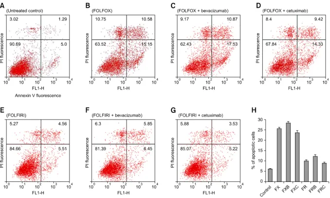
Fig. 1. Flowcytometric detection of apoptosis in the SW480 cell line. Cells treated with the respective drug regimens and untreated controls were dual-labeled with propidium iodide (PI) and Annexin V fluorescence and analyzed by flow cytometry. (A) Untreated control; (B) FOLFOX (FX), 5-fluorouracil (5-FU) + leucovorin + oxaliplatin; (C) FOLFOX + bevacizumab (FXB); (D) FOLFOX + cetuximab (FXC); (E) FOLFIRI (FR), 5-FU + leucovorin + irinotecan; (F) FOLFIRI + bevacizumab (FRB); (G) FOLFIRI + cetuximab (FRC). Dual-parameter dot-plot of fluorescein isothiocyanate-fluorescence (x-axis) versus PI-fluorescence (y-axis) showing fluorescence intensity (log scale). Lower left quadrants, live cells; lower right quadrants, apoptotic cells; upper left quadrants, necrotic cells; upper right quadrants, apoptotic and necrotic cells. The percentage of apoptotic cells is indicated on the plots. (H) Bar diagram showing the percentage of apoptotic cells after different treatments. The percentage of apoptotic cells increased after treatment with bevacizumab- containing regimens, but decreased after treatment with combinations containing cetuximab.
sented as means ± standard errors. Comparisons among the values for respective drug regimens were performed using analysis of variance with least-squares deviation verification. In each case, a P-value < 0.05 was considered statistically significant, and all calculations were per- formed using IBM SPSS ver. 18.0 (IBM Co., Armonk, NY, USA).
RESULTS
Effect of chemotherapeutic regimens on apoptosis The FOLFOX regimen was more effective in promoting apoptotic cell death, measured by flow cytometry, than FOLFIRI (Table 1). In particular, some cells showed stat- istically significant differences. The FOLFOX was sig-
nificantly more effective than FOLFIRI in HT29 and LoVo cells (P = 0.019 and 0.042, respectively). The bevacizumab- containing FOLFOX was significantly more effective than bevacizumab-containing FOLFIRI in LoVo, SW48, and SW480 cells (P = 0.010, 0.044, and 0.030, respectively).
Cetuximab-containing FOLFOX was also more effective than cetuximab-containing FOLFIRI in LoVo cells (P = 0.030). Chemosensitivities to the respective regimens var- ied greatly among cell lines. SW480 and HCT116 cells were significantly more sensitive to cell death induced by all drug regimens compared to other cell lines, especially AMC5 and Caco2 cells, which showed low sensitivity. In SW480 and HCT116 cells, bevacizumab-containing regi- mens exhibited greater apoptosis, whereas cetuximab- containing regimens showed reduced apoptosis (Fig. 1).
Unlike DLD cells, SW480 cells treated with oxaliplatin reg-
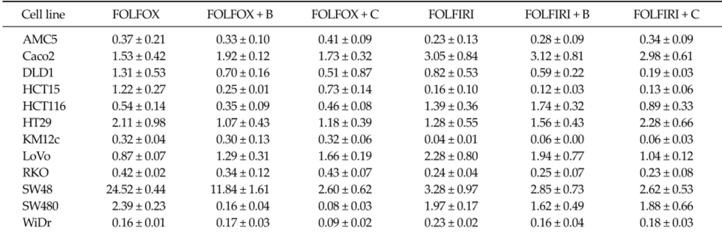
Table 2. Changes in VEGFA mRNA expression in colorectal cancer cell lines following treatment with the indicated drug regimens
Cell line FOLFOX FOLFOX + B FOLFOX + C FOLFIRI FOLFIRI + B FOLFIRI + C
AMC5 0.37 ± 0.21 0.33 ± 0.10 0.41 ± 0.09 0.23 ± 0.13 0.28 ± 0.09 0.34 ± 0.09 Caco2 1.53 ± 0.42 1.92 ± 0.12 1.73 ± 0.32 3.05 ± 0.84 3.12 ± 0.81 2.98 ± 0.61 DLD1 1.31 ± 0.53 0.70 ± 0.16 0.51 ± 0.87 0.82 ± 0.53 0.59 ± 0.22 0.19 ± 0.03 HCT15 1.22 ± 0.27 0.25 ± 0.01 0.73 ± 0.14 0.16 ± 0.10 0.12 ± 0.03 0.13 ± 0.06 HCT116 0.54 ± 0.14 0.35 ± 0.09 0.46 ± 0.08 1.39 ± 0.36 1.74 ± 0.32 0.89 ± 0.33 HT29 2.11 ± 0.98 1.07 ± 0.43 1.18 ± 0.39 1.28 ± 0.55 1.56 ± 0.43 2.28 ± 0.66 KM12c 0.32 ± 0.04 0.30 ± 0.13 0.32 ± 0.06 0.04 ± 0.01 0.06 ± 0.00 0.06 ± 0.03 LoVo 0.87 ± 0.07 1.29 ± 0.31 1.66 ± 0.19 2.28 ± 0.80 1.94 ± 0.77 1.04 ± 0.12 RKO 0.42 ± 0.02 0.34 ± 0.12 0.43 ± 0.07 0.24 ± 0.04 0.25 ± 0.07 0.23 ± 0.08 SW48 24.52 ± 0.44 11.84 ± 1.61 2.60 ± 0.62 3.28 ± 0.97 2.85 ± 0.73 2.62 ± 0.53 SW480 2.39 ± 0.23 0.16 ± 0.04 0.08 ± 0.03 1.97 ± 0.17 1.62 ± 0.49 1.88 ± 0.66 WiDr 0.16 ± 0.01 0.17 ± 0.03 0.09 ± 0.02 0.23 ± 0.02 0.16 ± 0.04 0.18 ± 0.03 Values present VEGFA mRNA levels after treatment with the respective regimen compared with untreated controls. Data are expressed as means ± errors of three independent experiments.
VEGFA, vascular endothelial growth factor A; FOLFOX, 5-fluorouracil (5-FU) + leucovorin + oxaliplatin; FOLFIRI, 5-FU + leucovorin + irinotecan; B, bevacizumab; C, cetuximab.
Fig. 2. Caspase-3 activities were analyzed by Western blot analysis.
SW480 cells (A) and DLD-1 cells (B) were treated with FOLFOX (FX), FOLFOX + bevacizumab (FXB), FOLFOX + cetuximab (FXC), FOLFIRI (FR), FOLFIRI + bevacizumab (FRB), or FOLFIRI + cetuximab (FRC) for 48 hours. Harvested-cell lysates were analy- zed by Western blotting with an anti-caspase-3 antibody. C, control;
FOLFOX (FX), 5-fluorouracil (5-FU) + leucovorin + oxaliplatin;
FOLFIRI (FR), 5-FU + leucovorin + irinotecan.
imens showed prominent activation of caspase-3, a preva- lent caspase that is ultimately responsible for the majority of apoptosis processes (Fig. 2).
Effect on angiogenesis
The effects of drug regimens on vascular endothelial growth factor A (VEGFA) mRNA levels were analyzed by quantitative real time RT-PCR. VEGFA mRNA levels were variably affected in the different cell lines (Table 2). The
FOLFOX regimen was more effective than FOLFIRI in HCT116 and WiDr cells (P = 0.026 and 0.049, respectively).
The bevacizumab-containing FOLFOX was significantly more effective than bevacizumab-containing FOLFIRI in HCT116 cells (P = 0.001). While in HCT15 cells, FOLFIRI ef- fect was more significant (P = 0.001). Bevacizumab, an an- ti-VEGF receptor antibody, produced a moderate angio- genesis-blocking effect in most cell lines when added to FOLFIRI/FOLFOX regimens. In particular, the effect of combination with FOLFOX was more statistically sig- nificant in HCT15 and SW480 cells (P = 0.003 and 0.021, re- spectively).
Invasion assay
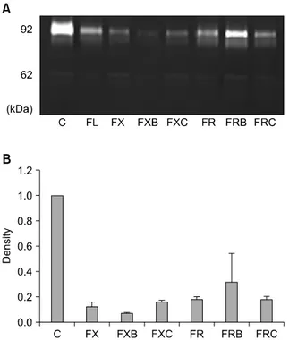
MMP-2 and MMP-9 activities were assayed by quantita- tive zymography. Both MMP-2 and MMP-9 activities were remarkably reduced in SW480 cells treated with all drug regimens, whereas the response of other cells varied ac- cording to regimens (Fig. 3). In addition, the FOLFOX regi- men more effectively reduced MMP-2 and MMP-9 activ- ities in SW480 cells than did the FOLFIRI regimen.
Autophagic cell death
Autophagic activity was detected by measuring changes in the electrophoretic mobility of LC3 protein in Western blots. This characteristic indicates a conversion of LC3
Fig. 3. Gelatinolytic matrix metalloproteinase (MMP) activity in the SW480 cell line detected by quantitative zymography. (A) Molecular markers indicate MMP-9 and MMP-2 as 92 and 62 kDa proteins, respectively. (B) Zymographic band densities were quantified by densitometry. For all panels, data are presented ± standard errors (error bars) of three independent experiments. C, control; FX, FOLFOX (5-fluorouracil [5-FU] + leucovorin + oxali- platin); FXB, FOLFOX + bevacizumab; FXC, FOLFOX + cetuximab;
FR, FOLFIRI (5-FU + leucovorin + irinotecan); FRB, FOLFIRI + bevacizumab; FRC, FOLFIRI + cetuximab.
protein from the non-autophagic form (LC3-I) to the auto- phagic membrane-recruited form (LC3-II). None of the drug regimens caused a change in LC3 conversion in any of the cell lines tested.
DISCUSSION
Several lines of evidence indicate that tumorigenesis in humans is a multistep process in which the various steps reflect genetic alterations that drive the progressive trans- formation of normal human cells into highly malignant derivatives. Six essential alterations in cell physiology col- lectively underline malignant growth: self-sufficiency with respect to growth signals, insensitivity to growth-in- hibitory signals, evasion of apoptosis, unlimited repli- cative potential, sustained angiogenensis, and tissue in-
vasion and metastasis. In each case, these physiologic changes correspond to successful efforts on the part of can- cer cells to surmount anticancer defense mechanisms that are “hardwired” into normal cells. In general, the effec- tiveness of conventional anticancer chemotherapy is thought to reflect direct cytostatic and cytotoxic effects on tumor cells [16].
Until the turn of the century, treatment options for CRC patients were limited, both in metastatic and adjuvant settings. For more than 40 years, 5-FU/LV was the standard care for metastatic CRC and node-positive CRC patients.
The addition of irinotecan to the 5-FU/LV backbone in the first-line setting improved outcomes in two randomized trials, one using bolus 5-FU and the other using combined bolus and infused 5-FU [17]. In both trials, the addition of irinotecan improved the response rate and the median overall survival and progression-free survival times; thus, the combination of irinotecan with a 5-FU-based regimen replaced 5-FU/LV as the standard first-line therapy for metastatic CRC. More recently, the use of biologically tar- geted drugs, such as bevacizumab and cetuximab, has been shown to provide additional clinical benefits for pa- tients with metastatic CRC [18,19]. In our study, mono- therapy using bevacizumab and cetuximab did not induce significant tumor inhibition, but treatment with these agents in combination with FOLFOX and FOLFIRI led to significantly higher response rates than was observed with FOLFOX and FOLFIRI alone.
Many chemotherapeutic agents mediate their cytotoxic effects by inducing apoptosis, which is generally thought to be a non-inflammatory, non-immunogenic process.
However, it has recently been suggested that apoptosis can follow biochemically distinct subroutines, some of which may result in immunogenic cell death, despite the morphological uniformity of apoptotic cell death [20,21].
In this context, oxaliplatin induces immunogenic apopto- sis accompanied by exposure of calreticulin; oxaliplatin implantation chemotherapy of CRC mice relies on an in- tact immune system including the presence of toll-like re- ceptor 4 (TLR4); and advanced CRC patients bearing a loss-of-function TLR4 allele have a lower progression-free survival after oxaliplatin-based chemotherapy than con- trol patients with a wild-type allele [22-24]. In the current
study, treatment combinations containing oxaliplatin led to significantly higher apoptosis rates with concurrent ac- tivation of caspase-3 than those containing irinotecan in the SW480 cells. These results suggest that oxaliplatin in- duces apoptotic cell death of CRC cells, and that this effect determines its therapeutic efficacy in CRC patients.
In our study, SW480 cells were the most sensitive to drug-induced apoptosis and invasion. The SW480 cell was derived from a primary Dukes’ stage B (colon adenocarci- noma) tumor from a 50-year-old Caucasian male [25,26].
SW480 cells are considered functionally p53-deficient be- cause the endogenous p53 protein contains two point mu- tations, R273H and P309S, which result in an abnormal p53 protein [27]. A study by Toscano et al. [28] reported that oxaliplatin enhances TRAIL-induced apoptosis in p53-mutant CRC cell lines, including SW480 cells [2]. In addition, SW480 cells show a microsatellite-stable and hMLH1-proficient profile. Fujita et al. [29] demonstrated that treatment of hMLH1-deficient cells with 5-FU re- sulted in 34 to 45% less apoptosis than treatment of hMLH1-proficient cells, and clinical investigations have shown that patients with microsatellite-stable cancers are more sensitive to 5-FU.
In this study, SW480 and HCT116 cells were more re- sistant to apoptosis when treated with regimens contain- ing cetuximab than when treated with regimens contain- ing bevacizumab. This finding is related to the fact that both cell lines harbor a KRAS mutation, with SW480 and HCT116 cells expressing G12V and G13D KRAS mutants, respectively. The finding that KRAS mutations are asso- ciated with resistance to cetuximab, reported here, is con- sistent with the results of previous clinical studies [30].
KRAS mutation status might allow the identification of pa- tients who are likely to benefit from cetuximab, and avoid the costly and potentially toxic administration of this treatment in nonresponder patients.
HDRA uses cancer tissue fragments in a three-dimen- sional matrix that maintains intercellular contact and in- teractions with stromal cells. The advantage of HDRA is its ability to assess the sensitivity of tumor cells to anti- cancer drugs under conditions similar to those in vivo.
However, HDRA is limited in that it relies solely on MTT assays to assess drug sensitivity. Therefore, we sought to
validate that conventional drug regimens, with and with- out molecular-targeted therapeutics, are associated with apoptotic and autophagic cell death pathways, as well as angiogenesis and invasion. In this study, all of the chemo- therapeutic regimens tested were associated with apop- totic cell death and invasion. Autophagy and anti-angio- genensis parameters were less closely linked to CRC cell death.
In conclusion, the present results suggest that apoptosis and invasion responses were the main indicators of drug sensitivity in CRC, highlighting the value of in vitro assays capable of assessing these parameters. Because HDRA maintains an in vivo-mimetic context, it is concurrently useful for evaluating the sensitivity of chemotherapeutic regimens. Thus, the MTT-based HDRA and apoptosis and invasion assays are valuable tools for assessing chemo- sensitivity in the context of CRC.
CONFLICTS OF INTEREST
No potential conflict of interest relevant to this article was reported.
REFERENCES
1. Kelly C, Cassidy J. Chemotherapy in metastatic colorectal cancer. Surg Oncol 2007;16:65-70.
2. Sabharwal A, Kerr D. Chemotherapy for colorectal cancer in the metastatic and adjuvant setting: past, present and future. Expert Rev Anticancer Ther 2007;7:477-87.
3. Tournigand C, Andre T, Achille E, Lledo G, Flesh M, Mery-Mignard D, et al. FOLFIRI followed by FOLFOX6 or the reverse sequence in advanced colorectal cancer: a randomized GERCOR study. J Clin Oncol 2004;22:229-37.
4. Hanahan D, Weinberg RA. The hallmarks of cancer. Cell 2000;100:57-70.
5. Ju JH, Chang SC, Wang HS, Yang SH, Jiang JK, Chen WC, et al. Changes in disease pattern and treatment outcome of colorectal cancer: a review of 5,474 cases in 20 years. Int J Colorectal Dis 2007;22:855-62.
6. Danese S, Sans M, de la Motte C, Graziani C, West G, Phillips MH, et al. Angiogenesis as a novel component of inflammatory bowel disease pathogenesis. Gastroentero- logy 2006;130:2060-73.
7. Hanrahan V, Currie MJ, Gunningham SP, Morrin HR, Scott PA, Robinson BA, et al. The angiogenic switch for vascular
endothelial growth factor (VEGF)-A, VEGF-B, VEGF-C, and VEGF-D in the adenoma-carcinoma sequence during colorectal cancer progression. J Pathol 2003;200:183-94.
8. Baker EA, Leaper DJ. Measuring gelatinase activity in col- orectal cancer. Eur J Surg Oncol 2002;28:24-9.
9. Baker EA, Leaper DJ. The plasminogen activator and ma- trix metalloproteinase systems in colorectal cancer: rela- tionship to tumour pathology. Eur J Cancer 2003;39:981-8.
10. Lubbe WJ, Zhou ZY, Fu W, Zuzga D, Schulz S, Fridman R, et al. Tumor epithelial cell matrix metalloproteinase 9 is a target for antimetastatic therapy in colorectal cancer. Clin Cancer Res 2006;12:1876-82.
11. Edinger AL, Thompson CB. Death by design: apoptosis, necrosis and autophagy. Curr Opin Cell Biol 2004;16:663-9.
12. West NJ, Courtney ED, Poullis AP, Leicester RJ. Apoptosis in the colonic crypt, colorectal adenomata, and manipu- lation by chemoprevention. Cancer Epidemiol Biomarkers Prev 2009;18:1680-7.
13. Baehrecke EH. Autophagy: dual roles in life and death?
Nat Rev Mol Cell Biol 2005;6:505-10.
14. Gozuacik D, Kimchi A. Autophagy and cell death. Curr Top Dev Biol 2007;78:217-45.
15. Kim JC, Kim DD, Lee YM, Kim TW, Cho DH, Kim MB, et al. Evaluation of novel histone deacetylase inhibitors as therapeutic agents for colorectal adenocarcinomas com- pared to established regimens with the histoculture drug response assay. Int J Colorectal Dis 2009;24:209-18.
16. Leman ES, Getzenberg RH. Nuclear structure as a source of cancer specific biomarkers. J Cell Biochem 2008;104:
1988-93.
17. Nannizzi S, Veal GJ, Giovannetti E, Mey V, Ricciardi S, Ottley CJ, et al. Cellular and molecular mechanisms for the synergistic cytotoxicity elicited by oxaliplatin and peme- trexed in colon cancer cell lines. Cancer Chemother Pharmacol 2010;66:547-58.
18. Kohne CH, Lenz HJ. Chemotherapy with targeted agents for the treatment of metastatic colorectal cancer. Oncolo- gist 2009;14:478-88.
19. Waldner MJ, Neurath MF. The molecular therapy of color- ectal cancer. Mol Aspects Med 2010;31:171-8.
20. Casares N, Pequignot MO, Tesniere A, Ghiringhelli F, Roux S, Chaput N, et al. Caspase-dependent immunogenicity of
doxorubicin-induced tumor cell death. J Exp Med 2005;
202:1691-701.
21. Sancho D, Mourao-Sa D, Joffre OP, Schulz O, Rogers NC, Pennington DJ, et al. Tumor therapy in mice via antigen targeting to a novel, DC-restricted C-type lectin. J Clin Invest 2008;118:2098-110.
22. Sun Q, Zheng Y, Liu Q, Cao X. Rapamycin reverses TLR4 signaling-triggered tumor apoptosis resistance by disrupt- ing Akt-mediated Bcl-xL upregulation. Int Immunophar- macol 2008;8:1854-8.
23. Tesniere A, Schlemmer F, Boige V, Kepp O, Martins I, Ghiringhelli F, et al. Immunogenic death of colon cancer cells treated with oxaliplatin. Oncogene 2010;29:482-91.
24. Lesterhuis WJ, de Vries IJ, Aarntzen EA, de Boer A, Scharenborg NM, van de Rakt M, et al. A pilot study on the immunogenicity of dendritic cell vaccination during ad- juvant oxaliplatin/capecitabine chemotherapy in colon cancer patients. Br J Cancer 2010;103:1415-21.
25. Leibovitz A, Stinson JC, McCombs WB 3rd, McCoy CE, Mazur KC, Mabry ND. Classification of human colorectal adenocarcinoma cell lines. Cancer Res 1976;36:4562-9.
26. Huerta S, Heinzerling JH, Anguiano-Hernandez YM, Huerta-Yepez S, Lin J, Chen D, et al. Modification of gene products involved in resistance to apoptosis in metastatic colon cancer cells: roles of Fas, Apaf-1, NFkappaB, IAPs, Smac/DIABLO, and AIF. J Surg Res 2007;142:184-94.
27. Rochette PJ, Bastien N, Lavoie J, Guerin SL, Drouin R.
SW480, a p53 double-mutant cell line retains proficiency for some p53 functions. J Mol Biol 2005;352:44-57.
28. Toscano F, Fajoui ZE, Gay F, Lalaoui N, Parmentier B, Chayvialle JA, et al. P53-mediated upregulation of DcR1 impairs oxaliplatin/TRAIL-induced synergistic anti-tumour potential in colon cancer cells. Oncogene 2008;27:4161-71.
29. Fujita H, Kato J, Horii J, Harada K, Hiraoka S, Shiraha H, et al. Decreased expression of hMLH1 correlates with re- duced 5-fluorouracil-mediated apoptosis in colon cancer cells. Oncol Rep 2007;18:1129-37.
30. Tol J, Koopman M, Cats A, Rodenburg CJ, Creemers GJ, Schrama JG, et al. Chemotherapy, bevacizumab, and cetux- imab in metastatic colorectal cancer. N Engl J Med 2009;
360:563-72.