326
KISEP Case Reports ••••••••••••••••••••••••••••••••••••••••••••••••••••••••••••••••••••••• 臨床耳鼻:臨床耳鼻:第臨床耳鼻:臨床耳鼻:第第 第11 卷卷卷 卷 第第第第 2 號號號號 2000
J Clinical Otolaryngol 2000;;;;11::::326-329
투시 촬영을 이용한 하인두 점막하 이물 제거
- 증 례 보 고 -
동아대학교 의과대학 이비인후과학교실
안영민·정성욱·강호정·배우용
Removal of Hypopharyngeal Submucosal Foreign Body Using Fluoroscopy
--
-- A Case Report --- -
Young-Min Ahn, MD, Sung-Uk Jung, MD, Ho-Jung Kang, MD and Woo-Yong Bae, MD
Department of Otolaryngology and Head & Neck Surgery, College of Medicine,Dong-A University, Pusan, Korea -
-
-- ABSTRACT ----
A 64-year-old woman visited to our hospital for foreign body sensation of throat and odynophagia following meal. A neck lateral radiograph revealed a wire-shaped metallic foreign body at the hypopharynx. Although we investigated the hypopharynx and esophagus throughout using rigid esophagoscope, we couldn’t find out it. Pharyx computed tomography showed the foreign body under the mucosal layer, and it was removed by submucosal dissection under fluoroscopic control. Repeated trial of rigid esophagoscopy raise the chance of complications such as esophageal mucosal injury, esophageal perforation, and cervical vertebrae injury. If foreign body cannot be found out despite repeated trial of rigid esophagoscopy, the possibility of submucosal migration should be considered. Fluoroscopically guided esophagoscopy is a safe and effective technique for the removal of submucosal foreign body. ((((J Clinical Otolaryngol 2000;11:326-329))))
KEY WORDS:Hypopharynx·Foreign body·Fluoroscopy.
서 론
하인두 이물은 이비인후과 영역에서 자주 경험하는 질 환으로, 대부분 순간적인 부주의로 인해 돌발적으로 발 생한다. 하인두 이물의 치료에 있어 가장 중요한 것은 이 물의 정확한 위치 확인이며, 이는 병력 청취와 이학적 검 사, 단순 X-선 촬영 등으로 어렵지 않게 확인할 수 있
다. 본 예의 이물은 점막하에 위치해 있어 위치 확인과 제거에 상당한 어려움이 있었기에 그 치험을 문헌 고찰 과 함께 보고하는 바이다.
증 례
64세 여자 환자가 내원 당일 오전 10시경 아침식사 로 비빔밥을 먹은 후 연하통 및 인후부 이물감이 발생 하여 인근 병원을 방문하였다. 경부 측면 X-선 검사에 서 하인두의 방사선 비투과성 이물을 확인하고, 이비인 후과와 내과에서 각각 후두경 검사와 식도내시경 검사를 시도하였으나 이물을 찾지 못해 본원으로 전원되었다.
내원 당시 실시한 이학적 검사상 하인두 및 후두의 논문접수일:2000년 9월 25일
심사완료일:2000년 11월 20일
교신저자:안영민, 602-715 부산광역시 서구 동대신동 3가 1번지 동아대학교 의과대학 이비인후과학교실
전화:(051) 240-5428・전송:(051) 253-0712 E-mail:ahnymin@shinbiro.com
안영민 외:투시 촬영을 이용한 하인두 점막하 이물 제거
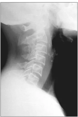
327 이물이나 점막 손상은 없었다. 경부 측면 X-선 검사에 서 하인두 이상와와 후인두벽에 걸쳐 있는 철사로 보이 는 금속성의 이물이 확인되어 전신 마취하에서 강직형 식 도경을 이용한 이물 제거술을 시도하였다(Fig. 1).
두 사람의 술자가 수차례에 걸쳐 강직형 식도경을 삽 입하였으나 이물을 찾을 수 없었으며 하인두와 식도의 점막 손상이나 다른 이상소견을 발견할 수 없었다. 이 물의 위치 확인 및 연하 운동에 의한 이물의 이동 등을 확인하기 위해 휴대용 경부 단순 촬영을 시행하였다. 이 물이 처음 위치에 그대로 있어 강직형 식도경을 다시 삽 입하여 확인하였으나 이물을 찾을 수 없었다. 이물이 점 막하에 들어 있을 가능성이 있어 수술을 중단하고 인두 전산화 단층촬영을 하였는데 이물은 하인두 후측벽의 점 막하에 위치해 있었다(Fig. 2). 다음날 식도경을 이용한 이물 제거술을 다시 시행하였다. 자보 직접 후두경으로 시야를 확보하고, 투시 촬영(fluoroscopy)의 연속 화면
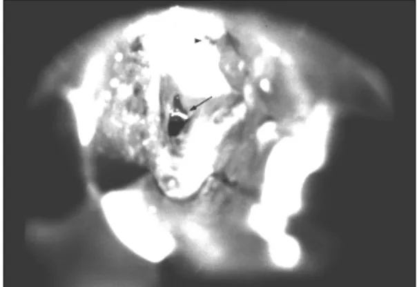
을 보면서(Fig. 3) 후두 미세 수술용 가위를 이물이 묻 혀 있는 점막 부위에 위치시킨 후 점막을 절개하여 이 물을 확인하고 이물겸자로 제거하였다(Figs. 4 and 5).
이물 제거후 식도 손상 및 다른 이물 유무 등을 확인하 기 위해 식도경을 다시 삽입하여 관찰하였으나 다른 이 상소견은 발견되지 않았다. 술후 경부 및 흉부 X-선 사 진상 방사선 비투과성 이물은 소실되었고 기흉이나 기 종격동 등의 소견도 없었다. 술후 환자는 수일간 전흉 부 동통을 호소하였으며 이는 잦은 식도경 삽입으로 인한 점막 손상때문으로 생각된다. 환자는 수일간의 입원 후 전흉부 동통이 사라졌으며 후유증없이 퇴원하였다.
고 찰
이비인후과 영역에서 이물의 개재 부위는 식도, 구강,
Fig. 1. Lateral radiograph of the neck showing a wire- shaped metallic foreign body at the level of the third
cervical vertebra. Fig. 3. Fluoroscopic finding showing foreign body (arrow) under the suction tip (arrow head).
Fig. 2. Axial CT scan of the neck showing a metallic fo- reign body (arrow) embedded in the hypopharyneal wall.
J Clinical Otolaryngol 2000;11:326-329
328 인두, 외이도, 비강, 기관 그리고 기관지 등이다. 이들 중 식도 이물이 가장 많으며 하인두 이물도 드물지 않게 발생한다.1)2) 식도 이물에 관해서는 많은 보고들이 있으 나 하인두 이물, 특히 주위 조직으로 침윤된 이물에 대 한 보고는 극히 드물다. Jemerin 등3)은 갑상선으로 침 윤된 생선 가시가 갑상선내 농양을 형성하였다고 보고 하였고, Muhanna 등4)도 갑상선으로 침윤된 생선 가시 를 갑상선 절제술을 통해 치료하였다고 보고하였다. Os- inubi 등5)은 경동맥에 접해있는 철재 이물을 외과적 수 술을 통해 제거하였다고 보고한 바 있다.
이와 같은 이물의 침윤은 연하운동이나 구역반사 등 에 의해 유발되는 하인두와 식도의 근수축,6) 이물에 의해 주위 조직으로 가해지는 압력 그리고 조직과 이물간의
국소반응7) 등의 몇 가지 요인에 의해 발생할 수 있다.
주위 조직으로 침윤된 이물은 식도 천공, 종격동염, 식 도대동맥루, 기관식도루8) 그리고 경부 척수염9) 등의 치 명적인 합병증을 야기할 수 있다. 본 예에서처럼 침윤성 이물은 그 자체로는 응급 상황이 아니더라도 이후 위중 한 합병증을 유발할 수 있으므로 즉각적인 제거가 필요 하다.
대부분의 식도 및 하인두 이물은 자세한 병력 청취와 이학적 검사, 그리고 단순 X-선 검사로 어렵지 않게 진 단할 수 있다.10-12) 단순 X-선 검사로도 진단이 되지 않을 때에는 수용성 조영제로 식도 조영술을 시행하기 도 한다.13)14) 이상의 방법으로도 진단이 되지 않을 경 우 전산화 단층촬영을 실시할 수 있는데 이는 단순 X- 선 검사에서 확인하기 힘든 크기가 작고 굵기가 가는 물 체나 방사선 투과성 물체의 진단을 가능하게 하고, 이물 의 위치를 좀더 정확하게 알 수 있게 해준다.14-16)
대다수의 이물은 내시경을 이용하여 제거가 가능하나, 식도 외강 이물의 경우 외과적 수술을 요할 수도 있
다.5)17) 외과적 수술로 식도 외강 이물을 제거할 때에는
술전 전산화 단층촬영을 통하여 이물의 위치를 정확히 파악하는 것이 무엇보다도 중요하며, 술중 투시 촬영을 함께 이용할 경우 수술 범위를 최소화할 수 있다.
투시 촬영은 매우 많은 질환의 진단과 치료에 이용되 고 있다. 이비인후과 영역에서는 예리하지 않은 식도 이 물을 투시 촬영하에서 Foley 카테터를 이용하여 제거하 는 방법이 이용되고 있으며 이는 효과와 안전성 면에서 전신 마취하 강직형 식도경을 이용한 이물 제거술과 동 일한 정도의 성적을 보이고 있다.18-20) 본 예에서도 투 시 촬영의 도움으로 단 0.5 cm 정도의 점막 절개만으로 2.5 cm 가량의 이물을 제거할 수 있었다.
침윤형 이물은 그 자체로 치명적인 합병증을 유발할 수 있으며, 이물 제거를 위한 강직형 식도경의 잦은 삽 입은 경추 손상, 식도 점막 손상, 식도 천공 등의 합병증 발생률을 높일 수 있다. 수차례의 식도경 삽입으로도 이 물이 확인되지 않을 경우 점막하 이물의 가능성을 고려 해야 하며, 이는 투시촬영을 이용한 이물 제거술로써 효 과적으로 치유될 수 있다.
중심 단어:하인두・이물・투시 촬영.
Fig. 4. The foreign body (arrow) revealed after submu- cosal dissection at the posterior hypopharyngeal wall.
Arrow head indicates esophageal inlet.
Fig. 5. The removed foreign body (wire).
안영민 외:투시 촬영을 이용한 하인두 점막하 이물 제거
329
REFERENCES
1) Kim SH, Lee CW, Cho JS. Clinical analysis of tracheo- esophageal foreign bodies. Korean J Otolaryngol 1989;32:
558-66.
2) Park SJ, Lee BD, Park JR, Choi HS, Chang HS, Kang JW.
A statistical analysis of foreign bodies in otolaryngolog- ical field. Korean J Otolaryngol 1986;29:848-57.
3) Jemarin EE, Aronoff JS. Foreign body in thyroid following perforation of esophagus. Surgery 1949;25:52-9.
4) Muhanna AA, Abu Chra KA, Dashti H. Thyroid lobecto- my for removal of a fish bone. J Laryngol Otol 1990;104:
511-2.
5) Osinubi OA, Osiname AI, Lonsdale RJ, Butcher C. Fore- ign body in the throat migrating through the common ca- rotid artery. J Laryngol Otol 1996;110:793-5.
6) Hammond VT. Penetrating foreign body of the pharynx.
J Laryngol Otol 1961;75:848-88.
7) Yu KF, Schild JA, Holinger PH. Extraluminal foreign bo- dies (coins) in the food and air passage. Ann Otol 1975;
84:619-23.
8) Stolz JL, Chamorro H, Arger PH. Fish bone fistulae. Arch Otolaryngol 1975;101:252-3.
9) Gupta KR, Kakao PK, Saharia PS. Impacted foreign body of retropharyngeal space. J Laryngol Otol 1972;86:519-21.
10) Jones NS, Lannigan FJ, Salama NY. Foreign bodies in the throat: aprospective study of 388 cases. J Laryngol Otol 1991;105:104-8.
11) Macpherson RI, Hill JG, Othersen HB, Tagge EP, Smith
CD. Esophageal foreign bodies in children: diagnosis, tr- eatment, and complications. Am J Roentgenol 1996;166:
919-24.
12) Al-Qudah A, Daradkeh S, Abu-Khalaf M. Esophageal foreign bodies. Eur J Cardiothorac Surg 1998;13:494-99.
13) Chon KM, Wang SG, Oh IJ, Park BI. A rare case of eso- phageal foreign body. Clin Otol 1993;4:193-6.
14) Braverman I, Gomori JM, Polv O, Saah D. The role of CT imaging in the evaluation of cervical esophageal foreign bodies. J Otolaryngol 1993;22:311-4.
15) Kobayashi T. Esophageal foreign bodies in children. Int J Pediatr Otorhinolaryngol 1984;7:193-8.
16) Eliashar R, Dano I, Dangoor E, Braverman I, Sichel JY.
Computed tomography diagnosis of esophageal bone im- paction: a prospective study. Ann Otol Rhinol Laryngol 1999;108:708-10.
17) Murthy PS, Bipin TV, Ranjit R, Murty KD, George V, Mathew KJ. Extraluminal migration of swallowed fore- ign body into the neck. Am J Otolaryngol 1995;16:213-5.
18) Harned RK 2nd, Strain JD, Hay TC, Douglas MR. Esop- hageal foreign bodies: safety and efficacy of Foley cathe- ter extraction of coins. Am J Roentgenol 1997;168:443-6.
19) Morrow SE, Bickler SW, Kennedy AP, Snyder CL, Sharp RJ, Ashcraft KW. Balloon extraction of esophageal for- eign bodies in children. J Pediatr Surg 1998;33:266-70.
20) Schunk JE, Harrison AM, Corneli HM, Nixon GW. Fluo- roscopic foley catheter removal of esophageal foreign bo- dies in children: experience with 415 episodes. Pediatrics 1994;94:709-14.

