D I A B E T E S & M E T A B O L I S M J O U R N A L D I A B E T E S & M E T A B O L I S M J O U R N A L
This is an Open Access article distributed under the terms of the Creative Commons Attribution Non-Commercial License (http://creativecommons.org/licenses/by-nc/4.0/) which permits unrestricted non-commercial use, distribution, and reproduction in any medium, provided the original work is properly cited.
Serum Levels of PCSK9 Are Associated with Coronary Angiographic Severity in Patients with Acute Coronary Syndrome
Kwi-Hyun Bae1,*, Sung Woo Kim2,*, Yeon-Kyung Choi1, Jung Beom Seo1, Namkyun Kim1, Chang-Yeon Kim2, Won Kee Lee3, Sungwoo Lee4, Jung Guk Kim1, In-Kyu Lee1, Jang Hoon Lee1, Keun-Gyu Park1
1Department of Internal Medicine, School of Medicine, Kyungpook National University, Daegu,
2Department of Internal Medicine, Daegu Catholic University Medical Center, Catholic University of Daegu School of Medicine, Daegu,
3Biostatistics Center, School of Medicine, Kyungpook National University, Daegu,
4New Drug Development Center, Daegu-Gyeongbuk Medical Innovation Foundation, Daegu, Korea
Background: Proprotein convertase subtilisin/kexin type 9 (PCSK9) is a circulating protein that promotes degradation of the low density lipoprotein receptor. PCSK9 has emerged as a target for lipid-lowering therapy, but the predictive value of the serum level of PCSK9 for the severity of coronary disease is largely unknown.
Methods: From December 2009 to July 2012, 121 individuals who underwent coronary angiography (CAG) because of clinically suspected acute coronary syndrome were enrolled in this study. Serum levels of PCSK9 and metabolic parameters were measured.
SYN TAX (SYNergy between percutaneous coronary intervention with [paclitaxel-eluting] TAXUS stent and cardiac surgery) and GRACE (Global Registry of Acute Coronary Events) scores were calculated.
Results: Individuals with CAG lesions (n=100) had significantly higher levels of PCSK9 than those without lesions (n=21). The study population was stratified into three groups according to serum levels of PCSK9. The odds radio for occurrence of one or more CAG lesions was significantly higher in the group with the highest level of PCSK9 (odds ratio, 7.468; P=0.011) than in the group with the lowest level of PCSK9. Serum PCSK9 was positively associated with the number of involved coronary arteries.
Multivariable linear regression indicated that levels of PCSK9 were positively correlated with GRACE risk scores and SYNTAX scores.
Conclusion: Serum PCSK9 concentrations are higher in patients with coronary artery lesions, and are associated with SYNTAX and GRACE scores, suggesting that PCSK9 is a potential biomarker of the severity of coronary artery disease.
Keywords: Cardiovascular diseases; Coronary angiography; Proprotein convertase 9
Corresponding authors: Keun-Gyu Park https://orcid.org/0000-0002-8403-1298 Department of Internal Medicine, School of Medicine, Kyungpook National University, 680 Gukchaebosang-ro, Jung-gu, Daegu 41944, Korea
E-mail: [email protected]
Jang Hoon Lee https://orcid.org/0000-0002-7101-0236
Department of Internal Medicine, School of Medicine, Kyungpook National University, 680 Gukchaebosang-ro, Jung-gu, Daegu 41944, Korea
E-mail: [email protected]
INTRODUCTION
Evaluation of individuals who present with acute chest pain, or other clinical features suggestive of acute coronary syndrome
(ACS), is often a clinical challenge. Biomarkers reflecting dis- tinct pathophysiological features have become fundamental tools for improvement of traditional diagnostic and risk-strati- fication methods for patients with ACS [1-3]. Multi-marker https://doi.org/10.4093/dmj.2017.0081
pISSN 2233-6079 · eISSN 2233-6087
strategies employing biomarkers from different pathological pathways can contribute to successful methodologies [4]. Tro- ponin I, N-terminal pro-B type natriuretic peptide (NT-proB- NP), and high-sensitivity C-reactive protein (hs-CRP) are bio- markers for which consistent and reliable data are available on their diagnostic and prognostic values for ACS [5,6]. Evidence also suggests that proprotein convertase subtilisin/kexin type 9 (PCSK9) could have value as a potential biomarker for ACS [7].
PCSK9 is a liver-derived circulating protein that has an im- portant role in the homeostasis of low density lipoprotein (LDL) via enhancement of degradation of the LDL receptor;
thereby, reducing clearance of LDL-cholesterol (LDL-C) [8].
PCSK9 loss-of-function mutations are associated with reduc- tions in circulating levels of LDL-C and in the risk of coronary heart disease [9]. Therefore, PCSK9 has received considerable attention as a promising target for lipid-lowering therapy [10].
A high plasma level of LDL-C is a major risk factor for cardio- vascular diseases, including ACS, and results from genetic, ex- perimental, and epidemiological studies show clear associa- tions between PCSK9 function and cardiovascular risk [11- 13]. Moreover, levels of PCSK9 are driven up by cardiac isch- emia during ACS events and may contribute to plaque vulner- ability in coronary vessels [14]. Evidence suggests that PCSK9 antibodies have a potential role in the management of ACS.
However, no data are available on the predictive value of serum PCSK9 concentrations for coronary angiographic severity.
In this study, our aim was to investigate whether a high level of serum PCSK9 is associated with a high risk and severity of ACS in individuals with coronary angiographic lesions. We also assessed the association of serum PCSK9 levels with SYN- TAX (SYNergy between percutaneous coronary intervention with [paclitaxel-eluting] TAXUS stent and cardiac surgery) and GRACE (Global Registry of Acute Coronary Events) scores, which are indicators of the severity of coronary artery occlusion.
METHODS
Study population and data collection
A retrospective, cross-sectional, observational study was con- ducted involving individuals who attended the Kyungpook National University Hospital from December 2009 to July 2012. Screening involved 444 individuals with suspected ACS, including unstable angina and both ST-segment elevation myocardial infarction and non-ST-segment elevation myocar-
dial infarction. Exclusion criteria were as follows: use of lipid- lowering drugs (statins, fibrates, nicotinic acid, or ezetimibe) within 3 months; infectious or systematic inflammatory dis- eases, significant hematological disorders, thyroid dysfunction, and severe hepatic and/or renal insufficiency; and female sex.
The final study population consisted of 121 individuals, 100 of whom had one or more narrowing lesions of the coronary ar- teries (with ≥50% stenosis) on coronary angiography (CAG), and 21 of whom had no such lesions. The study protocol was approved by the Institutional Review Board of Kyungpook Na- tional University Hospital (IRB no.: 2015-10-019-001), and all subjects provided written informed consent before participa- tion in the study.
Measurements of clinical and laboratory parameters Details of age, sex, body mass index (BMI), past history of dia- betes and hypertension, history of tobacco smoking, and fa- milial history of ACS were retrospectively collected from elec- tronic medical records. Laboratory data were acquired on the day of admission, before the CAG procedure. Serum levels of NT-proBNP were measured by an electrochemiluminescence immunoassay method (Modular Analytics E170; Roche Diag- nostics, Mannheim, Germany). Other biochemical variables were measured by standard laboratory techniques. Levels of serum total cholesterol, high density lipoprotein cholesterol (HDL-C), LDL-C, triglycerides, hs-CRP, creatinine, white blood cells, aspartate transaminase, and alanine transaminase were determined from blood samples collected after an over- night fast (of ≥8 hours).
Measurement of PCSK9
Blood samples were collected into EDTA (ethylenediaminetet- raacetic acid)-containing tubes in the morning after an over- night fast (of ≥8 hours), and centrifuged at 3,000 rpm for 10 minutes at 4°C to obtain serum. All the samples for the mea- surement of PCSK9 levels were stored at −80°C until analysis.
Serum PCSK9 levels were measured in duplicate by enzyme- linked immunosorbent assay with a commercial kit (R&D Sys- tems, Minneapolis, MN, USA) according to the manufacturer’s instructions, with a 1:1 dilution of samples in dilution buffer before the assay.
Measurement of the severity of coronary artery occlusion The severity of coronary artery occlusion was evaluated by cal- culation of SYNTAX scores and GRACE scores. The detailed
methodologies for calculating these scores have been de- scribed elsewhere.
SYNTAX score
SYNTAX is an anatomically based scoring system that quanti- tatively characterizes coronary vessels according to the num- ber, complexity, location, and functional aspect of obstructive lesions on CAG. Vessels with diameters >1.5 mm that contain lesions with stenosis diameters ≥50% are assessed. Each coro- nary segment is given a weight factor that is determined by the lesion location and severity. Lesion characteristics, including total occlusion, trifurcation, bifurcation, calcification, tortuosi- ty, length >20 mm, thrombus, and diffuse or small-vessel dis- ease, are combined to give a final score via the SYNTAX calcu- lator (www.syntaxscore.com). The SYNTAX score is an objec- tive angiography-based tool for grading of the complexity and severity of coronary artery disease (CAD).
GRACE risk score
The GRACE score is a prognostic risk-assessment tool for use in patients with ACS. Calculation of the score takes account of age, Killip class, systolic blood pressure, ST-segment deviation, cardiac arrest during presentation, serum creatinine levels, heart rate, and elevated cardiac enzyme or marker levels. An online calculator is used for derivation of GRACE scores (http:
//www.outcomes-umassmed.org/grace).
Statistical analysis
Continuous variables are presented as the mean±standard de- viation, and categorical variables are presented as the frequen- cies (and percentages) in each category. Student t-tests were used for comparison of the means of continuous variables and chi-square tests were used for comparison of the distributions of categorical variables. Analysis of covariance was used for identification of differences in serum levels of PCSK9 in groups of patients with different numbers of involved coronary arteries. The models were adjusted for age, BMI, past history of diabetes and hypertension, smoking, and familial history of ACS. The study population was divided into tertiles according to serum levels of PCSK9. Multivariable logistic regression models were used for calculation of the odds ratios (ORs) for the presence of one or more coronary angiographic lesions in patients in tertile 2 (T2) and tertile 3 (T3) compared with ter- tile 1 (T1). The models were also adjusted for multiple con- founding factors. Multivariable linear regression models were
applied for characterization of the association between serum levels of PCSK9 and SYNTAX and GRACE scores. Results of statistical tests were considered significant for P<0.05. Statisti- cal analysis was performed with SPSS version 18.0 software (SPSS Inc., Chicago, IL, USA).
RESULTS
Baseline characteristics
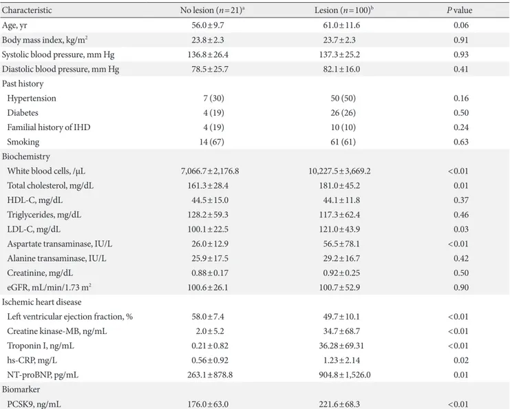
The baseline characteristics of study participants are shown in Table 1. The means or frequencies of age, BMI, HDL-C, tri- glycerides, creatinine, systolic and diastolic pressure, past his- tory of hypertension and diabetes, familial history of ACS, and history of tobacco smoking showed no significant differences between patients with no CAG lesions (n=21) and those with lesions (n=100). Mean levels of total cholesterol and LDL-C were significantly higher in patients with CAG lesions than in those without lesions. Levels of white blood cells, aspartate transaminase, creatine kinase-MB, troponin I, hs-CRP, and NT-proBNP were significantly higher (as a result of cardiac damage) in patients with CAG lesions than in those without lesions. Serum levels of PCSK9 were significantly higher in pa- tients with CAG lesions than in those without CAG lesions.
Baseline characteristics in tertiles stratified by levels of PCSK9
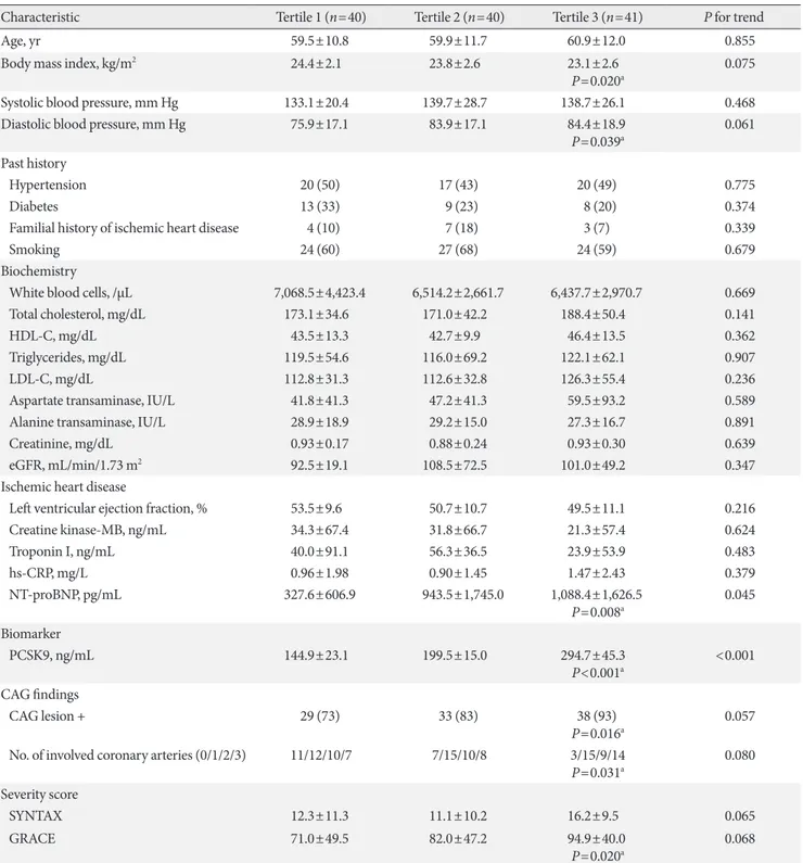
To assess the associations between serum levels of PCSK9 and different variables related to ACS, we stratified the study popu- lation into tertiles according to PCSK9 levels (Table 2). With the exception of NT-proBNP (and PCSK9), the baseline char- acteristics did not show statistical trends related to PCSK9 stratification. However, in comparisons of T1 with T3, proBNP, positive findings of CAG lesions, the number of involved coro- nary arteries, and GRACE scores were all significantly higher in T3 than in T1, whereas diastolic blood pressure was signifi- cantly lower in T3 than in T1. Although LDL-C levels and SYNTAX scores showed numerical increases from T1 to T3, statistical significance was not observed.
Association between PCSK9 levels and the occurrence of CAG lesions
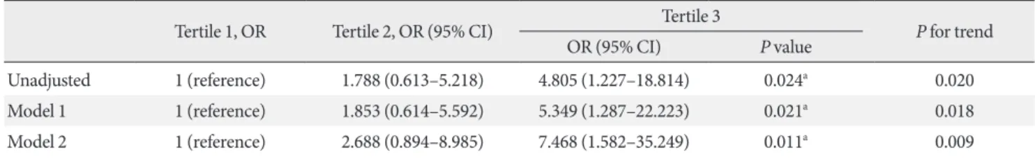
The proportion of individuals with CAG lesions was signifi- cantly higher in T3 than in T1 (unadjusted OR, 4.805; 95%
confidence interval [CI], 1.227 to 18.814; P=0.024) (Table 3).
The difference between T3 and T1 was enhanced by adjust-
ment for age, BMI, past history of diabetes and hypertension, smoking, and familial history of ACS (OR, 7.468; 95% CI, 1.582 to 35.249; P=0.011). The P for trend of increasing pro- portions of individuals with CAG lesions from T1 to T3 were significant (P=0.009 for the adjusted model 2).
Associations between serum PCSK9 concentrations and numbers of involved coronary arteries
Serum levels of PCSK9 were measured in 121 patients who
were assessed for coronary angiographic lesions. Mean PCSK9 levels in patients with 0 (n=21), 1 (n=42), 2 (n=29), and 3 (n=
29) involved arteries were compared by analysis of covariance.
After adjustment for age, BMI, past history of diabetes and hy- pertension, smoking, and familial history of ACS, individuals with one or more coronary artery lesions had significantly higher mean serum PCSK9 levels than individuals without coronary artery lesions (Fig. 1).
Table 1. Baseline clinical characteristics of study participants with and without coronary angiographic lesions
Characteristic No lesion (n=21)a Lesion (n=100)b P value
Age, yr 56.0±9.7 61.0±11.6 0.06
Body mass index, kg/m2 23.8±2.3 23.7±2.3 0.91
Systolic blood pressure, mm Hg 136.8±26.4 137.3±25.2 0.93
Diastolic blood pressure, mm Hg 78.5±25.7 82.1±16.0 0.41
Past history
Hypertension 7 (30) 50 (50) 0.16
Diabetes 4 (19) 26 (26) 0.50
Familial history of IHD 4 (19) 10 (10) 0.24
Smoking 14 (67) 61 (61) 0.63
Biochemistry
White blood cells, /μL 7,066.7±2,176.8 10,227.5±3,669.2 <0.01
Total cholesterol, mg/dL 161.3±28.4 181.0±45.2 0.01
HDL-C, mg/dL 44.5±15.0 44.1±11.8 0.37
Triglycerides, mg/dL 128.2±59.3 117.3±62.4 0.46
LDL-C, mg/dL 100.1±22.5 121.0±43.9 0.03
Aspartate transaminase, IU/L 26.0±12.9 56.5±78.1 <0.01
Alanine transaminase, IU/L 25.9±17.5 29.2±16.7 0.42
Creatinine, mg/dL 0.88±0.17 0.92±0.25 0.50
eGFR, mL/min/1.73 m2 100.6±26.1 100.7±52.9 0.90
Ischemic heart disease
Left ventricular ejection fraction, % 58.0±7.4 49.7±10.1 <0.01
Creatine kinase-MB, ng/mL 2.0±5.2 34.7±68.7 <0.01
Troponin I, ng/mL 0.21±0.82 36.28±69.31 <0.01
hs-CRP, mg/L 0.56±0.92 1.23±2.14 0.02
NT-proBNP, pg/mL 263.1±878.8 904.8±1,526.0 0.01
Biomarker
PCSK9, ng/mL 176.0±63.0 221.6±68.3 <0.01
Values are presented as mean±standard deviation (which were compared by Student t-tests) or number (%) in each category (which were com- pared by chi-square tests).
IHD, ischemic heart disease; HDL-C, high density lipoprotein cholesterol; LDL-C, low density lipoprotein cholesterol; eGFR, estimated glomer- ular filtration rate; hs-CRP, high-sensitivity C-reactive protein; NT-proBNP, N-terminal pro-B type natriuretic peptide; PCSK9, proprotein con- vertase subtilisin/kexin type 9.
aNo lesion, no coronary arterial lesion at coronary angiography, bLesion, one or more coronary arterial lesions at coronary angiography.
Table 2. Baseline clinical characteristics of study participants stratified into tertiles according to serum levels of PCSK9
Characteristic Tertile 1 (n=40) Tertile 2 (n=40) Tertile 3 (n=41) P for trend
Age, yr 59.5±10.8 59.9±11.7 60.9±12.0 0.855
Body mass index, kg/m2 24.4±2.1 23.8±2.6 23.1±2.6
P=0.020a 0.075
Systolic blood pressure, mm Hg 133.1±20.4 139.7±28.7 138.7±26.1 0.468
Diastolic blood pressure, mm Hg 75.9±17.1 83.9±17.1 84.4±18.9
P=0.039a 0.061
Past history
Hypertension 20 (50) 17 (43) 20 (49) 0.775
Diabetes 13 (33) 9 (23) 8 (20) 0.374
Familial history of ischemic heart disease 4 (10) 7 (18) 3 (7) 0.339
Smoking 24 (60) 27 (68) 24 (59) 0.679
Biochemistry
White blood cells, /μL 7,068.5±4,423.4 6,514.2±2,661.7 6,437.7±2,970.7 0.669
Total cholesterol, mg/dL 173.1±34.6 171.0±42.2 188.4±50.4 0.141
HDL-C, mg/dL 43.5±13.3 42.7±9.9 46.4±13.5 0.362
Triglycerides, mg/dL 119.5±54.6 116.0±69.2 122.1±62.1 0.907
LDL-C, mg/dL 112.8±31.3 112.6±32.8 126.3±55.4 0.236
Aspartate transaminase, IU/L 41.8±41.3 47.2±41.3 59.5±93.2 0.589
Alanine transaminase, IU/L 28.9±18.9 29.2±15.0 27.3±16.7 0.891
Creatinine, mg/dL 0.93±0.17 0.88±0.24 0.93±0.30 0.639
eGFR, mL/min/1.73 m2 92.5±19.1 108.5±72.5 101.0±49.2 0.347
Ischemic heart disease
Left ventricular ejection fraction, % 53.5±9.6 50.7±10.7 49.5±11.1 0.216
Creatine kinase-MB, ng/mL 34.3±67.4 31.8±66.7 21.3±57.4 0.624
Troponin I, ng/mL 40.0±91.1 56.3±36.5 23.9±53.9 0.483
hs-CRP, mg/L 0.96±1.98 0.90±1.45 1.47±2.43 0.379
NT-proBNP, pg/mL 327.6±606.9 943.5±1,745.0 1,088.4±1,626.5
P=0.008a 0.045
Biomarker
PCSK9, ng/mL 144.9±23.1 199.5±15.0 294.7±45.3
P<0.001a <0.001 CAG findings
CAG lesion + 29 (73) 33 (83) 38 (93)
P=0.016a 0.057
No. of involved coronary arteries (0/1/2/3) 11/12/10/7 7/15/10/8 3/15/9/14
P=0.031a 0.080
Severity score
SYNTAX 12.3±11.3 11.1±10.2 16.2±9.5 0.065
GRACE 71.0±49.5 82.0±47.2 94.9±40.0
P=0.020a 0.068
Values are presented as mean±standard deviation or number (%).
PCSK9, proprotein convertase subtilisin/kexin type 9; HDL-C, high density lipoprotein cholesterol; LDL-C, low density lipoprotein cholesterol;
eGFR, estimated glomerular filtration rate; hs-CRP, high-sensitivity C-reactive protein; NT-proBNP, N-terminal pro-B type natriuretic peptide;
CAG, coronary angiography; SYNTAX, SYNergy between percutaneous coronary intervention with (paclitaxel-eluting) TAXUS stent and car- diac surgery; GRACE, Global Registry of Acute Coronary Events.
aP value of tertile 3 (highest PCSK9) compared with tertile 1 (lowest PCSK9), determined by Student t-tests for continuous variables or chi- square tests for categorical variables.
Associations between PCSK9 levels and scores indicating the severity of coronary artery occlusion
Multivariable linear regression models revealed that the serum PCSK9 concentration was positively associated with the SYN- TAX score, which is an angiographic grading tool to determine the complexity of CAD (βstandard=0.191; P=0.035) (Table 4).
The GRACE risk score, which is a risk-prediction tool for use in patients with ACS, was also significantly associated with PCSK9 concentrations (βstandard=0.266; P=0.003). Further- more, after adjusting for age, BMI, past history of diabetes and hypertension, smoking, and familial history of ACS, PCSK9 levels were still positively correlated with SYNTAX scores (βstandard=0.204; P=0.037) and GRACE scores (βstandard=0.214;
P=0.007).
DISCUSSION
In this study, we demonstrated that serum PCSK9 concentra- tions were significantly associated with the proportions of pa- tients with CAG lesions. This association was still significant after adjustment for multiple variables. In addition, we found that PCSK9 levels were significantly associated with the num- ber of affected coronary arteries, SYNTAX scores, and GRACE scores.
Evidence suggests that circulating PCSK9 levels are associat- ed with the incidence of CAD [12-14]. In one study involving patients who were not receiving treatment with statins or fi- brates at the time of recruitment, higher serum PCSK9 levels were seen in one group of patients with angiographic CAD in- cluding ACS (385.0±146.9 ng/mL) than in controls without CAD (340.4±125.2 ng/mL, P<0.001) [15]. However, there was Table 3. Odds ratios for the presence of one or more coronary angiographic lesions in patients in different PCSK9 tertiles
Tertile 1, OR Tertile 2, OR (95% CI) Tertile 3
P for trend
OR (95% CI) P value
Unadjusted 1 (reference) 1.788 (0.613–5.218) 4.805 (1.227–18.814) 0.024a 0.020
Model 1 1 (reference) 1.853 (0.614–5.592) 5.349 (1.287–22.223) 0.021a 0.018
Model 2 1 (reference) 2.688 (0.894–8.985) 7.468 (1.582–35.249) 0.011a 0.009
Odds ratios were calculated for the presence of one or more coronary angiographic lesions in patients in tertile 2 and tertile 3 compared with tertile 1. Multivariable logistic regression model 1 included adjustment for age and body mass index (BMI). Multivariable logistic regression model 2 included adjustment for age, BMI, past history of hypertension and diabetes mellitus, familial history of ischemic heart disease, and smoking.
PCSK9, proprotein convertase subtilisin/kexin type 9; OR, odds ratio; CI, confidence interval.
aP value of tertile 3 (highest PCSK9) compared with tertile 1 (lowest PCSK9).
Table 4. Associations between serum PCSK9 concentrations and scores indicating the severity of coronary artery occlusion
βstandard P value
SYNTAX score
Unadjusted 0.191 0.035
Model 1 0.196 0.041
Model 2 0.204 0.037
GRACE score
Unadjusted 0.266 0.003
Model 1 0.203 0.009
Model 2 0.214 0.007
Multivariable regression model 1 included adjustment for age and body mass index (BMI). Multivariable regression model 2 included adjustment for age, BMI, past history of hypertension and diabetes mellitus, familial history of ischemic heart disease, and smoking.
PCSK9, proprotein convertase subtilisin/kexin type 9; SYNTAX, SYNergy between percutaneous coronary intervention with (paclitax- el-eluting) TAXUS stent and cardiac surgery; GRACE, Global Regis- try of Acute Coronary Events.
Fig. 1. Associations between serum proprotein convertase subtilisin/kexin type 9 (PCSK9) concentrations and numbers of involved arteries identified by coronary angiography. NS, not significant. aP<0.05.
300 200 100 0
PCSK9 (ng/mL)
0 1 2 3
No. of involved coronary arteries P=0.007a
P=0.001a P=NS
no association of PCSK9 with the number of lesions in the Ot- tawa group of participants [15]. In a prospective cohort study of 4,232 men and women 60 years-of-age at the time of recruit- ment, baseline serum levels of PCSK9 were associated with fu- ture risk of cardiovascular disease, even after adjustment for established risk factors [16]. In a study involving 2,030 patients with ACS undergoing CAG, mean levels of serum PCSK9 were found to increase over time [7]. In our study population, se- rum PCSK9 levels were positively associated with the propor- tion of patients with CAG lesions, and this association was still significant after adjustment for multiple variables. Our results, therefore, contribute to the evidence of a relationship between circulating PCSK9 levels and the development and progress of CAD including ACS.
It is well known that PCSK9 decreases hepatic clearance of plasma LDL-C by degrading the LDL receptor. Although plas- ma PCSK9 levels are associated with the abundance of the LDL receptor in hepatocytes, it is not clear whether its levels directly reflect LDL receptor activity. Recent several studies demon- strated the direct effects of PCSK9 on vascular cells [14,17,18].
High circulating PCSK9 levels are associated with inflamma- tion, a higher amount of necrotic component in coronary plaque, and a higher thrombotic substrate level, suggesting that circulating PCSK9 levels are associated with the severity of anatomical vascular disease, as measured by angiography [14].
Moreover, mitochondrial reactive oxygen species, generated during stressful states, may be an importance initiator of PCSK9 expression in vascular cells [17,18]. Therefore, it is pos- sible that PCSK9 has a direct role in the development of ath- erosclerosis. To our knowledge, our study is the first to charac- terize the association of serum PCSK9 concentrations with coronary angiographic severity using SYNTAX scoring sys- tem, and show the definite association between PCSK9 levels and coronary angiographic severity. Nonetheless, the prognos- tic value of the PCSK9 level in patients with ACS has not yet been conclusively demonstrated, and further investigation will be needed to compare PCSK9 and other prognostic biomark- ers of CAD.
The present study has several limitations. First, this study is a retrospective and cross-sectional study. Therefore, causal rela- tionship between PCSK9 and CAG finding could not be deter- mined. Second, relatively small number of participants re- duced the clarity of the results in our study. Third, to exclude variable hormonal effect, we only included male participants.
This did not show the utility of PCSK9 in general population.
In conclusion, we found that high serum PCSK9 levels were associated with high proportions of patients with CAG lesions.
Moreover, in our population, PCSK9 concentrations were as- sociated with SYNTAX and GRACE scores. Large-scale, pro- spective studies will be required to validate our findings and determine the potential utility of PCSK9 measurements in routine clinical practice.
CONFLICTS OF INTEREST
No potential conflict of interest relevant to this article was re- ported.
ACKNOWLEDGMENTS
This work was supported by grants (NRF-2017M3A9G 7073086 and NRF-2015R1A2A1A10052745) from the Nation- al Research Foundation of Korea funded by the Ministry of Science, ICT and Future Planning, and grants (HI16C1501 and HI15C0001) from the Korea Health Technology R&D Project through the Korea Health Industry Development Insti- tute, funded by the Ministry of Health and Welfare, Republic of Korea.
REFERENCES
1. del Val Martin D, Sanmartin Fernandez M, Zamorano Gomez JL. Biomarkers in acute coronary syndrome. IJC Metab Endocr 2015;8:20-3.
2. Ding Z, Liu S, Wang X, Mathur P, Dai Y, Theus S, Deng X, Fan Y, Mehta JL. Cross-talk between PCSK9 and damaged mtDNA in vascular smooth muscle cells: role in apoptosis. Antioxid Redox Signal 2016;25:997-1008.
3. Navarese EP, Kolodziejczak M, Kereiakes DJ, Tantry US, O’Connor C, Gurbel PA. Proprotein convertase subtilisin/kex- in type 9 monoclonal antibodies for acute coronary syndrome:
a narrative review. Ann Intern Med 2016;164:600-7.
4. Leander K, Malarstig A, Van’t Hooft FM, Hyde C, Hellenius ML, Troutt JS, Konrad RJ, Ohrvik J, Hamsten A, de Faire U.
Circulating proprotein convertase subtilisin/kexin type 9 (PCSK9) predicts future risk of cardiovascular events indepen- dently of established risk factors. Circulation 2016;133:1230-9.
5. Gencer B, Montecucco F, Nanchen D, Carbone F, Klingenberg R, Vuilleumier N, Aghlmandi S, Heg D, Raber L, Auer R, Juni P, Windecker S, Luscher TF, Matter CM, Rodondi N, Mach F.
Prognostic value of PCSK9 levels in patients with acute coro- nary syndromes. Eur Heart J 2016;37:546-53.
6. Ding Z, Liu S, Wang X, Deng X, Fan Y, Shahanawaz J, Shmook- ler Reis RJ, Varughese KI, Sawamura T, Mehta JL. Cross-talk between LOX-1 and PCSK9 in vascular tissues. Cardiovasc Res 2015;107:556-67.
7. Navarese EP, Kolodziejczak M, Schulze V, Gurbel PA, Tantry U, Lin Y, Brockmeyer M, Kandzari DE, Kubica JM, D’Agostino RB Sr, Kubica J, Volpe M, Agewall S, Kereiakes DJ, Kelm M. Ef- fects of proprotein convertase subtilisin/kexin type 9 antibod- ies in adults with hypercholesterolemia: a systematic review and meta-analysis. Ann Intern Med 2015;163:40-51.
8. Sabatine MS, Giugliano RP, Wiviott SD, Raal FJ, Blom DJ, Rob- inson J, Ballantyne CM, Somaratne R, Legg J, Wasserman SM, Scott R, Koren MJ, Stein EA; Open-Label Study of Long-Term Evaluation against LDL Cholesterol (OSLER) Investigators. Ef- ficacy and safety of evolocumab in reducing lipids and cardio- vascular events. N Engl J Med 2015;372:1500-9.
9. Robinson JG, Farnier M, Krempf M, Bergeron J, Luc G, Averna M, Stroes ES, Langslet G, Raal FJ, El Shahawy M, Koren MJ, Lepor NE, Lorenzato C, Pordy R, Chaudhari U, Kastelein JJ;
ODYSSEY LONG TERM Investigators. Efficacy and safety of alirocumab in reducing lipids and cardiovascular events. N Engl J Med 2015;372:1489-99.
10. Almontashiri NA, Vilmundarson RO, Ghasemzadeh N, Dan- dona S, Roberts R, Quyyumi AA, Chen HH, Stewart AF. Plas- ma PCSK9 levels are elevated with acute myocardial infarction in two independent retrospective angiographic studies. PLoS One 2014;9:e106294.
11. Aseri ZA, Habib SS, Alhomida AS, Khan HA. Relationship of high sensitivity C-reactive protein with cardiac biomarkers in patients presenting with acute coronary syndrome. J Coll Phy- sicians Surg Pak 2014;24:387-91.
12. Seidah NG, Awan Z, Chretien M, Mbikay M. PCSK9: a key modulator of cardiovascular health. Circ Res 2014;114:1022-36.
13. Zdravkovic V, Mladenovic V, Colic M, Bankovic D, Lazic Z, Petrovic M, Simic I, Knezevic S, Pantovic S, Djukic A, Zdravkovic N. NT-proBNP for prognostic and diagnostic eval- uation in patients with acute coronary syndromes. Kardiol Pol 2013;71:472-9.
14. Thygesen K, Alpert JS, Jaffe AS, Simoons ML, Chaitman BR, White HD; Joint ESC/ACCF/AHA/WHF Task Force for the Universal Definition of Myocardial Infarction, Katus HA, Lin- dahl B, Morrow DA, Clemmensen PM, Johanson P, Hod H, Underwood R, Bax JJ, Bonow RO, Pinto F, Gibbons RJ, Fox KA, Atar D, Newby LK, Galvani M, Hamm CW, Uretsky BF, Steg PG, Wijns W, Bassand JP, Menasche P, Ravkilde J, Ohman EM, Antman EM, Wallentin LC, Armstrong PW, Simoons ML, Januzzi JL, Nieminen MS, Gheorghiade M, Filippatos G, Luep- ker RV, Fortmann SP, Rosamond WD, Levy D, Wood D, Smith SC, Hu D, Lopez-Sendon JL, Robertson RM, Weaver D, Ten- dera M, Bove AA, Parkhomenko AN, Vasilieva EJ, Mendis S.
Third universal definition of myocardial infarction. Circulation 2012;126:2020-35.
15. McKenney JM, Koren MJ, Kereiakes DJ, Hanotin C, Ferrand AC, Stein EA. Safety and efficacy of a monoclonal antibody to proprotein convertase subtilisin/kexin type 9 serine protease, SAR236553/REGN727, in patients with primary hypercholes- terolemia receiving ongoing stable atorvastatin therapy. J Am Coll Cardiol 2012;59:2344-53.
16. Giannitsis E, Kurz K, Katus HA. Multimarker strategy in acute coronary syndrome: pro-multimarker. Acute Card Care 2007;
9:6-9.
17. Jaffe AS, Babuin L, Apple FS. Biomarkers in acute cardiac dis- ease: the present and the future. J Am Coll Cardiol 2006;48:1- 11.
18. Cohen JC, Boerwinkle E, Mosley TH Jr, Hobbs HH. Sequence variations in PCSK9, low LDL, and protection against coronary heart disease. N Engl J Med 2006;354:1264-72.