THE KOREAN JOURNAL OF HEMATOLOGY O R I G I N A L A R T I C L E
Clinical utility of FISH analysis in addition to G-banded karyotype in hematologic malignancies and proposal of a practical approach
Won Kyung Kwon
1, Jin Young Lee
1, Yeung Chul Mun
2, Chu Myong Seong
2, Wha Soon Chung
1, Jungwon Huh
1Departments of 1Laboratory Medicine, 2Internal Medicine, Ewha Womans University School of Medicine, Seoul, Korea
p-ISSN 1738-7949 / e-ISSN 2092-9129 DOI: 10.5045/kjh.2010.45.3.171 Korean J Hematol 2010;45:171-6.
Received on June 14, 2010 Revised on August 24, 2010 Accepted on September 10, 2010
Background
Fluorescence in situ hybridization (FISH) analysis can provide important information in the management of patients with hematologic malignancies. However, FISH performed in addition to G-banded karyotype can be labor-intensive and expensive. The aim of this study was to evaluate whether FISH gives additional information in the setting of adequate conventional cytogenetics in cases of hematologic malignancies.
Methods
Bone marrow aspirates were obtained from 135 patients at diagnosis (56 AML, 32 MDS, 20 ALL, and 27 MM) between 2005 and 2010. Interphase FISH was performed using the following probes: BCR/ABL1, AML1/ETO, PML/RARA, CBFB, MLL, EGR1, CEP8, and D7S486 for AML; CEP8, D20S108, EGR1, and D7S486 for MDS; BCR/ABL1, MLL, CDKN2A (p16), ETV6, and 6q21/c-myc for ALL; IgH, TP53, D13S25, IgH/CCND1, IgH/MAF, IgH/FGFR3, and 1q21/8p21 for MM. We compared the results of FISH with the corre- sponding aberrations identified by G-banded karyotype.
Results
Additional genetic aberrations detected by FISH (which were not identified by G-banded karyotype) were 4%, 9%, 50%, and 67% in AML, MDS, ALL, and MM, respectively. In ALL, CDKN2A and ETV6 FISH revealed additional genetic aberrations in 33% and 28% of cases, respectively. In MM, FISH was of benefit in detecting IgH, D13S25, TP53, and 1q21 re- arrangements, not detected by G-banded karyotype (31%, 36%, 20%, and 40%, re- spectively).
Conclusion
These results suggest that performing FISH in addition to G-banded karyotype may con- tribute little additional genetic information in AML and MDS, whereas routine FISH analy- sis appears to be an efficient screening method in ALL and MM.
Key Words FISH, Karyotype, Acute myeloid leukemia, Myelodysplastic syndrome, Acute lymphoblastic leukemia, Multiple myeloma
*Kwon WK and Lee JY contributed equally to this work and each is considered as first author.
Correspondence to Jungwon Huh, M.D., Ph.D.
Department of Laboratory Medicine, Ewha Womans Universitiy School of Medicine, Mokodong Hospital, 911-1, Mokdong, Yangcheon-gu, Seoul 158-710, Korea Tel: +82-2-2650-5320
Fax: +82-2-2650-5091 E-mail: JungWonH@ewha.ac.kr
Ⓒ 2010 Korean Society of Hematology
INTRODUCTION
Karyotypic investigations, including fluorescence in situ hybridization (FISH), have become increasingly important in the detection of hematologic malignancies [1, 2]. In cases where cytogenetic analysis is hampered by low in vitro mi- totic activity of cancer cells, poor chromosome morphology, considerable complexity, or a normal karyotype, FISH analy- sis has provided a rapid and reliable detection of specific
abnormalities in both mitotic and interphase cells [1, 2].
Both conventional G-banded karyotype and FISH analyses are currently integral components in the management of patients with hematologic malignancies.
However, in view of limited laboratory and health care resources, FISH can be a labor-intensive, time-consuming, and expensive procedure, particularly if a specific abnormal- ity has already been detected by G-banded karyotype.
Therefore, the FISH approach should be strategically planned in order to contribute information additional to that provided
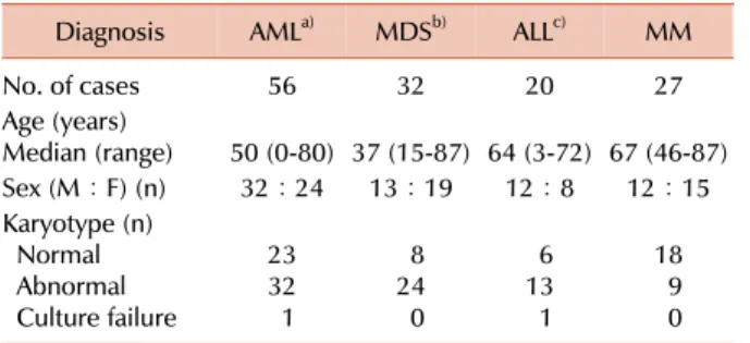
Table 1. Patient characteristics.
Diagnosis AMLa) MDSb) ALLc) MM
No. of cases 56 32 20 27
Age (years)
Median (range) 50 (0-80) 37 (15-87) 64 (3-72) 67 (46-87)
Sex (M:F) (n) 32:24 13:19 12:8 12:15
Karyotype (n) Normal Abnormal Culture failure
2332 1
248 0
136 1
189 0
a)Included 18 AML with recurrent chromosomal abnormalities [t(8;21) (n=6), t(15;17) (n=8), inv(16) (n=3), MLL (n=1)], 4 AML with myelodysplasia-related changes, and 34 AML, NOS, b)In- cluded refractory cytopenia with unilineage dysplasia (n=16), refractory cytopenia with multilineage dyaplasia (n=13), refractory anemia with excess of blasts (n=2), MDS-unclassifiable (n=1),
c)Included B-lineage (n=15), T-lineage (n=4), mixed phenotype acute leukemia (n=1).
Abbreviations: AML, acute myeloid leukemia; MDS, myelody- splastic syndrome; ALL, acute lymphoblastic leukemia; MM, multiple myeloma.
by conventional G-banded karyotype.
The aim of this study was to evaluate the clinical utility of FISH in addition to G-banded karyotype and to propose a practical approach for FISH in the detection of hematologic malignancies, including acute myeloid leukemia (AML), myelodysplastic syndrome (MDS), acute lymphoblastic leu- kemia (ALL), and multiple myeloma (MM).
MATERIALS AND METHODS
1. Patients
The study group included 135 patients with hematologic malignancies (56 AML, 32 MDS, 20 ALL, and 27 MM) be- tween 2005 and 2010. The characteristics of the patients are shown in Table 1.
2. Conventional cytogenetics
Cytogenetic studies were performed with unstimulated 24- and 48-hour cultures, using fresh bone marrow aspirates obtained from the 135 patients at diagnosis. When possible, at least 20 metaphases per sample were analyzed, and kar- yotypes were described according to the International System for Human Cytogenetic Nomenclature (ISCN, 2009) [3].
3. FISH
FISH studies of the 135 patients at diagnosis were per- formed on fresh bone marrow aspirates or fixed cells obtained from bone marrow cultures for conventional cytogenetics.
Commercially available probes (Abbott/Vysis, Downers Grove, IL, USA and Kreatech, Amsterdam, Netherlands) were used.
The AML panel included BCR/ABL1 dual color, dual fusion translocation probe; AML1/ETO dual color, dual fusion trans- location probe; PML/RARA dual color, dual fusion trans-
location probe; CBFB dual color, break-apart rearrangement probe; MLL (11q23) dual color, break-apart rearrangement probe; EGR1 (5q31)/D5S23, D5S721 dual color probe; CEP8 SpectrumOrange probe; and D7S486 (7q31) SpectrumOrange/
CEP7 SpectrumGreen probe. The MDS panel included CEP8 SpectrumOrange probe; D20S108 (20q12) SpectrumOrange probe; EGR1 (5q31)/D5S23, D5S721 dual color probe; and D7S486 (7q31) SpectrumOrange/CEP7 SpectrumGreen probe.
The ALL panel included BCR/ABL1 dual color, dual fusion translocation probe; MLL dual color, break-apart rearrange- ment probe; CDKN2A (9p21, p16) SpectrumOrange/CEP9 SpectrumGreen probe; ETV6 (12p13) dual color, break-apart rearrangement probe; and 6q21/c-myc dual color probe. The MM panel included, IgH dual color, break-apart rearrange- ment probe; TP53 SpectrumOrange probe; D13S25 (13q14.3) SpectrumOrange probe; IgH/CCND1 dual color, dual fusion translocation probe; IgH/MAF dual color, dual fusion trans- location probe; IgH/FGFR3 dual color, dual fusion trans- location probe; and 1q21/8p21 dual color probe. All probes except 6q21/c-myc and 1q21/8p21 (Kreatech) were provided by Abbott/Vysis. At least 200 interphase cells were scored for each probe by two independent experienced examiners.
RESULTS
1. AML (Table 2)
FISH studies confirmed the corresponding abnormalities identified by G-banded karyotype in all of the AML samples.
Additional abnormalities were detected by FISH in only two cases (4%). In 1 case with unsuccessful culture, AML1 (RUNX1) gain was identified in 66% of interphase cells by FISH. In the other case, characterized by a complex karyotype with marker chromosomes and abnormalities of chromosome 5, 17, 18, and 19, FISH detected MLL gain in 80% of inter- phase cells.
2. MDS (Table 2)
FISH confirmed the results of both normal and abnormal karyotype identified by G-banded karyotype. Abnormalities additional to those identified by G-banded karyotype were identified by FISH in 3 patients with normal karyotype (2 patients, 20q deletion in 5-6% of interphase cells; 1 patient, 7q deletion in 3% of interphase cells).
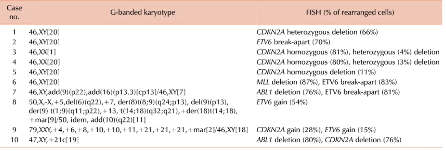
3. ALL (Tables 2 and 3)
Clonal abnormalities were found in 65% and 80% of pa- tients by G-banded karyotype and FISH, respectively.
Additional abnormalities were identified by FISH in 50%
of patients. Compared with G-banded karyotype, CDKN2A and ETV6 FISH revealed additional genetic aberrations in 33% and 28% of cases, respectively. Two patients showed ETV6 gain using FISH, corresponding to the hyperdiploidy or hypertriploidy by G-banded karyotype. In 2 patients, ABL1 deletions unassociated with t(9;22) were identified by FISH.
Table 2. Abnormalities detected by FISH in addition to G-banded karyotype.
N Additional
detection rate (%) Abnormal rate (%) AML
Total 56 4 50
BCR/ABL1 48 0 2
AML1/ETO 48 2 23
PML/RARA 48 0 19
CBFB 45 0 7
MLL 48 2 4
EGR1 17 0 6
D7S486 22 0 5
CEP8 2 0 50
MDS
Total 32 9 34
CEP8 32 0 16
D20S108 32 6 13
EGR1 32 0 3
D7S486 32 3 6
ALL
Total 20 50 80
BCR/ABL1 20 10 35
MLL 19 5 5
CDKN2A 18 33 44
ETV6 18 28 33
6q21/c-myc 11 0 9
MM
Total 27 67 93
IgH 26 31 50
TP53 25 20 20
D13S25 25 36 52
IgH/CCND1 27 19 37
IgH/FGFR3 19 11 21
IgH/MAF 19 0 11
1q21 20 40 55
8p21 20 15 15
Abbreviation: FISH, fluorescence in situ hybridization. see Table 1.
Table 3. Additional genetic aberrations identified by FISH in ALL.
Case
no. G-banded karyotype FISH (% of rearranged cells)
1 46,XY[20] CDKN2A heterozygous deletion (66%)
2 46,XY[20] ETV6 break-apart (70%)
3 46,XX[1] CDKN2A homozygous (81%), heterozygous (4%) deletion
4 46,XX[20] CDKN2A homozygous (80%), heterozygous (3%) deletion
5 46,XY[20] CDKN2A homozygous deletion (11%)
6 46,XY[20] MLL deletion (87%), ETV6 break-apart (83%)
7 46,XY,add(9)(p22),add(16)(p13.3)[cp13]/46,XY[7] ABL1 deletion (76%), ETV6 break-apart (81%) 8 50,X,-X,+5,del(6)(q22),+7, der(8)t(8;9)(q24;p13), del(9)(p13),
der(9) t(1;9)(q11;p22),+13, t(14;18)(q32;q21),+der(18)t(14;18), +mar[9]/50, idem, add(10)(q22)[11]
ETV6 gain (54%)
9 79,XXY,+4,+6,+8,+10,+10,+11,+21,+21,+21,+mar[2]/46,XY[18] CDKN2A gain (28%), ETV6 gain (15%)
10 47,XY,+21c[19] ABL1 deletion (80%), CDKN2A deletion (76%)
Abbreviations: FISH, fluorescence in situ hybridization; ALL, acute lymphoblastic leukemia.
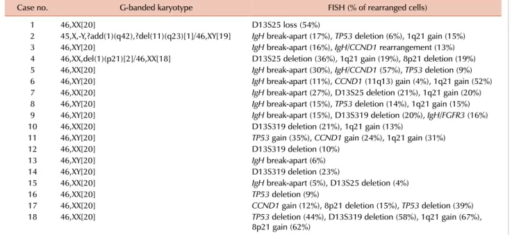
4. MM (Tables 2 and 4)
Clonal abnormalities were detected in 33% and 93% of patients by G-banded karyotype and FISH, respectively.
Additional abnormalities were identified by FISH in 67%
of patients, and among these, 89% had a normal karyotype as determined by G-banded karyotype. FISH was of benefit in detecting IgH, D13S25, TP53, and 1q21 rearrangements that were not detected by G-banded karyotype (31%, 36%, 20%, and 40%, respectively).
DISCUSSION
In this study, the percentages of additional genetic aberra- tions identified by FISH (which were not detected by G-banded karyotype) were 4%, 9%, 50%, and 67% in AML, MDS, ALL, and MM, respectively.
In AML and MDS, FISH did not add any relevant in- formation to that already provided by G-banded karyotype with regard to specific chromosomal abnormalities. Similar to our result, a previous study showed that AML FISH profile tests revealed additional genetic abnormalities only in 8%
of cases [4]. Another study showed that the discrepancy between G-banded karyotype and FISH for the diagnosis of AML is 7% [5]. Previous studies evaluating the utility of FISH in cases of MDS reported the detection of 6% or fewer abnormalities by FISH in addition to those detected by G-banded karyotype [6-13]. Other studies, however, have detected up to approximately 15% of additional abnormalities using FISH [14-16]. One study suggested that FISH testing may be informative only in MDS cases with culture failure or intermediate- to high-grade MDS cases with normal kar- yotype, indicating that cases with low-grade MDS and normal karyotypes do not appear to benefit from FISH testing [13].
Taken together, in the setting of adequate cytogenetic study, the sensitivity of the two techniques in detecting clinically significant chromosomal abnormalities seems to be similar, and FISH may have limited utility in the cases of AML
Table 4. Additional genetic aberrations identified by FISH in MM.
Case no. G-banded karyotype FISH (% of rearranged cells)
1 46,XX[20] D13S25 loss (54%)
2 45,X,-Y,?add(1)(q42),?del(11)(q23)[1]/46,XY[19] IgH break-apart (17%), TP53 deletion (6%), 1q21 gain (15%)
3 46,XY[20] IgH break-apart (16%), IgH/CCND1 rearrangement (13%)
4 46,XX,del(1)(p21)[2]/46,XX[18] D13S25 deletion (36%), 1q21 gain (19%), 8p21 deletion (19%)
5 46,XX[20] IgH break-apart (30%), IgH/CCND1 (57%), TP53 deletion (9%)
6 46,XY[20] IgH break-apart (11%), CCND1 (11q13) gain (4%), 1q21 gain (52%)
7 46,XX[20] IgH break-apart (27%), D13S25 deletion (21%), 1q21 gain (20%)
8 46,XY[20] IgH break-apart (15%), TP53 deletion (14%), 1q21 gain (15%)
9 46,XY[20] IgH break-apart (15%), D13S319 deletion (20%), IgH/FGFR3 (16%)
10 46,XX[20] D13S319 deletion (21%), 1q21 gain (13%)
11 46,XY[20] TP53 gain (35%), CCND1 gain (24%), 1q21 gain (31%)
12 46,XX[20] D13S319 deletion (10%)
13 46,XY[20] IgH break-apart (6%)
14 46,XY[20] D13S319 deletion (23%)
15 46,XX[20] IgH break-apart (5%), D13S25 deletion (4%)
16 46,XX[20] TP53 deletion (9%)
17 46,XX[20] CCND1 gain (12%), 8p21 deletion (15%), TP53 deletion (39%)
18 46,XX[20] TP53 deletion (44%), D13S319 deletion (58%), 1q21 gain (67%),
8p21 gain (62%) Abbreviations: FISH, fluorescence in situ hybridization; MM, multiple myeloma.
or MDS.
In contrast, FISH, particularly in the cases of lymphoid malignancies, becomes an invaluable tool for identifying spe- cific genetic changes other than G-banded karyotype. One study showed that ALL FISH profile tests revealed additional genetic aberrations not detected by G-banded karyotype in up to 49% of cases, and among these, ETV6/RUNX1 (TEL/ AML1) abnormalities were frequently detected (44%), fol- lowed by the abnormal CDKN2A (25%) and hyperdiploidy (18%) [4]. A further study also demonstrated that G-banded karyotype failed to detect a considerable part of the ETV6/RUNX1 (TEL/AML1) translocation (sensitivity 6%) [17]. As expected, FISH is of benefit in the detection of ETV6 and CDKN2A rearrangements, since these rearrange- ments are cytogenetically cryptic and invisible. On the other hand, the sensitivity of G-banded karyotype for the detection of BCR/ABL1 and MLL rearrangements in ALL is relatively high (80% and 85%, respectively) [17]. Consistent with pre- vious studies, our study showed that in ALL, ETV6 and CDKN2A genetic abnormalities, which were not identified by G-banded karyotype, could be additionally detected by FISH (28% and 33%, respectively).
As reported previously, FISH is useful for improving the detection rate of genetic abnormalities in MM, whereas con- ventional cytogenetics detects only 30-50% of abnormalities due to the low in vitro mitotic index of abnormal clones [18-25]. In this study, the detection rate of genomic aberra- tions in MM increased from 67% to 93% using FISH, com- pared with G-banded karyotype.
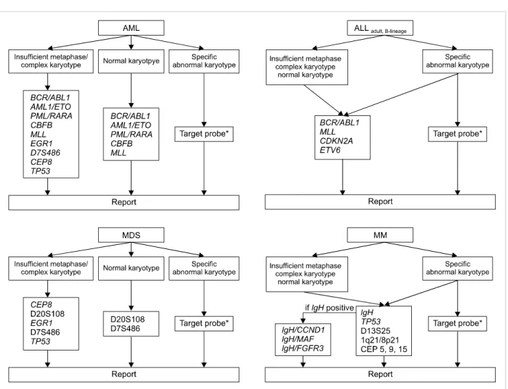
FISH has been performed extensively to detect genomic aberrations in hematologic malignancies; however, in view of limited laboratory resources, it may be an expensive
procedure. Here, we propose a strategy for cost-effective FISH utilization based on our results (Fig. 1). Importantly, this strategy should be based on the premise of cytogenetic adequacy (analyzing more than 20 consecutive, well-stained, well-spread metaphases). If a sufficient number of meta- phases with morphology good enough to detect microscopic abnormalities cannot be analyzed, this strategy should not be applied.
With respect to ALL and MM, routine FISH analysis is needed, irrespective of karyotypic results (normal or abnor- mal), since, as shown in this study, FISH can provide relevant information additional to that provided by G-banded kar- yotype (Tables 2-4). In order to detect aneuploidy, it may be necessary to include centromere probes for chromosome 5, 9, and 15 in the MM FISH panel [20, 22]. Here, we propose a FISH panel for adult B-lineage ALL; a FISH panel for children was not considered in this study.
In contrast to ALL and MM, an appropriate strategy should be selected in AML and MDS, depending on the results of G-banded karyotype. We recommend routine FISH analy- sis in cases with a complex karyotype, because FISH can identify details of aberrations that cannot be resolved by G-banded karyotype alone. In addition, in cases with few or no mitotic cells, routine FISH analysis would be of benefit for obtaining clinically relevant information regarding aberrations. In AML or MDS with a normal karyotype, cer- tain specific probes could be used to detect cryptic aberrations undetectable by G-banded karyotype. Among FISH probes that we did not evaluate in the present study, the TP53 probe may be needed in cases with complex karyotypes or insufficient metaphases; this is because the TP53 deletion is known to be associated with a poor prognosis [26, 27].
Fig. 1. Proposal for a cost-effective utilization of FISH in hematologic malignancies (*corresponded to specific chromosomal abnormalities identified by G-banded karyotype).
Of particular relevance, FISH analysis may be a superior method for disease monitoring, considering that G-banded karyotype could be hampered by a low in vitro mitotic activ- ity of cancer cells after treatment. Therefore, if specific chro- mosomal abnormalities are detected by G-banded karyotype at diagnosis, FISH analysis using target probes corresponding to the specific chromosomal abnormalities is needed to inves- tigate specific abnormal FISH signal patterns for use of mon- itoring markers during follow-up. An advantage of FISH over G-banded karyotype is that it requires considerably less time and effort. Therefore, apart from our proposal, FISH using specific target probes could be utilized in the initial assessment as an adjunct to G-banded karyotype, when critical genetic aberrations (such as PML/RARA rearrange- ment) should be rapidly identified to determine the best therapeutic approaches.
In addition to G-banded karyotype and FISH, PCR techni- ques are currently being employed. Of particular interest, multiplex reverse-transcription PCR can be used to simulta- neously identify numerous different translocations or chro- mosomal rearrangements. To detect fusion transcripts or translocations, either PCR or FISH could be used. However,
PCR techniques are unable to identify deletions or amplifica- tions that can be readily identified by FISH, and therefore it is unlikely that these techniques will serve as a substitute for FISH. Further study is needed to develop efficient and cost-effective strategies that combine the use of G-banded karyotype, FISH, and PCR techniques.
In conclusion, this study suggests that in the setting of an adequate karyotype of AML and MDS, routine FISH test- ing contributes little, if any, further genetic information.
In contrast, FISH panel testing for ALL and MM appears to be an efficient screening method, and routine FISH analysis should remain the method of choice. Finally, a consensus needs to be reached between laboratories as to the practical strategies for cost-effective utilization of FISH combined with G-banded karyotype.
REFERENCES
1. Swerdlow SH, Campo E, Harris NL, et al, eds. WHO classification of tumours of haematopoietic and lymphoid tissues. 4th ed. Lyon, France: IARC press, 2008:87-213.
2. Tibiletti MG. Interphase FISH as a new tool in tumor pathology.
Cytogenet Genome Res 2007;118:229-36.
3. Shaffer LG, Slovak ML, Campbell LJ, eds. An international system for human cytogenetic nomenclature (2009): recommendations of the international standing committee on human cytogenetic nomenclature. Basel, Switzerland: S. Karger AG, 2009.
4. Kim SR, Kim HJ, Kim SH. Clinical utility of fluorescence in situ hybridization profile test in detecting genetic aberrations in acute leukemia. Korean J Lab Med 2009;29:371-8.
5. Lee DY, See CJ, Hwang CD, Cho HI, Lee DS. Analysis of discrep- ancies between G-banding and FISH in hematologic abnorma- lities. Korean J Clin Pathol 2001;21:445-50.
6. Pitchford CW, Hettinga AC, Reichard KK. Fluorescence in situ hybridization testing for -5/5q, -7/7q, +8, and del(20q) in primary myelodysplastic syndrome correlates with conventional cytoge- netics in the setting of an adequate study. Am J Clin Pathol 2010;133:260-4.
7. Ketterling RP, Wyatt WA, VanWier SA, et al. Primary myelodys- plastic syndrome with normal cytogenetics: utility of ‘FISH panel testing’ and M-FISH. Leuk Res 2002;26:235-40.
8. Cherry AM, Brockman SR, Paternoster SF, et al. Comparison of interphase FISH and metaphase cytogenetics to study myelodys- plastic syndrome: an Eastern Cooperative Oncology Group (ECOG) study. Leuk Res 2003;27:1085-90.
9. Beyer V, Castagné C, Mühlematter D, et al. Systematic screening at diagnosis of -5/del(5)(q31),-7, or chromosome 8 aneuploidy by interphase fluorescence in situ hybridization in 110 acute myelo- cytic leukemia and high-risk myelodysplastic syndrome patients:
concordances and discrepancies with conventional cytogenetics.
Cancer Genet Cytogenet 2004;152:29-41.
10. Panani AD, Pappa V. Hidden chromosome 8 abnormalities de- tected by FISH in adult primary myelodysplastic syndromes. In Vivo 2005;19:979-81.
11. Mallo M, Arenillas L, Espinet B, et al. Fluorescence in situ hybrid- ization improves the detection of 5q31 deletion in myelodysplastic syndromes without cytogenetic evidence of 5q-. Haematologica 2008;93:1001-8.
12. Costa D, Valera S, Carrió A, et al. Do we need to do fluorescence in situ hybridization analysis in myelodysplastic syndromes as of- ten as we do? Leuk Res 2010 [Epub ahead of print].
13. Yang W, Stotler B, Sevilla DW, et al. FISH analysis in addition to G-band karyotyping: utility in evaluation of myelodysplastic syn- dromes? Leuk Res 2010;34:420-5.
14. Rigolin GM, Bigoni R, Milani R, et al. Clinical importance of inter- phase cytogenetics detecting occult chromosome lesions in mye- lodysplastic syndromes with normal karyotype. Leukemia 2001;15:1841-7.
15. Romeo M, Chauffaille Mde L, Silva MR, Bahia DM, Kerbauy J.
Comparison of cytogenetics with FISH in 40 myelodysplastic syn- drome patients. Leuk Res 2002;26:993-6.
16. Bernasconi P, Cavigliano PM, Boni M, et al. Is FISH a relevant prognostic tool in myelodysplastic syndromes with a normal chromosome pattern on conventional cytogenetics? A study on 57 patients. Leukemia 2003;17:2107-12.
17. Olde Nordkamp L, Mellink C, van der Schoot E, van den Berg H.
Karyotyping, FISH, and PCR in acute lymphoblastic leukemia:
competing or complementary diagnostics? J Pediatr Hematol Oncol 2009;31:930-5.
18. Yuregir OO, Sahin FI, Yilmaz Z, Kizilkilic E, Karakus S, Ozdogu H. Fluorescent in situ hybridization studies in multiple myeloma.
Hematology 2009;14:90-4.
19. Chen L, Li J, Xu W, et al. Molecular cytogenetic aberrations in pa- tients with multiple myeloma studied by interphase fluorescence in situ hybridization. Exp Oncol 2007;29:116-20.
20. Christensen JH, Abildgaard N, Plesner T, et al. Interphase fluo- rescence in situ hybridization in multiple myeloma and mono- clonal gammopathy of undetermined significance without and with positive plasma cell identification: analysis of 192 cases from the Region of Southern Denmark. Cancer Genet Cytogenet 2007;174:89-99.
21. Sáez B, Martín-Subero JI, Odero MD, et al. Interphase FISH for the detection of breakpoints in IG loci and chromosomal changes with adverse prognostic impact in multiple myeloma with normal karyotypes. Cancer Genet Cytogenet 2006;167:183-5.
22. Schmidt-Wolf IG, Glasmacher A, Hahn-Ast C, et al. Chromoso- mal aberrations in 130 patients with multiple myeloma studied by interphase FISH: diagnostic and prognostic relevance. Cancer Genet Cytogenet 2006;167:20-5.
23. Chen Z, Issa B, Huang S, et al. A practical approach to the detection of prognostically significant genomic aberrations in multiple myeloma. J Mol Diagn 2005;7:560-5.
24. Huang SY, Yao M, Tang JL, et al. Clinical significance of cytoge- netics and interphase fluorescence in situ hybridization analysis in newly diagnosed multiple myeloma in Taiwan. Ann Oncol 2005;16:1530-8.
25. Wiktor A, Van Dyke DL. Combined cytogenetic testing and fluo- rescence in situ hybridization analysis in the study of chronic lym- phocytic leukemia and multiple myeloma. Cancer Genet Cyto- genet 2004;153:73-6.
26. Haferlach C, Rieder H, Lillington DM, et al. Proposals for stand- ardized protocols for cytogenetic analyses of acute leukemias, chronic lymphocytic leukemia, chronic myeloid leukemia, chronic myeloproliferative disorders, and myelodysplastic synd- romes. Genes Chromosomes Cancer 2007;46:494-9.
27. Sreekantaiah C. FISH panels for hematologic malignancies.
Cytogenet Genome Res 2007;118:284-96.