This rare disease is characterized by a symmetrical sclerosis at the diametaphyseal portions of the lower extremities with extraskeletal involvement. Many internal organs and tissue sites, including the kidney and retroperitoneum, lung, pericardium, skin, orbit, and brain may occur. The symptoms and clinical manifestations depend on the organ involved. The prognosis is poor with progressive disease with resultant organ dysfunction. We report a case of Erdheim Chester disease. The patient was informed that the data concerning this case would be submitted for publication.
Case Report
A 69-year-old female patient was referred for both knees and lower extremities pains which began 6 years ago and were recently serious.
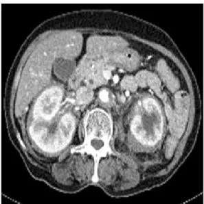
She has taken medication for diabetes and hypertension for 20 years and was in the hospital for polyuria and pyuria in the internal medi- cine department of this hospital. Soft tissue invasion around both kidneys (Fig. 1) and soft-tissue lesions around right coronary artery were found on abdominal CT (computed tomography) and their as- sessment was in progress.
Biopsy was preformed for soft-tissue lesions around both kidneys
Case Report
J Korean Bone Joint Tumor Soc 2013; 19: 28-32 • http://dx.doi.org/10.5292/jkbjts.2013.19.1.28 www.kbjts.or.krErdheim Chester Disease (ECD): A Case Report
Jin-Ho Lee, Sung-Taek Jung, and Yoo Duk Choi*
Department of Orthopedic Surgery, Chonnam National University College of Medicine, *Department of Pathology, Chonnam National University Medical School, Gwangju, Korea
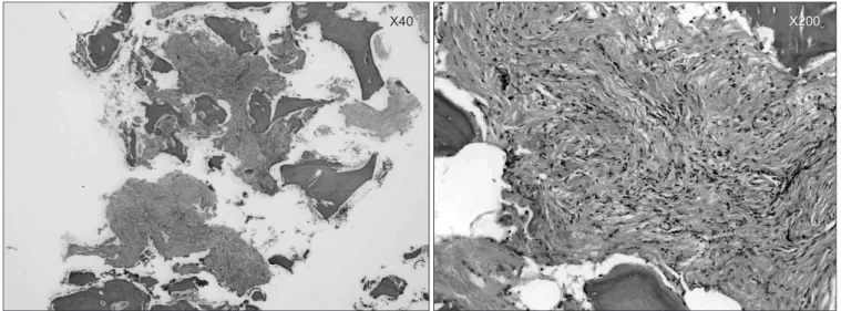
under the ultrasonography guide and the pathologic features revealed collagenous fibroadipose tissue with lymphoplasma and histiocytes.
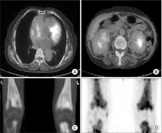
The result immunohistochemical staining were a positive for CD68, and negative for S-100, CD1a (Fig. 2). PET CT (Positron Emission Tomography) showed high metabolic lesions of SUV 4.7 in heart, kidney, bilateral distal femoral, and proximal tibia (Fig. 3) and X- ray also showed osteosclerotic and osteolytic lesions (Fig. 4). Thus, this department and the departments of hematology-oncology and cardiology implemented the integrated treatment under the suspicion of hematologic malignancy or multiple bone metastasis. While the
Received March 19, 2013 Revised May 30, 2013 Accepted May 31, 2013
Correspondence to: Sung-Taek Jung
Department of Orthopedic Surgery, Chonnam National University Hospital, 8, Hak- dong, Dong-gu, Gwangju 501-757, Korea
TEL: 82-62-227-1640
FAX: 82-62-225-7794
E-mail: [email protected]
Erdheim Chester disease (ECD) is very rare non-Langerhans cell histiocytosis (LCH) which occurs in the skeletal system and multiple organs.
As it is progressive, sometimes it causes fatal results. However, it is often misdiagnosed as LCH or multiple bone metastasis and, thus, is very difficult to diagnose. In Korea, only 10 cases were first reported in 1999. In particular, there have been a few orthopedic approaches or reports in English-speaking literatures, and no report has been issued in Korea. The authors performed bone biopsy in patients with knee and lower extremity pain who were referred for the integrated treatment. We attempts to report this diagnosis experience with literature review.
Key word: Langerhans cell histiocytosis
Copyrights © 2013 by The Korean Bone and Joint Tumor Society
“This is an Open Access article distributed under the terms of the Creative Commons Attribution Non-Commercial License (http://creativecommons.org/licenses/by-nc/3.0/) which permits unrestricted non-commercial use, distribution, and reproduction in any medium, provided the original work is properly cited.”
대한골관절종양학회지:제19권 제1호 2013
Figure 1. On computed tomography, there are soft tissue invasions around both kidney.
pISSN : 1226-4962 eISSN : 2233-9841
29
Erdheim Chester Disease (ECD)
department of hematology-oncology considered bone marrow ex- amination and the department of cardiology considered biopsy, this department performed biopsy under the suspicion of multiple bone metastasis and lymphoma. The bone biosy revealed an intraosseous fibrosis (Fig. 5). We diagnosed the patient with ECD based on the above clinical and biopsy findings. She is under internal treatment.
Discussion
Erdheim Chester disease (ECD) is very rare non-Langerhans cell histiocytosis which invade the skeletal system and multiple organs.
It was first introduced by Chester1) and is histologically characterized by the accumulation of lipid laden foamy macrophage.2,3) Although the cause of disease has not been established yet, the high turnover of LDL seems to play an important role. In Korea, 10 cases4) were first reported. There have been a few orthopedic approaches or re-
ports in English-speaking literatures, and no report has been issued in Korea.
Since the clinical characteristics of ECD are much unknown and it is difficult to diagnose, clinical suspicion is important. Most cases are developed after forty years of age and are characterized by os- teosclerotic lesions which are well marked by bilateral symmetrical boundary of metaphysis of long bone. It may occur in ribs, sacrum, head, neck, and spine. It is rare to occur in the spine in case of ex- tremities3) and its occurrence in head and neck is associated with diabetes insipidus. Patients clinically have arthralgia, keloid forma- tion, and a systemic symptom which invades several organs such as heart, liver, lung, and kidney.5,6) It is known to be accompanied by extraskeletal symptoms2) in about its 50%. A clinical prognosis is de- termined by invaded organs and if it invades the blood vessel system, it shows a bad prognosis.7,8)
ECD is progressive disease. Generally, patients are often seen in Figure 2. On histologic finding, pathologic features revealed collagenous fibroadipose tissue with lymphoplasma and histiocytes. The result immunohistochemical staining were a positive for CD68, and negative for S-100, CD1a.
30
Jin-Ho Lee, et al.
Figure 3. On PET CT, it showed high metabolic lesions of SUV 4.7 in heart (A), kidney (B), bilateral distal femoral, and proximal tibia (C, D).
Figure 4. X-ray also showed osteosclerotic and osteolytic lesions on both distal femur and proximal tibia.
31
Erdheim Chester Disease (ECD)
the hospital with arthralgia or lower extremity pain caused by ortho- pedic osteoarthritis. Thus, if the disease is not recognized, it is im- possible to prevent its progress and the condition will become worse.
Generally bony invasion is characterized by osteosclerotic lesions which are well marked by the bilateral symmetrical boundary of iliac metapre or are patch-shaped.3) Our patient had mixed lesions of osteosclerotic and osteolytic lesions, resulting in authors' misdiagno- sis with multiple bone metastasis.
The patient was positive for CD68 but negative for CDla and S-100 on the immunohistochemical stain. Based on this, LCH which shows similar clinical conditions could be excluded.
The patient could be diagnosed with ECD based on orthopedical bone biopsy and clinical characteristics. Then, she could be systemi- cally treated and invasive diagnosis testing which is more dangerous could be prevented.
Although ECD is progressive and fetal disease and its prognosis is determined by invaded organs or its degree, the condition often becomes serious by patients often seen in the hospital with arthralgia who cannot recognize their disease. In other words, it is considered that clinical suspicion of ECD is needed based on the above radio- logical and clinical findings.
As ECD is very rare and fatal disease which is not easy to diag- nose, correct diagnosis and treatment is essential. If middle-aged people seen with arthralgia and lower extremity pain show symmet- ric and bilateral bone lesions, the clinical suspicion of ECD is neces- sary. Thus, the authors reported the diagnosis and caution of ECD which has few orthopedic reports and is often misdiagnosed through our experience and literature review.
References
1. Chester W. Über Lipoidgranulomatose (Over lipoid granuloma- tosis). Virchows Arch Pathol Anat Physiol. 1930:279:561-602.
2. Veyssier-Belot C, Cacoub P, Caparros-Lefebvre D, et al. Erd- heim-Chester disease. Clinical and radiologic characteristics of 59 cases. Medicine (Baltimore). 1996;75:157-69.
3. Schmidt HH, Gregg RE, Shamburek R, Brewer BH Jr, Zssssech LA. Erdheim-Chester disease: low low-density lipoprotein lev- els due to rapid catabolism. Metabolism. 1997;46:1215-9.
4. Park YK, Ryu KN, Huh B, Kim JD. Erdheim-Chester disease: a case report. J Korean Med Sci. 1999;14:323-6.
5. Resnick D, Greenway G, Genant H, Brower A, Haghighi P, Emmett M. Erdheim-Chester disease. Radiology. 1982;142:
289-95.
6. Atkins HL, Klopper JF, Ansari AN, Iwai J. Lipid (cholesterol) granulomatosis (Chester-Erdheim disease) and congenital megacalices. Clin Nucl Med. 1978;3:324-7.
7. Haroche J, Amoura Z, Dion E, et al. Cardiovascular involve- ment, an overlooked feature of Erdheim-Chester disease:
report of 6 new cases and a literature review. Medicine (Balti- more). 2004;83:371-92.
8. Lachenal F, Cotton F, Desmurs-Clavel H, et al. Neurological manifestations and neuroradiological presentation of Erd- heim-Chester disease: report of 6 cases and systematic review of the literature. J Neurol. 2006;253:1267-77.
Figure 5. The bone biopsy revealed an intraosseous fibrosis. H & E (Hematoxylin & Eosin) staining.
Erdheim Chester Disease (ECD): 증례 보고
이진호
•
정성택•
최유덕*전남대학교 의과대학 전남대학교병원 정형외과교실, *병리학교실
Erdheim chester disease (ECD)은 골격계 및 다발성 장기에 발생하는 매우 드문 비 랑거한스세포 조직구증(Langerhans cell histiocytosis,LCH)으로 구분되는 질환으로 진행성이며 때론 치명적인 결과를 야기한다. 하지만 이는 LCH 혹은 다발성 골 전이로 오인 되기 쉬우며 진단 또한 극히 어렵다. 국내에서는 1999년에 10예가 처음으로 보고되었을 뿐이며 특히 정형외과적으로 접근 및 보고는 문헌상 영어권에서 극히 소수의 문헌만이 존재하며 국내에는 보고된 예가 없다. 저자들은 슬관절 및 하지통은 주소로 본과에 협진된 환 자에 대해 골 조직검사 시행하였으며 이에 대한 진단적 경험을 문헌 고찰과 함께 보고하고자 한다.
색인단어: 랑거한스세포 조직구증
접수일 2013년 3월 19일 심사수정일 2013년 5월 30일 게재확정일 2013년 5월 31일 교신저자 정성택
광주시 동구 학동 8, 전남대학교병원 정형외과
TEL 062-227-1640, FAX 062-225-7794, E-mail [email protected]
Case Report
J Korean Bone Joint Tumor Soc 2013; 19: 28-32 • http://dx.doi.org/10.5292/jkbjts.2013.19.1.28 www.kbjts.or.krCopyrights © 2013 by The Korean Bone and Joint Tumor Society
“This is an Open Access article distributed under the terms of the Creative Commons Attribution Non-Commercial License (http://creativecommons.org/licenses/by-nc/3.0/) which permits unrestricted non-commercial use, distribution, and reproduction in any medium, provided the original work is properly cited.”
The Journal of the Korean Bone and Joint Tumor Society Vol. 19, No. 1 (June 2013)
pISSN : 1226-4962 eISSN : 2233-9841