Korean J Gastroenterol Vol. 63 No. 6, 366-368 http://dx.doi.org/10.4166/kjg.2014.63.6.366 pISSN 1598-9992 eISSN 2233-6869
CASE REPORT
Korean J Gastroenterol, Vol. 63 No. 6, June 2014 www.kjg.or.kr
양잿물에 의한 식도협착 환자에서 편평세포암종으로 오인된 거짓상피종 증식
한장수, 이상우, 서강흠, 김승영, 현종진, 정성우, 구자설, 임형준
고려대학교 의과대학 내과학교실
Pseudoepitheliomatous Hyperplasia Mimicking Esophageal Squamous Cell Carcinoma in a Patient with Lye-induced Esophageal Stricture
Jang Soo Han, Sang Woo Lee, Kang Heum Suh, Seung Young Kim, Jong Jin Hyun, Sung Woo Jung, Ja Seol Koo and Hyung Joon Yim Department of Internal Medicine, Korea University College of Medicine, Seoul, Korea
Pseudoepitheliomatous hyperplasia is a benign condition that may be caused by prolonged inflammation, chronic infection, and/or neoplastic conditions of the mucous membranes or skin. Due to its histological resemblance to well-differentiated squamous cell carcinoma, pseudoepitheliomatous hyperplasia may occasionally be misdiagnosed as squamous cell carcinoma.
The importance of pseudoepitheliomatous hyperplasia is that it is a self-limited condition that must be distinguished from squamous cell carcinoma before invasive treatment. We report here on a rare case of esophageal pseudoepitheliomatous hyperplasia in a 67-year-old Korean woman with a lye-induced esophageal stricture. Although esophageal pseudoepitheliomatous hyperplasia is infrequently encountered, pseudoepitheliomatous hyperplasia should be considered in the differential diagnosis of esophageal lesions. (Korean J Gastroenterol 2014;63:366-368)
Key Words: Pseudoepitheliomatous hyperplasia; Squamous cell carcinoma; Esophagus
Received October 29, 2013. Revised November 18, 2013. Accepted November 18, 2013.
CC This is an open access article distributed under the terms of the Creative Commons Attribution Non-Commercial License (http://creativecommons.org/licenses/
by-nc/3.0) which permits unrestricted non-commercial use, distribution, and reproduction in any medium, provided the original work is properly cited.
교신저자: 이상우, 425-707, 안산시 단원구 적금로 123, 고려대학교 안산병원 내과
Correspondence to: Sang Woo Lee, Department of Internal Medicine, Korea University Ansan Hospital, 123 Jeokgeum-ro, Danwon-gu, Ansan 425-707, Korea.
Tel: +82-31-412-5580, Fax: +82-31-412-5582, E-mail: [email protected] Financial support: None. Conflict of interest: None.
INTRODUCTION
Pseudoepitheliomatous hyperplasia (PEH) is a benign condition that may be caused by prolonged inflammation, chronic infection, and/or neoplastic conditions of the mu- cous membranes or skin.1,2 Due to its histological resem- blance to well-differentiated squamous cell carcinoma (SCC), PEH may occasionally be misdiagnosed as SCC.1,2 PEH generally involves the skin or mucous membranes.1,2
However, no case of an esophageal lesion associated with PEH has been reported. We report here on a rare case of esophageal PEH in a 67-year-old Korean woman with a lye-in-
duced esophageal stricture.
CASE REPORT
A 67-year-old woman presented to our hospital with dys- phagia that had developed approximately one month previously. She had a history of an esophageal stricture caused by lye ingestion approximately 50 years previously and had undergone esophageal resection and esophagogas- trostomy seven years previously. Her medical history in- cluded hypertension and diabetes mellitus, and her family history was unremarkable. She was a non-smoker and social
Han JS, et al. Pseudoepitheliomatous Hyperplasia Mimicking SCC in a Patient with Esophageal Stricture 367
Vol. 63 No. 6, June 2014
Fig. 1. Endoscopic view of the eso- phagus showing an intraluminal protruding mass 25 cm from the incisor teeth during: (A) an initial esophagogastroduodenoscopy; (B) a surveillance esophagogastroduode- noscopy performed after two months.
Fig. 2. Lugol chromoscopy showing the mucosal lesion stained with Lugol’s solution.
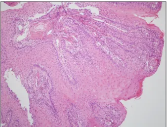
Fig. 3. Histological view of the esophageal mucosal lesion showing prominent hyperplasia of the epithelium (H&E, ×100).
drinker. She had recently reported a weight loss of 18 kg. An initial esophagogastroduodenoscopy showed an intra- luminal protruding lesion 25 cm from the incisor teeth (Fig.
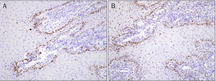
1A). The endoscope could not pass through the lesion due to the esophageal stricture. Lugol chromoscopy and biopsy were performed in order to exclude esophageal cancer. The mucosal lesion was stained with Lugol’s solution (Fig. 2), and histological examination showed prominent irregular hyper- plasia of the epithelium (Fig. 3). On immunohistochemical staining, Ki67 and p53 expression was observed only in the basal layer of the epithelium (Fig. 4). After two months, the lesion showed significant improvement without specific treatment (Fig. 1B).
DISCUSSION
The histological findings of PEH, characterized by the prominent proliferation of epithelium, are similar to those of well-differentiated SCC; therefore, PEH may occasionally be misdiagnosed as SCC.1,2 The pathogenesis of PEH remains unclear and there is no standard treatment. Although dis- tinguishing PEH from SCC can be difficult, the lack of atypical features, such as nuclear atypia and mitosis, and recognition of the associated underlying entities, are usually helpful in ex- cluding SCC.3 In addition, immunohistological staining meth- ods, including staining for Ki67 and p53 expression, have been used for differentiation of PEH from SCC. SCC shows nu- clear reactivity for Ki67 and p53 with staining of nuclei throughout the entire thickness of the epithelium, whereas PEH usually shows increased nuclear staining only in the bas- al layer of the epithelium.4,5 In our case, the esophageal le-
368 한장수 등. 편평세포암종으로 오인된 식도협착 환자의 거짓상피종 증식
The Korean Journal of Gastroenterology
Fig. 4. Immunohistochemical view of the esophageal mucosal lesion showing staining limited to the basal layer of benign epithelium with: (A) Ki67 immunostain (×200); (B) p53 immunostain (×200).
sion was stained with Lugol’s solution, and p53 and Ki67 ex- pression was observed only in the basal layer. The im- portance of PEH is that it is a self-limited condition that must be distinguished from SCC before invasive treatment. PEH usually involves the skin or mucous membranes.1,2 However, no case of an esophageal lesion associated with PEH has been reported. To the best of our knowledge, this is the first case report of esophageal PEH in a female patient with a his- tory of lye-induced esophageal stricture. Although esoph- ageal PEH is infrequently encountered, PEH should be con- sidered in the differential diagnosis of esophageal lesions.
REFERENCES
1. Fu X, Jiang D, Chen W, Sun Bs T, Sheng Z. Pseudoepithelioma- tous hyperplasia formation after skin injury. Wound Repair Regen 2007;15:39-46.
2. Zayour M, Lazova R. Pseudoepitheliomatous hyperplasia: a review. Am J Dermatopathol 2011;33:112-122.
3. Biswas A, Gey van Pittius D, Stephens M, Smith AG. Recurrent primary cutaneous lymphoma with florid pseudoepithelioma- tous hyperplasia masquerading as squamous cell carcinoma.
Histopathology 2008;52:755-758
4. Lee YS, Teh M. p53 expression in pseudoepitheliomatous hyper- plasia, keratoacanthoma, and squamous cell carcinoma of skin. Cancer 1994;73:2317-2323.
5. Zarovnaya E, Black C. Distinguishing pseudoepitheliomatous hyperplasia from squamous cell carcinoma in mucosal biopsy specimens from the head and neck. Arch Pathol Lab Med 2005;129:1032-1036.