pISSN 1598-9992 eISSN 2233-6869
ORIGINAL ARTICLE
위 점막 연관 림프조직 림프종에서 PET/CT의 유용성
황진원, 지삼룡, 이상헌, 김지현, 설상영, 이석모1
인제대학교 의과대학 부산백병원 내과학교실, 핵의학교실1
Efficacy of Positron Emission Tomography/Computed Tomography in Gastric Mucosa-associated Lymphoid Tissue Lymphoma
Jin Won Hwang, Sam Ryong Jee, Sang Heon Lee, Ji Hyun Kim, Sang Yong Seol, and Seok Mo Lee1
Departments of Internal Medicine and Nuclear Medicine1, Busan Paik Hospital, Inje University College of Medicine, Busan, Korea
Background/Aims: This study evaluated the diagnostic efficacy of fluorine-18 fluorodeoxyglucose PET/CT (F-18 FDG PET/CT) for patients with gastric mucosa-associated lymphoid tissue (MALT) lymphoma and examined the association between FDG avidity and the clinical factors affecting lesions.
Methods: Among the patients diagnosed with gastric MALT lymphoma, 16 who underwent a PET/CT for gastric MALT lymphoma were semi-quantitatively and qualitatively tested for FDG avidity of lesions in the stomach. Retrospectively collected data was analyzed to investigate the clinicoradiological factors and endoscopic findings between the patients with positive F-18 FDG PET/CT scans and those with negative scans.
Results: Eight of the 16 patients showed FDG avidity. When comparing the size of lesions in the stomach, the patients with FDG avidity had significantly larger lesions than those without (28.8 mm vs. 15.0 mm, p=0.03). The FDG-avid group has a significantly higher rate of positive CT scans than the non-avid group (75% vs. 13%, p=0.03). According to the endoscopic finding of the lesions, FDG avidity was pronounced with 75% of the protruding tumors, and 100% of the erosive-ulcerative types, which are a type of depressed tumors.
Conclusions: When gastric MALT lymphoma is large, when lesions are found using abdominal CT scans, and the macroscopic appearance of a lesion is that of a protruding tumor or erosive-ulcerative type of depressed tumor, there is a high probability that such patients may have a positive F-18 FDG PET/CT scan. (Korean J Gastroenterol 2016;67:183-188)
Key Words: Positron emission tomography; Stomach; Lymphoma, B-cell, marginal zone
Received February 22, 2016. Revised March 25, 2016. Accepted March 31, 2016.
CC This is an open access article distributed under the terms of the Creative Commons Attribution Non-Commercial License (http://creativecommons.org/licenses/
by-nc/4.0) which permits unrestricted non-commercial use, distribution, and reproduction in any medium, provided the original work is properly cited.
Copyright © 2016. Korean Society of Gastroenterology.
교신저자: 지삼룡, 47392, 부산시 부산진구 복지로 75, 인제대학교 부산백병원 소화기내과
Correspondence to: Sam Ryong Jee, Department of Gastroenterology, Inje University Busan Paik Hospital, 75 Bokji-ro, Busanjin-gu, Busan 47392, Korea. Tel: +82-51- 890-6536, Fax: +82-51-890-6341, E-mail: [email protected]
Financial support: None. Conflict of interest: None.
INTRODUCTION
Gastric mucosa-associated lymphoid tissue (MALT) lym- phoma represents nearly 35% of all gastric lymphomas and the stomach is the most common site of extranodal MALT lymphoma.1-3 It is possible to diagnose a gastric MALT lym- phoma by endoscopy and biopsy, but more tests are always needed.4,5 EUS-guided fine needle aspiration can be help-
ful6,7 and EUS can reduce the false negative error rate of en- doscopy and biopsy.8,9 When a patient is diagnosed with gas- tric MALT lymphoma, a variety of tests are needed to de- termine the stage of lymphoma. CT is useful to examine lymph nodes above and below the diaphragm but is not as useful in detecting lymph nodes near the stomach.10 Fluorine-18 fluorodeoxyglucose PET (F-18 FDG PET) is widely used in determining the stage of lymphoma and in evaluating
treatment responses and recurrence.11-14 Little research us- ing F-18 FDG PET has been conducted on MATL lymphoma;
although there have been some studies on the clinical use of F-18 FDG PET for MALT lymphoma, most investigated the characteristics of MALT lymphoma in various non-gastro- intestinal sites and only a few investigated the clinical validity of F-18 FDG PET for gastric MALT lymphoma.15-21 Unlike the F-18 FDG PET, F-18 FDG PET/CT evaluates the glucose me- tabolism of a tumor and the anatomical changes of a lesion, thereby facilitating the differentiation of physiological activ- ities and lesions on a PET scan. As a result, it can help localize lesions from the physiological activities of F-18 FDG in the stomach. This study examines clinical differences in the PET/CT findings of patients who underwent an F-18 FDG PET/CT after being diagnosed with gastric MALT lymphoma to determine the stage of lymphoma and to evaluate the effi- cacy of a PET/CT scan in diagnosing gastric MALT lymphoma.
SUBJECTS AND METHODS
1. Patients
The retrospective data of 71 patients diagnosed with gas- tric MALT lymphoma between September 2008 and October 2014 at Inje University Busan Paik Hospital (Busan, Korea) was analyzed. For this study, clinical data and radiological outcomes obtained from the medical records of the patients who underwent PET/CT scans were analyzed. The study ex- cluded patients who were under 18 years of age, did not have an endoscopic diagnosis and biopsy results, did not have PET/CT scans before gastric MALT lymphoma treatment, and those whose post-treatment observation period was less than six months or whose clinical data was missing.
This study was carried out in compliance with The World Medical Association Declaration of Helsinki and ethical prin- ciples for medical research involving human subjects. We an- alyzed treatment records and clinical outcomes of the pa- tients’ medical data by adhering to the deliberation require- ments of the Inje University Busan Paik Hospital’s clinical eth- ics committee (IRB No. INJE 15-0088).
2. Clinicoradiological factors
The study examined the following factors: age, gender, blood test results, infection status, and eradication rate of Helicobacter pylori, treatment modalities other than erad-
ication, improvement with gastric MALT lymphoma treat- ment, Ann Arbor stage, positive CT findings of lesions in the stomach, bone marrow invasion, extragastric lesions, and endoscopic and radiological diagnoses of gastric MALT lymphoma. The endoscopic macroscopic findings were cate- gorized as chronic gastritis-like tumors, depressed tumors, or protruding tumors according to the classification of lesions by Hirose et al.22 In addition, the number, location, and size of gastric lesions were investigated by endoscopy.
Associations between the aforementioned clinical factors and positive F-18 FDG PET/CT results were studied, as was the connection between the lesions’ maximum standardized uptake value (SUVmax) among the patients with positive find- ings and their clinical factors.
3. FDG PET/CT imaging procedure
All patients fasted for at least six hours and had a blood glu- cose level of less than 120 mg/dL before receiving an intra- venous injection of 481 MBq (13 mCi) of FDG. PET and CT im- ages from the skull base to the proximal thigh were obtained using a dedicated PET/CT scanner (Discovery STE; GE Healthcare, Milwaukee, WI, USA) 60 minutes post-injection.
Just before taking the PET/CT images, all patients were en- couraged to drink at least 500 mL of water to ensure stomach dilation. CT images were first acquired with acquisition pa- rameters of 140 Kvp, 80 mA, 0.8 sec per CT rotation and a pitch of 6, followed by torso emission scans for 2.5 to 3 mi- nutes per bed position. The acquired PET data were re- constructed to volumetric images using a 2D-OSEM algo- rithm with two iterations and 16 subsets.
4. Analysis of F-18 FDG PET/CT image
The PET/CT images of every patient were first examined by nuclear medicine physicians, after which the SUVmax of tumors seen on the images was measured. In order to differentiate physiological activity from a lesion, we had to locate the gastric lesion discovered by endoscopy using PET/CT scans. The le- sions are considered as follows; 1) the FDG uptake in the stom- ach can be macroscopically detected when compared to nor- mal physiological activities, 2) the diffuse uptake in a dilated stomach is greater than the liver uptake. Therefore, the SUVmax
of tumors was measured. The SUVmax was defined as de- cay-corrected FDG tissue concentration (Bq/g)/(injected dose of F-18 FDG [Bq]/patient’s body weight [g]).
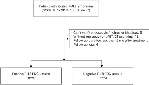
Fig. 1. Flowchart shows patient in- clusion.
MALT, mucosa-associated lymphoid tissue; F-18 FDG, fluorine-18 fluoro- deoxyglucose.
5. Statistical analysis
A statistical analysis was conducted using IBM SPSS Statistics software version 20.0 (IBM Co., Armonk, NY, USA).
Based on the F-18 FDG uptake in PET/CT scan, a Mann-Whitney test was used for continuous variables, whereas a Fisher’s exact test was used in categorical variables. Null hypotheses of no difference were rejected if p-values were less than 0.05.
RESULTS
Of the 71 patients who received gastric MALT lymphoma treatments from September 2008 to October 2014, 55 pa- tients were excluded from the study. Thus, 16 patients were enrolled (Fig. 1). Of these, eight had lesions with positive F-18 FDG uptake, while the rest had lesions with negative F-18 FDG uptake. The average SUVmax of the lesions with positive F-18 FDG uptake when scanned by PET/CT was 4.9 (range, 2.3-8.7).
When the two groups were compared in terms of age, gen- der, blood test results, infection status and eradication rate of H. pylori, treatment modalities other than eradication, im- provement with gastric MALT lymphoma treatment, Ann Arbor stage, positive CT findings of the lesions in the stom- ach, bone marrow invasion, and extragastric lesions, there were no significant differences in the F-18 FDG uptake be- tween them (Table 1). Upon CT scans, the group that had le- sions with positive F-18 FDG uptake was more likely to have positive CT findings in the stomach than the group that had
lesions with negative F-18 FDG uptake (75% vs. 13%, p=
0.03).
1. The relationship between endoscopic findings and F-18 FDG uptake patterns
Based on the endoscopic macroscopic findings, the 16 pa- tients were classified as having chronic gastritis-like tumors (n=5), depressed tumors (n=7), and protruding tumors (n=4). The association between F-18 FDG uptake patterns and macroscopic classifications of gastric MALT lymphoma is shown in Table 2. F-18 FDG uptake was observed in 75%
(3/4) of the protruding tumors, 57% (4/7) of the depressed tumors, and 20% of the chronic gastritis-like tumors (1/5).
Every patient with an erosive-ulcerative type tumor, a kind of depressed tumor, had positive F-18 FDG uptake (Fig. 2), whereas all patients with discolored-scar type tumors had negative F-18 FDG uptake (Fig. 3). There was no significant difference in F-18 FDG uptake patterns, in agreement with the number and location of MALT lymphoma. Except for four cases in which the size was difficult to measure as the lesions were diffuse, the lesions in the group with positive gastric F-18 FDG uptake (28.8±10.9 mm) were significantly larger than that of the group with negative gastric F-18 FDG uptake (15.0±7.1 mm) (p=0.03).
DISCUSSION
The benefits of F-18 FDG PET scan are recognized in de- termining the stages of and treatment regimens for diffuse large B-cell lymphoma and Hodgkin’s lymphoma.11-14
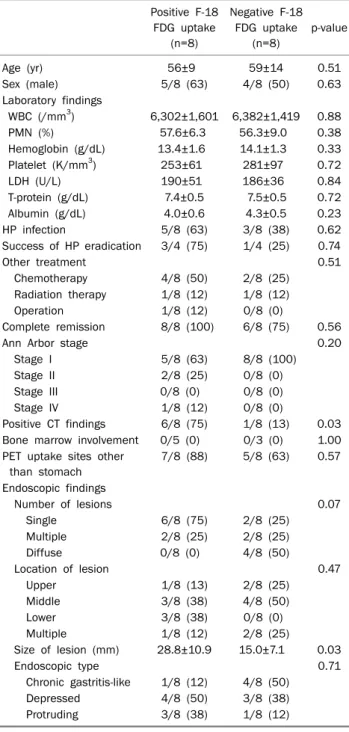
Table 1. Comparison between Patients with Positive F-18 FDG Uptake and Patients with Negative F-18 FDG Uptake
Positive F-18 FDG uptake
(n=8)
Negative F-18 FDG uptake
(n=8)
p-value
Age (yr) 56±9 59±14 0.51
Sex (male) 5/8 (63) 4/8 (50) 0.63
Laboratory findings
WBC (/mm3) 6,302±1,601 6,382±1,419 0.88
PMN (%) 57.6±6.3 56.3±9.0 0.38
Hemoglobin (g/dL) 13.4±1.6 14.1±1.3 0.33
Platelet (K/mm3) 253±61 281±97 0.72
LDH (U/L) 190±51 186±36 0.84
T-protein (g/dL) 7.4±0.5 7.5±0.5 0.72
Albumin (g/dL) 4.0±0.6 4.3±0.5 0.23
HP infection 5/8 (63) 3/8 (38) 0.62
Success of HP eradication 3/4 (75) 1/4 (25) 0.74
Other treatment 0.51
Chemotherapy 4/8 (50) 2/8 (25)
Radiation therapy 1/8 (12) 1/8 (12)
Operation 1/8 (12) 0/8 (0)
Complete remission 8/8 (100) 6/8 (75) 0.56
Ann Arbor stage 0.20
Stage I 5/8 (63) 8/8 (100)
Stage II 2/8 (25) 0/8 (0)
Stage III 0/8 (0) 0/8 (0)
Stage IV 1/8 (12) 0/8 (0)
Positive CT findings 6/8 (75) 1/8 (13) 0.03 Bone marrow involvement 0/5 (0) 0/3 (0) 1.00 PET uptake sites other
than stomach
7/8 (88) 5/8 (63) 0.57
Endoscopic findings
Number of lesions 0.07
Single 6/8 (75) 2/8 (25)
Multiple 2/8 (25) 2/8 (25)
Diffuse 0/8 (0) 4/8 (50)
Location of lesion 0.47
Upper 1/8 (13) 2/8 (25)
Middle 3/8 (38) 4/8 (50)
Lower 3/8 (38) 0/8 (0)
Multiple 1/8 (12) 2/8 (25)
Size of lesion (mm) 28.8±10.9 15.0±7.1 0.03
Endoscopic type 0.71
Chronic gastritis-like 1/8 (12) 4/8 (50)
Depressed 4/8 (50) 3/8 (38)
Protruding 3/8 (38) 1/8 (12)
Values are presented as mean±SD or n (%).
F-18 FDG, fluorine-18 fluorodeoxyglucose; WBC, white blood cell;
PMN, polymorphonuclear neutrophil; HP, Helicobacter pylori.
Table 2. Endoscopic Classification of Gastric MALT Lymphoma and F-18 FDG Uptake Patterns
Endoscopic classification Subtype Positive Chronic gastritis-like
tumors (n=5)
Atrophic type (n=4) 1 Nodular arthritis type (n=1) 0 Depressed tumors (n=7) Erosive-ulcerative type (n=4) 4 Discolored-scar type (n=3) 0 Protruding tumors (n=4) Thickened-fold type (n=3) 2 Single-protrusion type (n=1) 1 Multiple-protrusion type (n=0) 0 MALT, mucosa-associated lymphoid tissue; F-18 FDG, fluorine-18 fluorodeoxyglucose.
However, there have been only a few studies investigating the clinical utility of F-18 FDG PET for gastric MALT lymphoma.
This study’s value is in its use of F-18 FDG PET/CT rather than just PET to accurately locate the lesions discovered by endos- copy on a CT image and evaluate them. Of the 16 patients confirmed to have gastric MALT lymphoma, eight patients
(50%) had lesions that had higher FDG uptake compared to their normal physiological gastric activity, similar to the pos- itivity (62.5%, 10/16) reported by Hirose et al.22
According to the findings of this study, a CT scan showed more positive findings for lesions among the group with pos- itive F-18 FDG uptake than the group with negative F-18 FDG uptake. Moreover, the size of gastric MALT lymphoma was significantly larger among the group with positive F-18 FDG uptake. However, two patients were positive on FDG PET/CT scans, although no lesion was discovered on a CT scan (one protruding tumor, one depressed tumor in endoscopic find- ing). This indicates that not only the size of a lesion but the metabolic activity affects a positive FDG PET/CT finding.
Therefore, a large population study is needed to investigate the role of F-18 FDG PET/CT for the prognostic stratification of gastric MALT lymphoma.
Hirose et al.22 reported the benefit of F-18 FDG PET in find- ing the protruding type of gastric MALT lymphoma in partic- ular after comparing gastric MALT lymphoma between 16 pa- tients and 16 controls, despite some overlap between the pa- tients’ FDG uptake patterns and the control group’s FDG up- take patterns. According to the authors, 50% of patients found to have chronic gastritis-like tumors using endoscopy had positive F-18 FDG uptake, 40% of patients with de- pressed tumors had positive F-18 FDG uptake and 100% of patients with protruding tumors had positive F-18 FDG uptake. In the present study, one of five patients (20%) with chronic gastritis-like tumors, four of seven patients (57%) with depressed tumors, and three of four patients (75%) with protruding tumors had positive F-18 FDG uptake. This con- firmed earlier findings that F-18 FDG PET scan is more effec- tive in diagnosing protruding tumors than in diagnosing chron- ic gastritis-like tumors or depressed tumors. Hirose et al.22
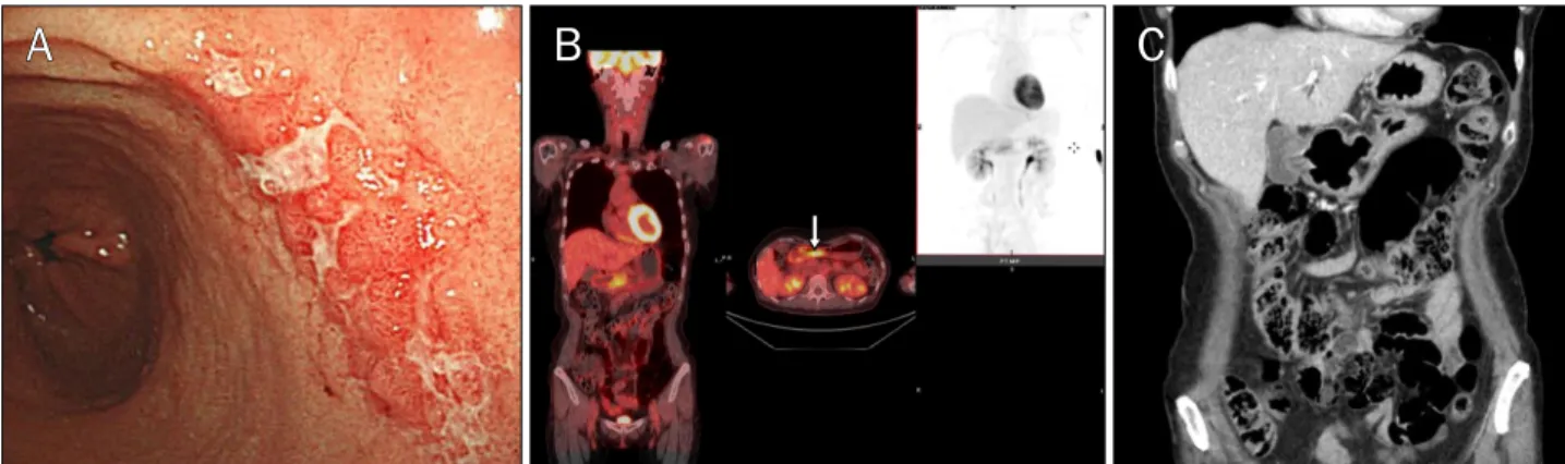
Fig. 2. This case shows the fluorine-18 fluorodeoxyglucose (F-18 FDG) uptake in an erosive-ulcerative type of gastric mucosa-associated lymphoid tissue lymphoma. (A) A 56-year-old female patient had an erosive-ulcerative lesion on the antral posterior wall of the stomach. (B) Maximum standardized uptake value of F-18 FDG uptake (arrow) was 6.2. (C) Abdomen CT shows irregular wall thickening at the gastric antrum.
Fig. 3. This case shows no fluorine-18 fluorodeoxyglucose (F-18 FDG) uptake in a discolored-scar type of gastric mucosa-associated lymphoid tissue lymphoma. (A) A 53-year-old male patient had a discolored-scar lesion on the greater curvature in lower body of the stomach. (B) F-18 FDG PET revealed no significant F-18 FDG uptake in the stomach. (C) Abdomen CT shows no wall thickening at the gastric body.
found that F-18 FDG uptake was positive in nodular antritis type (100%, 1/1), a type of chronic gastritis-like tumors and in 50% (2/4) of the erosive-ulcerative type, a type of the de- pressed tumors. In this study, positive F-18 FDG uptake was not observed in nodular antritis type but in 100% (4/4) of the erosive-ulcerative type. This finding confirmed that a PET scan would be useful in determining the stages of gastric MALT lym- phoma with erosive-ulcerative type tumors. Discolored-scar type, a form of the depressed tumors, did not have positive F-18 FDG uptake in this study or in the study by Hirose et al.
Although the number of lesions did not differ significantly, there was positive F-18 FDG uptake when there was a single lesion and negative F-18 FDG uptake when lesions were diffuse. There needs to be a large population study in the fu- ture in order to investigate F-18 FDG uptake patterns of endo- scopic macroscopic findings in gastric MALT lymphoma.
To allow for the endoscopist’s possible subjectivity when
evaluating gastric lesions, additional indices such as the as- sociation between EUS findings and F-18 FDG uptake pattern in gastric MALT lymphoma can be used. However, among 16 patients of this study, two subjects with F-18 FDG uptake16 and two subjects without F-18 FDG uptake had taken EUS.
The insufficient number of the subjects did not allow stat- istical testing of EUS differences.
In this study, three of eight patients who had positive F-18 FDG uptake on a PET/CT scan before gastric MALT lymphoma treatment had PET/CT scans after the treatment. Because all of them showed negative FDG uptake in the PET/CT scan af- ter the treatment, the F-18 FDG PET/CT is thought to be effec- tive in determining the treatment effects on gastric MALT lym- phoma patients who have positive F-18 FDG uptake on PET/CT scans. Perry et al.16 reported that nine of 12 patients had a positive PET/CT scan initially at diagnosis and achieved a complete response after therapy, according to follow-up
scan. Beal et al.17 reported that eight patients obtained post-treatment FDG-PET scans. In five of those eight, the re- peated FDG-PET scan indicated a complete response, and in three there was an indeterminate or mixed response.
However, additional research with more subjects is needed.
There are a few limitations of the study. Firstly, the re- search findings cannot be generalized due to the small scale of the research with a small number of research subjects at a single institution. Secondly, there is bias in selecting the subjects as the data were collected retrospectively from pa- tients’ medical charts and radiological diagnoses. To over- come such limitations, research involving more institutions and prospective approach is needed.
In conclusion, when gastric lesions are found on abdomi- nal CT scans in patients with gastric MALT lymphoma, when the lesions are large by endoscopy, and their macroscopic ap- pearance shows they are either the protruding tumors or ero- sive-ulcerative type, the lesions are likely to have positive F-18 FDG uptake. Therefore, PET/CT is potentially useful in diagnosing the patients with these lesions and evaluating the effects of their treatment in gastric MALT lymphoma.
REFERENCES
1. Radan L, Fischer D, Bar-Shalom R, et al. FDG avidity and PET/CT patterns in primary gastric lymphoma. Eur J Nucl Med Mol Imaging 2008;35:1424-1430.
2. Radaszkiewicz T, Dragosics B, Bauer P. Gastrointestinal malig- nant lymphomas of the mucosa-associated lymphoid tissue: fac- tors relevant to prognosis. Gastroenterology 1992;102:
1628-1638.
3. Taal BG, Burgers JM, van Heerde P, Hart AA, Somers R. The clin- ical spectrum and treatment of primary non-Hodgkin's lympho- ma of the stomach. Ann Oncol 1993;4:839-846.
4. Taal BG, Boot H, van Heerde P, de Jong D, Hart AA, Burgers JM.
Primary non-Hodgkin lymphoma of the stomach: endoscopic pattern and prognosis in low versus high grade malignancy in re- lation to the MALT concept. Gut 1996;39:556-561.
5. Fung CY, Grossbard ML, Linggood RM, et al. Mucosa-associated lymphoid tissue lymphoma of the stomach: long term outcome after local treatment. Cancer 1999;85:9-17.
6. Toyoda H, Ono T, Kiyose M, et al. Gastric mucosa-associated lym- phoid tissue lymphoma with a focal high-grade component diag- nosed by EUS and endoscopic mucosal resection for histologic evaluation. Gastrointest Endosc 2000;51:752-755.
7. Queneau PE, Helg C, Brundler MA, et al. Diagnosis of a gastric mucosa-associated lymphoid tissue lymphoma by endoscopic
ultrasonography-guided biopsies in a patient with a parotid gland localization. Scand J Gastroenterol 2002;37:493-496.
8. Fujishima H, Misawa T, Maruoka A, Chijiiwa Y, Sakai K, Nawata H. Staging and follow-up of primary gastric lymphoma by endo- scopic ultrasonography. Am J Gastroenterol 1991;86:719-724.
9. Lévy M, Hammel P, Lamarque D, et al. Endoscopic ultra- sonography for the initial staging and follow-up in patients with low-grade gastric lymphoma of mucosa-associated lymphoid tis- sue treated medically. Gastrointest Endosc 1997;46:328-333.
10. Grau E, Gomez A, Cuñat A, Oltra C. Computed tomography in stag- ing of primary gastric lymphoma. Lancet 1996;347:1261.
11. Delbeke D, Martin WH, Morgan DS, et al. 2-deoxy-2-[F-18]fluoro- D-glucose imaging with positron emission tomography for initial staging of Hodgkin's disease and lymphoma. Mol Imaging Biol 2002;4:105-114.
12. Kostakoglu L, Coleman M, Leonard JP, Kuji I, Zoe H, Goldsmith SJ. PET predicts prognosis after 1 cycle of chemotherapy in ag- gressive lymphoma and Hodgkin's disease. J Nucl Med 2002;
43:1018-1027.
13. Spaepen K, Stroobants S, Dupont P, et al. Early restaging posi- tron emission tomography with (18)F-fluorodeoxyglucose pre- dicts outcome in patients with aggressive non-Hodgkin's lymphoma. Ann Oncol 2002;13:1356-1363.
14. Guay C, Lépine M, Verreault J, Bénard F. Prognostic value of PET using 18F-FDG in Hodgkin's disease for posttreatment evalua- tion. J Nucl Med 2003;44:1225-1231.
15. Ambrosini V, Rubello D, Castellucci P, et al. Diagnostic role of 18F-FDG PET in gastric MALT lymphoma. Nucl Med Rev Cent East Eur 2006;9:37-40.
16. Perry C, Herishanu Y, Metzer U, et al. Diagnostic accuracy of PET/CT in patients with extranodal marginal zone MALT lymphoma. Eur J Haematol 2007;79:205-209.
17. Beal KP, Yeung HW, Yahalom J. FDG-PET scanning for detection and staging of extranodal marginal zone lymphomas of the MALT type: a report of 42 cases. Ann Oncol 2005;16:473-480.
18. Hoffmann M1, Wöhrer S, Becherer A, et al. 18F-Fluoro-de- oxy-glucose positron emission tomography in lymphoma of mu- cosa-associated lymphoid tissue: histology makes the difference. Ann Oncol 2006;17:1761-1765.
19. Alinari L, Castellucci P, Elstrom R, et al. 18F-FDG PET in muco- sa-associated lymphoid tissue (MALT) lymphoma. Leuk Lymphoma 2006;47:2096-2101.
20. Enomoto K, Hamada K, Inohara H, et al. Mucosa-associated lym- phoid tissue lymphoma studied with FDG-PET: a comparison with CT and endoscopic findings. Ann Nucl Med 2008;22:
261-267.
21. Yi JH, Kim SJ, Choi JY, Ko YH, Kim BT, Kim WS. 18F-FDG uptake and its clinical relevance in primary gastric lymphoma. Hematol Oncol 2010;28:57-61.
22. Hirose Y, Kaida H, Ishibashi M, et al. Comparison between endo- scopic macroscopic classification and F-18 FDG PET findings in gastric mucosa-associated lymphoid tissue lymphoma patients.
Clin Nucl Med 2012;37:152-157.