INTRODUCTION
The extraocular muscle (EOM) is the most frequent site for the involvement of thyroid orbitopathy, followed in order by in- ferior, medial, superior, lateral rectus, and oblique muscles. The EOM is the rarest site for the involvement of extranodal mar- ginal zone B-cell lymphoma of mucosa-associated lymphoid tis- sue (MALT lymphoma) in the orbit. Several cases of MALT
lymphoma involving the EOM have appeared in the English lit- erature, and most of these are in the form of case reports (1-5).
The imaging findings of MALT lymphoma involving the EOM are limited to those of a single imaging modality only (1-3). As such, imaging features of this rare entity at CT, MRI, and 18F-flu- orodeoxyglucose positron emission tomography/CT (18F-FDG PET/CT) altogether in a single case have not appeared in the lit- erature.
J Korean Soc Radiol 2013;68(6):453-457 http://dx.doi.org/10.3348/jksr.2013.68.6.453
Received March 1, 2013; Accepted April 11, 2013 Corresponding author: Sang Kwon Lee, MD Department of Radiology, Dongsan Medical Center, Keimyung University School of Medicine, 56 Dalseong-ro, Jung-gu, Daegu 700-712, Korea.
Tel. 82-53-250-7735 Fax. 82-53-250-7766 E-mail: [email protected]
This is an Open Access article distributed under the terms of the Creative Commons Attribution Non-Commercial License (http://creativecommons.org/licenses/by-nc/3.0) which permits unrestricted non-commercial use, distri- bution, and reproduction in any medium, provided the original work is properly cited.
We report a case of mucosa-associated lymphoid tissue (MALT) lymphoma involving the medial rectus muscle in a 47-year-old man along with CT, MRI, 18F-fluorodeoxy- glucose positron emission tomography/CT (18F-FDG PET/CT), and pathologic features.
The lesion was manifested as a fusiform enlargement isolated to the right medial rec- tus muscle with involvement of its tendinous insertion. The lesion was isoattenuating to the brain on non-enhanced CT images, showing as isointense to gray matter on fast spin echo T1- and T2-weighted images with fat saturation, and showed homoge- neous enhancement on contrast-enhanced CT and MR images. The maximum stan- dardized uptake value on 18F-FDG PET/CT was 4.9 g/mL. The results of histological and immunohistochemical examinations of the specimen obtained by biopsy of the right medial rectus muscle were consistent with MALT lymphoma. It should be noted that the extraocular muscle (EOM) is a rare location for the involvement of MALT lympho- ma, and MALT lymphoma of the EOM may mimic thyroid orbitopathy.
Index terms
Lymphoma, B-Cell, Marginal Zone Eye Diseases
Tomography, X-Ray Computed Magnetic Resonance Imaging
Positron-Emission Tomography and Computed Tomography
CT, Magnetic Resonance, and
18F-Fluorodeoxyglucose Positron Emission Tomography/CT Imaging Features of Mucosa-Associated Lymphoid Tissue Lymphoma Involving Medial Rectus Muscle: A Case Report
1내직근을 침범한 점막 연관성 림프조직 림프종의 CT, MRI 및
18F-Fluorodeoxyglucose Positron Emission Tomography/CT 영상소견:
증례 보고1
Sang Kwon Lee, MD
1, Mi Sun Choe, MD
2Departments of 1Radiology, 2Pathology, Dongsan Medical Center, Keimyung University School of Medicine, Daegu, Korea
volvement were documented in the bone marrow biopsy. Frac- tionated external beam irradiation of a total dose of 3600 cGy was applied to the right orbit. Near complete resolution of the MALT lymphoma of the right medial rectus muscle was achieved at follow-up MRI 230 days after the initial study (Fig.
1I). The patient underwent endoscopic biopsy for a raised, ero- sive mucosal lesion of the stomach 250 days after initial diagno- sis of orbital MALT lymphoma, which revealed gastric MALT lymphoma. He underwent additional curative fractionated ex- ternal beam radiation therapy at a dose of 3060 cGy targeted to the stomach. Follow-up endoscopic examination with biopsy, performed 133 days after initial diagnosis of gastric MALT lym- phoma, demonstrated complete remission. No evidence of re- current disease was observed in the orbit, stomach, or elsewhere until 13 months after complete remission.
DISCUSSION
Thyroid orbitopathy is the most common cause of abnormal enlargement of the EOM. Enlargement of the EOM associated with thyroid orbitopathy is bilateral and symmetric in 70% of cases. The inferior rectus muscle is most frequently affected, and the muscle enlargement is characteristically fusiform with spar- ing of tendinous insertion. The most common nonthyroidal causes of EOM enlargement are inflammatory and vascular dis- orders, and neoplastic diseases (6). MALT lymphoma, an extra- nodal, monoclonal, small B-cell proliferation, is an indolent dis- ease that usually presents as a localized extranodal tumor.
MALT lymphoma arises from various mucosal and nonmuco- sal tissues. It most commonly occurs in the stomach, but can in- volve the orbital/ocular adnexa, salivary glands, and lungs (7).
Orbital MALT lymphoma most frequently involves the con- junctiva (51%), while the EOM is the rarest site of involvement (5%) (8). Several cases of MALT lymphoma involving the EOM have been presented in the English literature in the form of case reports or clinical studies (1-5). Of them, a few cases have pre- sented imaging features (1-3). However, none have described im- aging findings in detail. Rossman et al. (1) reported a case of an enlarged medial rectus muscle caused by MALT lymphoma that was misdiagnosed as thyroid orbitopathy for over 3 years, even tures at CT, MRI, and 18F-FDG PET/CT.
CASE REPORT
A 47-year-old man presented with a chief complaint of right ocular discomfort lasting for 1 year. He had not experienced oc- ular pain or diplopia. His past history was unremarkable. The ophthalmologic examination disclosed proptosis of the right eye, but the motion of the EOM was preserved. The laboratory tests, including blood cell count, biochemistry, and thyroid hor- mones, were normal. CT, performed by using a SOMATOM Definition Flash scanner (Siemens Healthcare, Forchheim, Ger- many) with a slice thickness of 3.0 mm revealed a fusiform en- largement isolated to the right medial rectus muscle, appearing as isoattenuating compared with the brain on non-enhanced CT images, and showed homogeneous, moderate enhancement on contrast-enhanced CT images, with involvement of the ten- dinous insertion (Fig. 1A, B). Orbital MRI was performed for better characterization of the lesion and for better assessment of the extent by using a 3.0-T unit (Signa Excite; GE Medical Sys- tem, Milwaukee, WI, USA). The enlarged medial rectus muscle was isointense to the gray matter on fast spin echo (FSE) T1- weighted images (T1WI) [repetition time (TR)/echo time (TE) = 650.0/13.6; echo train length (ETL) = 3; field of view (FOV) = 160
× 160; matrix number/number of excitation = 320 × 192/2; slice thickness = 3.0 mm] with fat saturation (Fig. 1C) and FSE T2- weighted images (T2WI) (TR/TE = 5000.0/111.9; ETL = 20;
FOV = 160 × 160; matrix number/number of excitation = 384 × 224/2; slice thickness = 3.0 mm) with fat saturation (Fig. 1D), and showed homogenous intense enhancement on gadolinium (Gd)-enhanced FSE T1WI with fat saturation (Fig. 1E). 18F- FDG PET/CT, performed by using Discovery STE (GE Health- care, Milwaukee, WI, USA), demonstrated increased FDG up- take [maximum standardized uptake value (SUVmax) = 4.9 g/
mL] in the right medial rectus muscle with fusiform enlarge- ment (Fig. 1F). No abnormally increased FDG uptake was not- ed in other parts of the body. In view of clinical and imaging findings, our tentative diagnosis was lymphoma and euthyroid Graves’ ophthalmopathy. The patient underwent biopsy of the right medial rectus muscle. The histological and immunohisto-
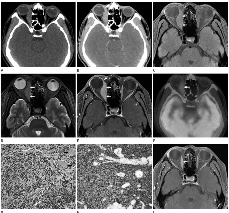
Fig. 1. CT, MRI, 18F-FDG PET/CT, and pathologic features of MALT lymphoma involving the medial rectus muscle in a 47-year-old man.
A. A non-enhanced CT image shows fusiform enlargement isolated to the right medial rectus muscle (arrows), which is isoattenuating compared with brain.
B. A contrast-enhanced CT image reveals homogeneous, moderate enhancement of the enlarged muscle (arrows) with involvement of its tendi- nous insertion (arrowhead).
C, D. Fast spin echo T1- (C) and T2-weighted MR images (D) with fat saturation show fusiform enlargement of the right medial rectus muscle (arrows), which is isointense to the gray matter.
E. A gadolinium-enhanced T1-weighted MR image with fat saturation demonstrates homogeneous intense enhancement of the enlarged muscle (arrows) with involvement of its tendinous insertion (arrowhead).
F. A 18F-FDG PET/CT image reveals moderate FDG uptake (SUVmax = 4.9 g/mL) within the enlarged right medial rectus muscle (arrow).
G. A photomicrograph of histological examination of the specimen obtained by biopsy of the right medial rectus muscle demonstrates diffuse infiltration of relatively small monomorphic atypical lymphoid cells which infiltrate into the skeletal muscle (arrows) (H&E stain, × 400).
H. A photomicrograph of immunohistochemical staining demonstrates diffuse strong positivity for CD20 (original magnification, × 400).
I. A follow-up gadolinium-enhanced MR image 230 days after initial MR study reveals near complete resolution of MALT lymphoma of the right medial rectus muscle (arrow).
Note.-MALT = mucosa-associated lymphoid tissue, SUVmax = maximum standardized uptake value, 18F-FDG PET = 18F-fluorodeoxyglucose posi- tron emission tomography
D
G A
E
H B
F
I C
REFERENCES
1. Rossman D, Michel R, Codere F. A case of an enlarged me- dial rectus muscle. Int Ophthalmol 2009;29:319-321 2. Byard SD, Chowdhury HR, Lee RM, Hyer J, Hart-George AL.
Unilateral isolated extraocular muscle lymphoma. BMJ Case Rep 2012;2012:bcr0220125750
3. Benetatos L, Alymara V, Asproudis I, Bourantas KL. Ritux- imab as first line treatment for MALT lymphoma of extra- ocular muscles. Ann Hematol 2006;85:625-626
4. Abalo-Lojo JM, Baleato-Gonzalez S, Abdulkader I, Gonza- lez F. Extraocular muscle involvement in MALT lympho- mas. Orbit 2011;30:186-188
5. Izambart C, Robert PY, Petellat F, Petit B, Gastaud P, Lagier J, et al. Extraocular muscle involvement in marginal zone B-cell lymphomas of the orbit. Orbit 2008;27:345-349 6. Lacey B, Chang W, Rootman J. Nonthyroid causes of ex-
traocular muscle disease. Surv Ophthalmol 1999;44:187- 213
7. Raderer M, Streubel B, Woehrer S, Puespoek A, Jaeger U, Formanek M, et al. High relapse rate in patients with MALT lymphoma warrants lifelong follow-up. Clin Cancer Res 2005;11:3349-3352
8. Lee JL, Kim MK, Lee KH, Hyun MS, Chung HS, Kim DS, et al. Extranodal marginal zone B-cell lymphomas of muco- sa-associated lymphoid tissue-type of the orbit and ocular adnexa. Ann Hematol 2005;84:13-18
9. Zhao K, Luo YZ, Zhou SH, Dai BL, Luo XM, Yan SX, et al. 18F- fluorodeoxyglucose positron emission tomography/com- puted tomography findings in mucosa-associated lymphoid tissue lymphoma of the larynx: a case report and literature review. J Int Med Res 2012;40:1192-1206
10. Pfeffer MR, Rabin T, Tsvang L, Goffman J, Rosen N, Symon Z. Orbital lymphoma: is it necessary to treat the entire or- bit? Int J Radiat Oncol Biol Phys 2004;60:527-530 placement of the globe. Byard et al. (2) reported a case of MALT
lymphoma isolated to a single extraocular muscle, presenting as an approximately 30 × 15-mm-sized mass arising from the re- gion of the left superior rectus muscle with no signs of bone ero- sion. Benetatos et al. (3) reported a case of MALT lymphoma in which Gd-enhanced MR imaging revealed tumefaction of the inferior rectus muscle with subsequent decrease of tumor extent on follow-up study 6 months after rituximab treatment.
Our case represents the first case in which imaging findings at CT, MRI, and 18F-FDG PET/CT were presented together in a single case of this rare entity. In our case, the medial rectus mus- cle which involved the MALT lymphoma was manifested as a fusiform enlargement of the muscle on CT and MRI, which re- sembled findings of thyroid orbitopathy. However, the differen- tiation from thyroid orbitopathy was established by involvement of the tendinous insertion in MALT lymphoma of our case. 18F- FDG PET/CT findings of MALT lymphoma involving the EOM have not appeared in the English literature. The SUVmax of MALT lymphoma involving the medial rectus in our case was 4.9 g/
mL, which was similar to that of MALT lymphoma elsewhere (9). We hypothesized that the low FDG uptake (SUVmax = 4.9 g/
mL) of MALT lymphoma involving the medial rectus muscle in our case might be related to the indolent course of this entity.
Although MALT lymphomas respond well to surgical remov- al, radiotherapy is the treatment of choice for localized disease in the orbit/ocular adnexa. A dose of 30-35 Gy has been report- ed to be sufficient to provide local control and cure of the dis- ease localized to the orbit/ocular adnexa and is associated with excellent survival (10).
In summary, it should be noted that the EOM is a rare location for the involvement of MALT lymphoma, and fusiform enlarge- ment of EOM in MALT lymphoma may mimic that of thyroid orbitopathy on CT or MRI. The differentiation can be established by the fact that the tendinous insertion of EOM is involved in MALT lymphoma, which is not the case in the thyroid orbitopa- thy. In cases with solitary muscle enlargement without features
내직근을 침범한 점막 연관성 림프조직 림프종의 CT, MRI 및
18F-Fluorodeoxyglucose Positron Emission Tomography/CT 영상소견: 증례 보고1
이상권
1· 최미선
2저자들은 47세 남자의 내직근을 침범한 점막 연관성 림프조직(mucosa-associated lymphoid tissue; MALT) 림프종 1예 를 CT, MRI, 18F-fluorodeoxyglucose positron emission tomography/CT (18F-FDG PET/CT) 및 병리학적 소견과 함께 보고하고자 한다. 병변은 힘줄부착부위의 침범을 동반한 우측 내직근 단독의 방추형 종대로 발현하였으며, 비조영증강 CT 영상에서 뇌와 비슷한 감쇠를, 지방포화 고속스핀에코 T1- 및 T2-강조영상에서 회질과 등신호강도를 보였으며, 조 영증강 CT 및 MR 영상에서 균질한 조영증강을 보였다. 18F-FDG PET/CT에서 maximum standardized uptake value는 4.9 g/mL였다. 우측 내직근의 생검에 의한 검체의 조직학적 및 면역조직화학적 검사의 결과는 MALT 림프종과 일치하였 다. 외안근은 MALT 림프종에 의해 드물게 침범될 수 있으며, 외안근의 MALT 림프종은 갑상선 안병증을 닮을 수 있다.
계명대학교 의과대학 동산의료원 1영상의학과, 2병리과