Donor Specific Antibody Negative Antibody-Mediated Rejection after ABO Incompatible Liver Transplantation
Boram Lee, M.D.
1, Soomin Ahn, M.D.
2, Haeryoung Kim, Ph.D.
3, Ho-Seong Han, Ph.D.
1, Yoo-Seok Yoon, Ph.D.
1, Jai Young Cho, Ph.D.
1and Young Rok Choi, M.D.
1Departments of Surgery
1and Pathology
2, Seoul National University Bundang Hospital, Seoul National University College of Medicine, Seongnam, Department of Pathology, Seoul National University Hospital, Seoul National
University College of Medicine
3, Seoul, Korea
Antibody-mediated rejection (AMR) is a major complication after ABO-incompatible liver transplantation. According to the 2016 Banff Working Group on Liver Allograft Criteria for the diagnosis of acute AMR, a positive serum donor specific antibody (DSA) is needed. On the other hand, the clinical significance of the histological findings of AMR in the absence of DSA is unclear. This paper describes a 57-year-old man (blood type, O+) who suffered from hepatitis B virus cirrhosis with hepatocellular carcinoma.
Pre-operative DSA and cross-matching were negative. After transplantation, despite the improvement of the liver function, acute AMR was observed in the protocol biopsy on postoperative day 7; the cluster of differentiation 19+ (CD19+) count was 0% and anti-ABO antibody titers were 1:2. This paper presents the allograft injury like AMR in the absence of DSA after ABOi living donor liver transplantation with low titers of anti-ABO antibody and depleted serum CD19+ B cells.
Key Words: Liver transplantation, Antibody-dependent cell cytotoxicity, Rejection, Living donors, HLA antigens
중심 단어: 간이식, 항체의존성세포매개성세포독성, 거부반응, 생체공여자, 인간백혈구항원Received July 6, 2018 Revised September 6, 2018 Accepted September 13, 2018
Corresponding author: Young Rok Choi
Department of Surgery, Seoul National University Bundang Hospital, Seoul National University College of Medicine, 82 Gumi-ro 173beon-gil, Bundang-gu, Seongnam 13620, Korea
Tel: 82-31-787-7111, Fax: 82-31-787-4055 E-mail: [email protected]
INTRODUCTION
After the first liver allo-transplantation was performed in the 1960s, the liver was considered an “immune tolerogenic organ”(1). However, in 1989, Gugenheim et al.(2) reported allograft rejection in ABO-incompatible liver transplantation (ABOi-LT). In a total of 17 ABOi-LT recipients, six showed graft failure due to impaired immune responses and anti- body-mediated rejection (AMR). To avoid AMR, various desensitization methods have been developed. The protocol for ABOi-LT consists of pre-operative intravenous ritux-
imab (RTX), multiple session of plasmapheresis (PP), local infusion therapy, or splenectomy. After administration of RTX, an anti-cluster of differentiation 20 (CD20) mono- clonal antibody, in ABOi-LT, Usuda et al.(3) showed that the incidence of AMR in ABOi-LT was markedly reduced;
indeed, recent outcomes were found to be comparable to those of ABO-compatible-LT(3,4). However, AMR is still a major complication after ABOi-LT. According to the 2016 Banff Working Group on Liver Allograft Criteria for the diagnosis of acute AMR requires the positive serum donor specific antibody (DSA)(5). However, clinical significance of histological findings of AMR in the absence of DSA is unclear. Here, we present the case of allograft injury like AMR under the absence of DSA after ABOi-LT with the low titers of anti-ABO antibody and depleted serum CD19+
B cells.
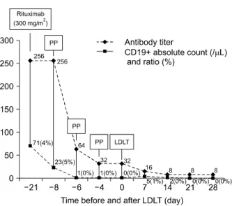
Fig. 1. Desensitization protocol and changes in the cluster of differentiation 19+ (CD19+) lymphocyte count and the titer of anti ABO antibody. The titers of anti-ABO antibodies decreased to 1:8 from 1:256, and serum CD19+ B cells were depleted before the operation. Abbreviations: PP, plasmapheresis; LDLT, living donor liver transplantation.
CASE REPORT
A 57-year-old man (O, Rh+) suffered from hepatitis B virus-related liver cirrhosis and hepatocellular carcinoma (Child-Pugh score and the model for end-stage liver disease [MELD] score were 7 and 9). His wife was the only poten- tial living donor (A, Rh+). The patient received RTX (300 mg/m2) 3 weeks before ABOi-LT and then underwent three sessions of PP. Pre-operative DSA and cross-matching were negative. The titers of anti-ABO antibodies decreased to 1:8 from 1:256, and serum CD19+ B cells were depleted before the operation (Fig. 1). There were no specific findings dur- ing the operation. Immunosuppression after transplantation consisted of tacrolimus (target level: 8 to 12 ng/mL), my- ophenoate mofetil (1,000 mg/day), and steroids. Although the laboratory data on the second post-operative day showed a slight abnormality in liver function; those values gradually decreased (Fig. 2). At 7 days postoperatively, there were no specific findings on computed tomography.
The protocol biopsy on postoperative day (POD) 7 showed mild portal inflammation with endotheliitis, and comple- ment 4d (C4d) positivity was also observed (2016 Banff Criteria H-score 1, C4d score 2) (Fig. 3A). Clinically, the recipient’s hepatic enzyme and condition were improved,
and follow-up liver biopsy was performed without any re- jection treatment. Follow-up biopsy on POD 14 (Fig. 3B) showed similar histologic feature to the previous biopsy (H-score 1, C4d score 2). The DSA test was negative at that time; however, because of persistent histopathological changes, we decided to start the rejection therapy. Three sessions of PP were performed, and a low dose of intra- venous immunoglobulin (0.8 g/kg) was administered for 3 days. Follow-up biopsy on POD 21 (Fig. 3C) revealed ag- gravated degree of portal inflammation and endotheliitis (H-score 2, C4d score 2). We performed additional steroid pulse therapy (500 mg hydrocortisone for 3 days, followed by tapering). The biopsy on POD 28 (Fig. 3D) showed im- provement (H-score 1, C4d score 0). After confirming that pathological symptoms were alleviated, the recipient was discharged. The follow-up biopsy on POD 9 months (Fig.
3E), the histological features showed that no inflammatory changes in the portal tract and the absence of portal endo- thelial and stromal C4d deposition. However, 1 year after, a weak positive finding was observed in the DSA test, sug- gesting
de novo
DSA production.Written informed consent was obtained from the patients for publication of this case report and any accompanying images.
DISCUSSION
AMR is a relatively uncommon complication of ABOi liv- ing donor liver transplantation, but can be fatal and result in graft failure. AMR occurs as primary AMR, which devel- ops in a recipient with preformed anti-ABO antibodies, or as secondary AMR, in which antibodies develop
de novo
following LT, resulting in the pathogenesis of acute and chronic liver allograft rejection(6). In our case, asympto- matic acute AMR occurred at 1 week after transplantation.Moreover, acute AMR developed despite the very low titers of anti-ABO antibodies and the presence of 0% serum CD19+ B cells with no DSA.
A recent study reported that high pre-operative antibody titers did not have a significant effect on AMR. Instead, it is important to prevent new antibody production after trans- plantation(7). However, in general, desensitization protocols have been directed toward the elimination of anti-ABO an-
Fig. 2. Changes in aspartate aminotransferase (AST)/alanine aminotransferase (ALT), total bilirubin, prothrombin time (INR), and tacrolimus (TAC) level after living donor liver transplantation (LDLT). We performed liver biopsy on postoperative day (POD) 7, 14, 21, and 28. After pathologic confirm the acute antibody-mediated rejection, we conducted rejection therapy (POD 16 to 20, intravenous immunoglobulin [IVIg, 0.8 g/kg] for 3 days and plasmapheresis [PP]; POD 23 to 25, steroid pulse therapy [500 mg of hydrocortisone 500 mg for 3 days then tapering]). Abbreviation: MMF, myophenoate mofetil.
tibody titers and suppression of B cell activity before and after transplantation. In our case, acute AMR occurred de- spite the fact that the patient showed low serum anti-ABO antibody titers during pre- and post-transplantation. In addi- tion, there were no increases in anti-ABO antibody titers, CD19+ lymphocyte counts, or hepatic enzyme levels at the time of diagnosis of acute AMR. Thus, the clinical course of our patient differed from that of other AMR cases after ABOi-LT.
Recent studies have evaluated the effects of DSA on short- and long-term prognosis. In the past, preformed DSA was generally considered to be clinically unrelated to liver allograft outcomes(8-11). However, recent studies have confirmed inferior clinical outcomes in some but not all
DSA-positive patients(12). For this reason, many trans- plantation centers have performed cross-matching tests with DSA before transplantation to reduce the possibility of rejection. In our patient, the status of DSA before and after transplantation was negative.
Because the factors affecting the development of AMR are unclear, we performed additional analyses to identify other risk factors. There are several reports regarding the role of MHC class I related chain A (MICA) antigens in solid organ transplantation. We conducted an additional test for antibodies against MICA antigens. The results showed that the recipient’s serum had no antibodies against MICA alleles. MICA molecules are glycoproteins that are expressed on the cellular membrane. The test for crossmatch can de-
Fig. 3. Liver histology: (A) postoperative day (POD) 7 (×400), (B) POD 14 (×400), (C) POD 21 (×400), (D) POD 28 (×400), (E) POD 9 months (×400) (HE stain and C4d deposition). (A) POD 7 (2016 Banff Criteria H-score 1, C4d score 2): mild portal infiltration of neutrophils and some eosinophils with the presence of portal endothelial and stromal complement component 4d (C4d) deposition, The pathologist reported that the possibility of acute antibody-mediated rejection (AMR) in ABO-incompatible grafts could not be excluded.
(B) POD 14 (2016 Banff Criteria H-score 1, C4d score 2): portal inflammation involving most of the portal tracts with portal venous endotheliitis with endothelial cell hypertrophy and mixed lymphocytes, neutrophils and eosiophils were as observed on postoperative day 14. Moreover, the immunofluorescence staining showed a linear pattern of C4d staining on the endothelial cell and stroma. Additional donor specific antibody results after transplantation were also negative. (C) POD 21 (2016 Banff Criteria H-score 2, C4d score 2): acute AMR findings were still observed on pathologic examination and aggravated degree of portal inflammation and endotheliitis. (D) POD 28 (2016 Banff Criteria H-score 1, C4d score 0): histological analysis on postoperative day 28 showed that inflammatory cell infiltration was localized in one portal tract and that hepatocellular swelling, a change induced by steroid pulse therapy, was observed in liver allografts.
(E) POD 9 months: the histological features showed that no inflammatory changes in the portal tract and the absence of portal endothelial and stromal C4d deposition.
tect alloantibodies against donor HLA-I and -II antigens, but neither test detects alloantibodies against MICA because the lymphocytes used in the test do not express MICA antigens on their cell surfaces(13). Several clinical studies have shown that MICA antibodies correlate with increased risk of rejection and decreased allograft survival rates following renal or heart transplantation. However, in case of liver transplantation, there are no relationships between AMR and MICA in LT.
We performed in total four liver biopsies once per week for accurate diagnosis and response to treatment. Liver bi- opsy is considered a practical diagnostic tool for identifying liver pathology, despite its possibly of morbid complications.
Alten et al.(14) reported of 703 cases of liver biopsy done for 409 liver transplant patients, complication occurred in 10 cases (1.4%), but were resolved with conservative treatment. They concluded that liver biopsy is a relatively safe and adequate diagnostic tool for liver transplant patients. Thereby we decided to periodically conduct fol- low-up liver biopsies whenever the patient did not follow a typical post-operative course or when we could not rule
out the possibility of rejection.
After considering all the factors, the recipient did not have the factor that caused acute AMR and did not show any clinical manifestations. After thorough examination, the patient did not possess any of the factors nor clinical mani- festations that may have caused acute AMR. Moreover, C4d staining did not show a diffuse intense pattern unlike in typical AMR, and hematoxylin and eosin staining (H&E) showed weak inflammatory findings. In adults, there find- ings can be observed in allograft dysfunction due to biliary obstruction, recurrent hepatitis B virus (HBV) or hepatitis C virus (HCV) and plasma cell hepatitis(5). In addition, combined AMR and T-cell mediated rejection is reported to be relatively common(15); therefore, it needs to clear distinction. However, according to the component lesion scoring for acute AMR in liver allograft(5), the recipient’s pathologic findings corresponded to C4d-score 3 (diffuse [>50% portal tracks] C4d deposition in >50% of the cir- cumference of portal microvascular endothelia) and histo- pathologys score 2 (monocytic, eosinophilic, or neutrophilic microvasculitis/capillaritis, defined as at least 5 to 10 leuko-
cytes marginated and/or intraluminal in the maximally in- volved capillary prominent portal and/or sinusoidal micro- vascular endothelial cell enlargement involving a majority of portal tracts or sinusoids, with variable but noticeable portal capillary and inlet venule dilatation and variable por- tal edema). Because the DSA was negative in pre-, post- transplant tests, the patients belongs to indeterminate for AMR according to Banff Working Group criteria for acute AMR in liver allografts. However, since the pathological changes of the tissues were evident, we treated the patient according to AMR.
Although, clinical significance of DSA (−) AMR is un- clear, it has been shown that the treatment improves histo- pathologic changes in this case. In ABOi-LT, investigations for immediate histologic allograft injury similar to AMR might be needed to know its clinical significance.
CONFLICTS OF INTEREST
No potential conflict of interest relevant to this article was reported.